Volume 2013, Article ID 312590,7pages http://dx.doi.org/10.1155/2013/312590
Research Article
Plasmacytoid Dendritic Cell Response to CpG ODN Correlates
with CXCL16 Expression and Is Inhibited by ox-LDL
Mayda Gursel,
1Dennis M. Klinman,
2and Ihsan Gursel
31Department of Biological Sciences, Middle East Technical University, 06800 Ankara, Turkey
2Cancer and Inflammation Program, National Cancer Institute, Frederick, MD 21702, USA
3Department of Molecular Biology and Genetics, Bilkent University, 06800 Ankara, Turkey
Correspondence should be addressed to Mayda Gursel; [email protected] Received 16 July 2013; Accepted 6 September 2013
Academic Editor: Ishak Tekin
Copyright © 2013 Mayda Gursel et al. This is an open access article distributed under the Creative Commons Attribution License, which permits unrestricted use, distribution, and reproduction in any medium, provided the original work is properly cited. Structurally distinct classes of synthetic CpG oligonucleotides (ODN) differentially activate human immune cells. K-type ODN trigger plasmacytoid dendritic cells (pDCs) to differentiate and produce TNF𝛼. In contrast, D-type ODN stimulate large amounts of IFN𝛼 secretion from pDCs. The cell-surface receptor CXCL16 was previously shown to influence the nature and specificity of CpG ODN-induced immune activation. Here, we evaluated the expression and function of CXCL16 on pDC from healthy volunteers. We report that increased CXCL16 expression correlated with enhanced in vitro response exclusively to D-type CpG ODN. Conversely, enzymatic digestion of the receptor resulted in a decrease in IFN𝛼 production. Moreover, ox-LDL presence significantly inhibited D-ODN mediated IFN𝛼 production by pDCs. Coculture of enriched pDCs with the CXCR6 expressing Jurkat T cells decreased the activation threshold of these cells responding to D-ODN, suggesting that CXCL16/CXCR6 interaction may play an important role in modifying the response of pDCs to environmental danger signals.
1. Introduction
Synthetic oligodeoxynucleotides (ODN) containing unmeth-ylated CpG motifs stimulate an innate immune response characterized by the production of cytokines, chemokines, and Ig by B cells, dendritic cells (DC), NK cells, and
macrophages [1–4]. In humans, these motifs are recognized
by B cells and plasmacytoid dendritic cells (pDC) that express
Toll-like receptor 9 (TLR9) [5,6]. Human PBMC recognize
and respond to structurally distinct classes of CpG motifs [7–
13]. Of these ODN classes, K type (also known as B class)
phosphorothioate ODN express multiple TCGTT and/or TCGTA motifs that stimulate B cells to proliferate and secrete IL-6 and IgM and promote the survival, activation, and
maturation of pDC in the absence of IFN𝛼 production [7,8,
10]. In contrast, D type ODN (also known as A class) contain
a phosphodiester purine/pyrimidine/CG/purine/pyrimidine motif capped at each end by a phosphorothioate polyG tail
are poor stimulators of B cells [8–10]. However, D type
ODN stimulate pDCs to produce high levels of IFN𝛼 [8–
10]. Previous work has established that D but not K type
ODN bind to the chemokine and scavenger receptor CXCL16
expressed on the surface of pDCs [14]. This interaction directs
D type ODN into the recycling endosomal compartment, where TLR9-MyD88-IRF7 signaling pathway is activated,
leading to robust IFN𝛼 production [15]. The 3rd class of CpG
ODN, designated as C-type, contain one or two CpG motifs
with a phosphodiester backbone at the 5end [11,12]. C ODN
also contains a palindromic sequence on a phosphorothioate
backbone at the 3end and can induce proliferation of B cells
and production of low amounts of IFN𝛼 from pDCs [11,12].
CXCL16 functions as a scavenger receptor [16], a
che-mokine [17], and an adhesion [18] molecule, playing a
prominent role in the pathogenesis of atherosclerosis [19]
and psoriasis [20]. CXCL16 binds to the chemokine receptor
CXCR6/Bonzo, expressed on the surface of CD4+ and CD8+
T cells [21]. Soluble to cell-surface expressed CXCL16 is
controlled by the metalloproteinase ADAM10 that actively
cleaves the membrane bound receptor [22]. This
activ-ity can be inhibited by the metalloproteinase inhibitor GM6001, thereby increasing the amount of the cell surface
expressed CXCL16 [14, 22]. Conversely, treatment with o-sialoglycoprotease selectively digests the membrane-bound
CXCL16 [14, 21]. In order to clarify the role of CXCL16
expression on human pDCs during CpG ODN-mediated immune activation, we modified the expression levels of this protein in human peripheral blood mononuclear cells prior to stimulation. Results indicate that preventing the cleavage of membrane-bound CXCL16 increased both the number of pDC expressing CXCL16 and their response to D-ODN. In contrast, digesting the membrane-bound CXCL16 reduced the number of pDC expressing CXCL16 and their response to D-ODN. Interestingly, our data indicated that circulating ox-LDL may have detrimental effect on pDC derived D-ODN mediated IFN𝛼 production, suggesting an adverse role during viral infection for individuals with elevated ox-LDL levels. Furthermore, we also show for the first time that coculture of purified pDCs with CXCR6 expressing Jurkat T cells decreased the threshold concentration of D-ODN mediated IFN𝛼 production. This effect was specific to CXCR6 as CCR5 expressing Jurkat cells proved to be ineffective. These results suggest an important role for CXCL16/CXCR6 interaction in modifying the response of pDCs to environmental danger signals.
2. Methods
2.1. Reagents. Endotoxin free ODN were purchased from
IDT. Sequences of ODN used (5 → 3) were K3 CpG
ODN, ATCGACTCTCGAGCGTTCTC; K3-flip control ODN, ATGCACTCTGCAGGCTTCTC; D35 CpG ODN, GGtgcatcgatgcaggggGG; D35-flip control ODN, GGtgcatgc-atgcaggggGG; C type CpG ODN, TCGTCGTTTTCGGCG-CGCGCCG. Bases shown in capital letters are phosphoroth-ioate, and those in lower case are phosphodiester. All FITC-, phycoerythrin (PE)-, and PE-Cy5 conjugated antibodies except for BDCA-2 were purchased from Biolegend (London, UK). FITC- or PE-conjugated BDCA-2 was from Miltenyi Biotech (CA, USA). BDCA-4 magnetic bead based pDC isolation kit was from Miltenyi Biotec. Polyclonal goat anti-human CXCL16 (purified and biotin labeled) and its isotype matched control were from R&D Systems.
2.2. Cells and Culture Conditions. PBMC (2–4 × 10⁶/mL)
from healthy volunteers were obtained following informed consent and were cultured in RPMI 1640 containing 5%
fetal calf serum (FCS), 50 U/mL penicillin, 50𝜇g/mL
strep-tomycin, 0.3𝜇g/mL L-glutamine, 1 𝜇M nonessential amino
acids, 1𝜇M sodium pyruvate, 10 mM HEPES, and 10−5M
2-mercaptoethanol. Cells were stimulated for 24–48 h with 1–
3𝜇M ODN depending on the assay. In some experiments,
magnetic bead enriched pDCs (100,000/well) were plated in 96-well flat-bottom plates. Cell-surface CXCL16
expres-sion was modified/blocked by treating cells with 50𝜇M
GM6001 or 25𝜇g/mL O-sialoglycoprotein endopeptidase
(from Pasteurella haemolytica), 10𝜇g/mL OxLDL, 10 𝜇g/mL
LDL, recombinant IFN𝛾 and TNF𝛼 (20 ng/mL each), or
PMA/ionomycin (250 pg/mL/100 pg/mL) for 30 min at 37∘C.
Jurkat cells stably expressing CCR5 or CXCR6 were a kind
gift from Dr. Keith Peden (FDA, CBER, Section of Retroviral Immunology) and were cocultured with enriched pDCs in 96-well U-bottom plates (1 : 1 ratio; 100,000 cells/well).
2.3. Flow Cytometric Analysis. Cultured cells were washed
in cold PBS, fixed, and stained as previously described [14].
Data was acquired (20,000–50,000 events) on a FACScalibur flow cytometer, and data were analyzed using the CELLQuest software (both from Beckton Dickenson, San Jose, CA). The following combination of antibodies were used in identifica-tion of pDCs: CD123(+)/BDCA-2(+).
2.4. ELISA. Ninety-six well microtiter plates (Millipore,
Bed-ford, MA) were coated with antibodies that recognize human IFN𝛼 (PBL Biomedical Laboratories, New Brunswick, NJ),
IP-10 (e-biosciences), or IL-6 (Biolegend) [14]. The plates
were blocked with PBS-5% BSA. Supernatants from cultured cells were added, and their cytokine content quantitated by the addition of biotin-labeled anti-cytokine antibody followed by conjugated avidin and phosphatase-specific colorimetric substrate. Standard curves were gener-ated using known amounts of recombinant human IFN𝛼2a IP-10 or IL-6. All assays were performed in duplicate.
3. Results and Discussion
3.1. Metalloproteinase Inhibitor GM-6001 Enhances Cytokine Production Induced by D-ODN. Membrane-bound CXCL16
is expressed by professional antigen presenting cells (APC)
such as pDCs and macrophages [14, 23]. Previous
stud-ies have demonstrated that the ratio of soluble to cell-surface expressed CXCL16 is controlled by the disintegrin-like metalloproteinase ADAM10 that actively cleaves the membrane bound receptor and that this activity can be inhibited by the metalloproteinase inhibitor GM6001, thereby increasing the amount of the cell surface expressed CXCL16
[22, 23]. To assess whether inhibition of ADAM10 would
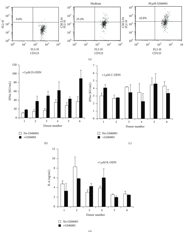
affect the response to the three classes of CpG ODN, PBMC from 6 donors were pretreated with GM6001 for 30 min and then stimulated with the D, C, or K type ODN. Exposure to the metalloproteinase inhibitor resulted in
increased CXCL16 expression on pDCs (Figure 1(a)), whereas
24.6 ± 2.3 of untreated pDCs expressed cell-surface associ-ated CXCL16 metalloproteinase inhibitor treatment caused
a∼72% increase in surface expression levels for the protein
(Table 1). GM6001 pretreatment also resulted in a significant increase in D-ODN responsiveness (∼2-fold for individual
donors, 𝑃 < 0.05) (Figure 1(b) and Table 1) but had no
effect on cells stimulated with C (Figure 1(c)) or K-ODN
(Figure 1(d)). These results support the previous findings [14] and strengthen the case for CXCL16 involvement in D-ODN specific cellular activation.
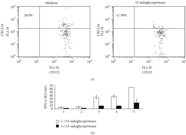
3.2. Cleavage of CXCL16 on the Surface of pDCs Reduces Cytokine Production Induced by D-ODN. Treatment of cells
with the enzyme O-sialoglycoprotease was shown to cleave
cell-surface expressed CXCL16 [22]. Treatment of PBMCs
104 103 102 101 100 104 103 102 101 100 100 101 102 103 104 100 101 102 103 104 104 103 102 101 100 104 103 102 101 100 Is o typ e CX CL16 CX CL16 Medium CD123 CD123 CD123 25.4% 0.6% 42.8% 50 𝜇M GM6001 FL3-H FL2-H FL2-H FL2-H FL3-H FL3-H (a) 0 20 40 60 80 100 120 1 2 3 4 5 6 Donor number No GM6001 +GM6001 IFN 𝛼 (KU/mL) +3 𝜇M D-ODN (b) 0 1 2 3 4 5 6 7 1 2 3 4 5 6 Donor number No GM6001 +GM6001 IFN 𝛼 (KU/mL) +1 𝜇M C-ODN (c) 0 2 4 6 8 10 12 1 2 3 4 5 6 Donor number No GM6001 +GM6001 IL -6 (n g/mL) +1 𝜇M K-ODN (d)
Figure 1: Metalloproteinase inhibitor GM-6001 enhances cytokine production induced by D-ODN. (a) PBMC (4 × 10⁶/mL) were preincubated in the absence or presence of GM6001 (50𝜇M) for 30 min, fixed, and stained for CXCL16 expression. Cells treated as in (a) were washed and then stimulated with 3𝜇M D-ODN (b), 1 𝜇M of C ODN (c), or 1 𝜇M of K-ODN (d) for 24 h. Cytokine production (IL-6 for K-ODN and IFN𝛼 for D and C ODN) was assessed from culture supernatants using ELISA. Response of 6 individual donors is shown.
Table 1: Altering CXCL16 expression influences “D” ODN induced cytokine production.
pDC expressing CXCL16 D-ODN induced IFN𝛼 production
Treatment % of all pDC % change KU/mL % change
Untreated 24.6± 2.3 26.3± 17.8
GM6001 43.1± 6.2∗ ↑ 72 ± 2∗ 50.3± 24.1∗ ↑ 115 ± 56∗
O-sialoglycoprotease 9.6± 3.7∗ ↓ 55 ± 1∗ 6.6± 4.0∗ ↓ 70 ± 12∗
PBMC from 5-6 different donors were treated with 50𝜇M of GM6001 or 25 𝜇g/mL of o-sialoglycoprotein endopeptidase (from Pasteurella haemolytica) for 30 min at 37∘C. The cells were then stimulated in vitro with 3𝜇M “D” ODN for 24 h, and the production of IFN𝛼 determined by ELISA. The fraction of CD123/BDCA-2 double positive pDC expressing CXCL16 was determined before and after Rx. Treatment-induced changes were calculated for each donor independently and then averaged.
∗𝑃 < 0.05. 26.0% 11.59% Medium O-sialoglycoprotease 104 104 103 103 102 102 101 101 100 100 100 101 102 103 104 FL2-H FL2-H FL2-H 104 103 102 101 100 FL2-H CX CL16 CX CL16 CD123 CD123 (a) 0 10 20 30 40 50 60 70 1 2 3 4 5 (−) O-sialoglycoprotease (+) O-sialoglycoprotease IFN-𝛼 (KU/mL) (b)
Figure 2: Digestion of CXCL16 on the surface of pDCs reduces cytokine production induced by D-ODN. (a) PBMC (4× 10⁶/mL) were preincubated in the absence or presence of O-sialoglycoprotein endopeptidase (25𝜇g/mL) for 30 min, fixed, and stained for CXCL16 expression. (b) Cells treated as in (a) were washed and then stimulated with 3𝜇M D-ODN for 24 h. Cytokine production was assessed from culture supernatants using ELISA. Response of 6 individual donors is shown.
surface-associated CXCL16 protein levels (Table 1,𝑃 < 0.05
andFigure 2(a)). This decrease correlated with a significant
reduction in D-ODN responsiveness (𝑃 < 0.05,Table 1and
Figure 2(b)).
These results indicate that factors that can modulate pDC associated cell-surface CXCL16 can affect the magnitude of the immune response to D-ODN. It remains to be seen whether CXCL16 also plays a role in modifying the immune response of pDCs during viral infections.
3.3. CXCL16/CXCR6 Interaction Reduces the Threshold of Activation in pDCs Responding to D-ODN. Transmembrane
CXCL16 is composed of three domains: the chemokine domain that interacts with the receptor CXCR6, the glycosy-lated mucin-like stalk, and the cytoplasmic domain that con-tains a potential tyrosine phosphorylation and
SH2-protein-binding site [21, 24]. Thus, the cytoplasmic domain may
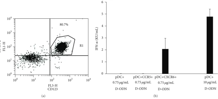
also play a role in cell signaling. To assess whether CXCL16 engagement on pDCs can modify the immune response to CpG ODN mediated immune activation, pDCs from 3 differ-ent donors were enriched using immunomagnetic separation (at least 80% pure as determined by pDC-specific marker
staining,Figure 3(a)). Enriched pDCs were then incubated
CD123 BD CA -2 104 103 102 101 100 104 103 102 101 100 FL3-H FL1-H R1 80.7% (a) 0 1 2 3 4 5 6 IFN-𝛼 (KU/mL) pDC+ 0.75 𝜇g/mL D-ODN 0.75 𝜇g/mL D-ODN 0.75 𝜇g/mL D-ODN pDC+ 10 𝜇g/mL D-ODN pDC+CCR5+ pDC+CXCR6+ (b)
Figure 3: CXCL16/CXCR6 interaction reduces the threshold of activation in pDCs responding to D-ODN. (a) pDCs were enriched from human PBMCs using the BDCA-4 isolation kit as recommended by the manufacturer. Purity of cells was established by flow cytometry following staining with pDC associated cell-surface markers. (b) Enriched pDCs (100,000 cells/well) were incubated with CXCR6 or CCR5 expressing Jurkat T cells (1 : 1 ratio) in 96-well U-bottom plates. Cocultures were then stimulated with a suboptimal concentration of D-ODN (0.75𝜇g/mL). pDC stimulated with optimal concentration of D-ODN (10 𝜇g/mL) served as a positive control. Cytokine production was assessed from culture supernatants 24 h later using ELISA. Average response of 3 different pDC preparations is shown (mean± S.D).
CXCL16 engagement. A separate set of pDCs were incubated with CXCR6 negative CCR5 positive Jurkat cells as negative control. The cocultures were then stimulated with subopti-mal concentration of D-ODN that did not yield detectable levels of IFN𝛼 production (0.75 𝜇g/mL as determined in preliminary experiments). Results showed that coculture of purified pDCs with CXCR6 expressing Jurkat cells decreased the threshold concentration of D-ODN mediated IFN𝛼 production, enabling the cells to respond to an otherwise
nonstimulating concentration of this ODN (Figure 3(b)).
This effect was specific to CXCR6 since coculture with CCR5
expressing cells showed no such effect (Figure 3(b)). This
result suggests that CXCL16/CXCR6 interaction may modify pDC responsiveness to environmental danger signals.
3.4. Oxidized LDL and Recombinant IFN𝛾/TNF𝛼 Modify
Cytokine Production Induced by D-ODN. We further
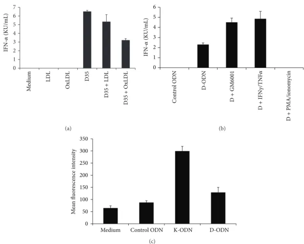
exam-ined the effect of scavenger receptor ligand oxidized low density lipoprotein (oxLDL) on D-ODN mediated immune activation. Preincubation of enriched pDCs with oxLDL
resulted in∼50% reduction in IFN𝛼 production in samples
stimulated with D-ODN, while native LDL showed very weak
or undetectable inhibitory effect (Figure 4(a)).
Hyperlipi-demia and hypercholesterolemia are two prime risk factors in the development of atherosclerosis, and accumulating
evidence suggests that DC functions may be hampered [25].
We demonstrated that increased levels of oxLDL may influ-ence signaling pathways of pDC, thereby altering immune responses against pathogens. This result suggests that oxLDL
levels may be of importance in modifying the pDC response during host resistance to microbial infections.
Expression of CXCL16 is induced by the inflammatory cytokines IFN-gamma and TNF-alpha. These two cytokines synergize to upregulate both the soluble and the
cell-surface associated CXCL16 [26]. To assess whether pDC
response to D-ODN would be affected under conditions replicating an ongoing chronic inflammation, enriched pDCs were stimulated with the CpG ODN in the absence or presence of the recombinant cytokines IFN𝛾/TNF𝛼. Results show that similar to GM6001, recombinant cytokine pre-treatment increased the D-ODN dependent IFN𝛼 production
∼2-fold (Figure 4(b),𝑃 < 0.05). In contrast, PMA/ionomycin
pretreatment which strongly downregulates CXCL16
expres-sion [27] failed to induce cytokine production following
D-ODN stimulation (Figure 4(c)).
K versus D-ODN differentially activate human cells to produce distinct cytokines (TNF𝛼 versus IFN𝛼). To assess whether CXCL16 expression is altered during CpG ODN stimulation, PBMC were stimulated with K or D-ODN for 48 h, followed by staining for cell-surface expressed CXCL16. Cells stimulated with K-ODN expressed 3.4-fold more membrane-associated CXCL16 when compared to
con-trol ODN treated cells (Figure 4(c),𝑃 < 0.05). Conversely,
D-ODN stimulated cells showed a modest increase (1.4-fold), suggesting that CXCL16 expression is not controlled by type I interferons.
In conclusion, we show that factors that can alter the extent of cell-surface CXCL16 expression can profoundly alter the immune response induced specifically by D-ODN
0 1 2 3 4 5 6 7 IFN-𝛼 (KU/mL) M edi um LDL O xLD L D35 D35 + LD L D35 + O xLD L (a) 0 1 2 3 4 5 6 IFN-𝛼 (KU/mL) C o n tr o l O D N D -ODN D + GM6001 D + PMA/io no m ycin D + IFN 𝛾/TNF 𝛼 (b) 0 50 100 150 200 250 300 350
Medium Control ODN K-ODN D-ODN
M ea n fl u o res cence in te n si ty (c)
Figure 4: Oxidized LDL and recombinant IFN𝛾/TNF𝛼 modify cytokine production induced by D-ODN. (a) Enriched pDCs (100,000 cells/well) were preincubated with LDL or OxLDL (10𝜇g/mL each) and then stimulated with 3 𝜇M of D-ODN. Cytokine production was assessed from culture supernatants 24 h later using ELISA. Average response of 3 different pDC preparations is shown (mean± S.D). (b) Enriched pDCs were preincubated in the absence or presence of GM6001 (50𝜇M), recIFN𝛾/TNF𝛼 (20 ng/mL each), or PMA/ionomycin (250 pg/mL/100 pg/mL) for 30 min at 37∘C. Cytokine production was assessed from culture supernatants 24 h later using ELISA. Average response of 3 different pDC preparations is shown (mean± S.D). (c) PBMC (4 × 10⁶/mL) were preincubated in the absence or presence of 1𝜇M Control ODN, 1 𝜇M K-ODN, or 3 𝜇M D-ODN for 48 h. Cells were then fixed and stained for CXCL16 expression. MFI of CXCL16 stained cells± S.D of 3 different donors is shown.
and that CXCL16/CXCR6 interaction decreases the threshold concentration of D-ODN mediated IFN𝛼 production. This D-ODN sensitivity probably depends on the synergistic action of CXCL16/CXCR6 signaling cascade and more effi-cient delivery of D-ODN to endosome where TLR9 resides.
Conflict of Interests
Mayda Gursel, Dennis M. Klinman, and Ihsan Gursel have patents related to the use of CpG ODN. All rights to such patents have been assigned to the U.S. Federal Government. The authors declare no further conflict of interests.
References
[1] H. Hemmi, O. Takeuchi, T. Kawai et al., “A toll-like receptor recognizes bacterial DNA,” Nature, vol. 408, no. 6813, pp. 740– 745, 2000.
[2] C. Bode, G. Zhao, F. Steinhagen, T. Kinjo, and D. M. Klinman, “CpG DNA as a vaccine adjuvant,” Expert Review of Vaccines, vol. 10, no. 4, pp. 499–511, 2011.
[3] A. M. Krieg, “CpG still rocks! update on an accidental drug,”
Nucleic Acid Therapeutics, vol. 22, no. 2, pp. 77–89, 2012.
[4] K. J. Ishii and S. Akira, “Innate immune recognition of, and regulation by, DNA,” Trends in Immunology, vol. 27, no. 11, pp. 525–532, 2006.
[5] F. Takeshita, C. A. Leifer, I. Gursel et al., “Cutting edge: role of toll-like receptor 9 in CpG DNA-induced activation of human cells,” Journal of Immunology, vol. 167, no. 7, pp. 3555–3558, 2001. [6] N. Kadowaki, S. Ho, S. Antonenko et al., “Subsets of human dendritic cell precursors express different toll-like receptors and respond to different microbial antigens,” Journal of
Experimen-tal Medicine, vol. 194, no. 6, pp. 863–869, 2001.
[7] G. Hartmann and A. M. Krieg, “Mechanism and function of a newly identified CpG DNA motif in human primary B cells,”
[8] D. Verthelyi, K. J. Ishii, M. Gursel, F. Takeshita, and D. M. Klinman, “Human peripheral blood cells differentially rec-ognize and respond to two distinct CpG motifs,” Journal of
Immunology, vol. 166, no. 4, pp. 2372–2377, 2001.
[9] A. Krug, S. Rothenfusser, V. Hornung et al., “Identification of CpG oligonucleotide sequences with high induction of IFN𝛼/𝛽 in plasmacytoid dendritic cells.,” European Journal of
Immunology, vol. 31, no. 7, pp. 2154–2163, 2001.
[10] M. G¨ursel, D. Verthelyi, I. G¨ursel, K. J. Ishii, and D. M. Klinman, “Differential and competitive activation of human immune cells by distinct classes of CpG oligodeoxynucleotide,” Journal of
Leukocyte Biology, vol. 71, no. 5, pp. 813–820, 2002.
[11] G. Hartmann, J. Battiany, H. Poeck et al., “Rational design of new CpG oligonucleotides that combine B cell activation with high IFN-𝛼 induction in plasmacytoid dendritic cells,”
European Journal of Immunology, vol. 33, no. 6, pp. 1633–1641,
2003.
[12] J. D. Marshall, K. Fearon, C. Abbate et al., “Identification of a novel CpG DNA class and motif that optimally stimulate B cell and plasmacytoid dendritic cell functions,” Journal of Leukocyte
Biology, vol. 73, no. 6, pp. 781–792, 2003.
[13] U. Samulowitz, M. Weber, R. Weeratna et al., “A novel class of immune-stimulatory cpg oligodeoxynucleotides unifies high potency in type i interferon induction with preferred structural properties,” Oligonucleotides, vol. 20, no. 2, pp. 93–101, 2010. [14] M. Gursel, I. Gursel, H. S. Mostowski, and D. M. Klinman,
“CXCL16 influences the nature and specificity of CpG-induced immune activation,” Journal of Immunology, vol. 177, no. 3, pp. 1575–1580, 2006.
[15] K. Honda, Y. Ohba, H. Yanai et al., “Spatiotemporal regulation of MyD88-IRF-7 signalling for robust type-I interferon induc-tion,” Nature, vol. 434, no. 7036, pp. 1035–1040, 2005.
[16] T. Shimaoka, N. Kume, M. Minami et al., “Molecular cloning of a novel scavenger receptor for oxidized low density lipoprotein, SR-PSOX, on macrophages,” The Journal of Biological
Chem-istry, vol. 275, no. 52, pp. 40663–40666, 2000.
[17] A. Wilbanks, S. C. Zondlo, K. Murphy et al., “Expression cloning of the STRL33/BONZO/TYMSTR ligand reveals ele-ments of CC, CXC, and CX3C chemokines,” Journal of
Immunology, vol. 166, no. 8, pp. 5145–5154, 2001.
[18] T. Shimaoka, T. Nakayama, N. Fukumoto et al., “Cell surface-anchored SR-PSOX/CXC chemokine ligand 16 mediates firm adhesion of CXC chemokine receptor 6-expressing cells,”
Jour-nal of Leukocyte Biology, vol. 75, no. 2, pp. 267–274, 2004.
[19] D. M. Wuttge, X. Zhou, Y. Sheikine et al., “CXCL16/SR-PSOX is an interferon-𝛾-regulated chemokine and scavenger receptor expressed in atherosclerotic lesions,” Arteriosclerosis,
Thrombosis, and Vascular Biology, vol. 24, no. 4, pp. 750–755,
2004.
[20] S. T. Oh, A. Schramme, W. Tilgen, P. Gutwein, and J. Reichrath, “Overexpression of CXCL16 in lesional psoriatic skin,” Dermato-Endocrinology, vol. 1, no. 2, pp. 114–118, 2009. [21] M. Matloubian, A. David, S. Engel, J. E. Ryan, and J. G. Cyster, “A
transmembrane CXC chemokine is a ligand for HIV-coreceptor Bonzo,” Nature Immunology, vol. 1, no. 4, pp. 298–304, 2000. [22] P. J. Gough, K. J. Garton, P. T. Wille, M. Rychlewski, P. J.
Dempsey, and E. W. Raines, “A disintegrin and metallopro-teinase 10-mediated cleavage and shedding regulates the cell surface expression of CXC chemokine ligand 16,” Journal of
Immunology, vol. 172, no. 6, pp. 3678–3685, 2004.
[23] S. Tabata, N. Kadowaki, T. Kitawaki et al., “Distribution and kinetics of SR-PSOX/CXCL16 and CXCR6 expression on
human dendritic cell subsets and CD4+ T cells,” Journal of
Leukocyte Biology, vol. 77, no. 5, pp. 777–786, 2005.
[24] A. Ludwig and C. Weber, “Transmembrane chemokines: versa-tile “special agents” in vascular inflammation,” Thrombosis and
Haemostasis, vol. 97, no. 5, pp. 694–703, 2007.
[25] M. Kiss, Z. Czimmerer, and L. Nagy, “The role of lipid-activated nuclear receptors in shaping macrophage and dendritic cell function: from physiology to pathology,” Journal of Allergy and
Clinical Immunology, vol. 132, no. 2, pp. 264–286, 2013.
[26] S. Abel, C. Hundhausen, R. Mentlein et al., “The transmem-brane CXC-chemokine ligand 16 is induced by IFN-𝛾 and TNF-𝛼 and shed by the activity of the disintegrin-like metallopro-teinase ADAM10,” Journal of Immunology, vol. 172, no. 10, pp. 6362–6372, 2004.
[27] P. Shashkin, D. Simpson, V. Mishin, B. Chesnutt, and K. Ley, “Expression of CXCL16 in human T cells,” Arteriosclerosis,
Thrombosis, and Vascular Biology, vol. 23, no. 1, pp. 148–149,
Submit your manuscripts at
http://www.hindawi.com
Stem Cells
International
Hindawi Publishing Corporationhttp://www.hindawi.com Volume 2014
Hindawi Publishing Corporation
http://www.hindawi.com Volume 2014
Hindawi Publishing Corporation
http://www.hindawi.com Volume 2014
Behavioural
Neurology
Endocrinology
International Journal ofHindawi Publishing Corporation
http://www.hindawi.com Volume 2014 Hindawi Publishing Corporation
http://www.hindawi.com Volume 2014
Disease Markers
Hindawi Publishing Corporation
http://www.hindawi.com Volume 2014
BioMed
Research International
Oncology
Journal of Hindawi Publishing Corporationhttp://www.hindawi.com Volume 2014
Hindawi Publishing Corporation
http://www.hindawi.com Volume 2014
Oxidative Medicine and Cellular Longevity
Hindawi Publishing Corporation
http://www.hindawi.com Volume 2014
PPAR Research
The Scientific
World Journal
Hindawi Publishing Corporationhttp://www.hindawi.com Volume 2014
Immunology Research
Hindawi Publishing Corporation
http://www.hindawi.com Volume 2014 Journal of
Obesity
Journal ofHindawi Publishing Corporation
http://www.hindawi.com Volume 2014
Hindawi Publishing Corporation
http://www.hindawi.com Volume 2014
Computational and Mathematical Methods in Medicine
Ophthalmology
Journal ofHindawi Publishing Corporation
http://www.hindawi.com Volume 2014
Diabetes Research
Journal of Hindawi Publishing Corporationhttp://www.hindawi.com Volume 2014
Hindawi Publishing Corporation
http://www.hindawi.com Volume 2014
Research and Treatment
AIDS
Hindawi Publishing Corporation
http://www.hindawi.com Volume 2014
Gastroenterology Research and Practice
Hindawi Publishing Corporation
http://www.hindawi.com Volume 2014