Case Report
Stimulus-induced myoclonus treated effectively with clonazepam in
genetically con
firmed Coffin–Lowry syndrome
☆
Elif Acar Arslan
a,⁎
, Serdar Ceylaner
b, Güzide Turanl
ı
c aDepartment of Pediatric Neurology, Hacettepe University Children's Hospital, Ankara, Turkey
bDepartment of Molecular Biology and Genetics, Intergen Genetics Center, Ankara, Turkey c
Department of Pediatric Neurology, Medipol University Medical School of Medicine, Istanbul, Turkey
a b s t r a c t
a r t i c l e i n f o
Article history:
Received 27 September 2014 Accepted 30 September 2014 Available online 25 November 2014 Keywords:
Clonazepam Coffin–Lowry syndrome Stimulus-induced myoclonus
Purpose: Coffin–Lowry syndrome (CLS) is a rare X-linked semidominant syndromic genetic disorder that is characterized by typical facial and radiologicfindings, psychomotor and growth retardation, and various skeletal anomalies. A distinctive paroxysmal disorder called stimulus-bound myoclonus is clinically heterogeneous and is generally characterized by a sudden loss of muscle tone that is regained within a few seconds and is induced by sudden auditory or tactile stimulus. As the pathophysiology of stimulus-induced drop episodes (SIDEs) is not well understood, there is no definite therapy for those episodes.
Methods: We report a 15-year-old female with stimulus-induced drop episodes occurring many times a day that resulted in failure to perform her daily activities. Because her SIDEs were misdiagnosed as atonic seizures, she was treated with several antiepileptic drugs, including valproic acid, levetiracetam, lamotrigine, primidone, car-bamazepine, and clobazam.
Results: We realized that her clinical and radiologicalfindings, together with SIDEs, are compatible with Coffin– Lowry syndrome. All of her medications were discontinued following the diagnosis of SIDE, and she was started on clonazepam. After treatment, she became more independent and was able to perform her daily activities. Subsequently, her episodes decreased from 3 times a day to 1–2 times a month. Sodium oxybate and fluoxetine were added to the treatment protocol without remarkable improvement. Her genetic analysis revealed a heterozygous variation of CLS.
Conclusion: We conclude that SIDE should be included in a differential diagnosis of epileptic seizures in patients with CLS and that clonazepam is an effective choice in the treatment of SIDEs.
© 2014 The Authors. Published by Elsevier Inc. This is an open access article under the CC BY-NC-ND license (http://creativecommons.org/licenses/by-nc-nd/3.0/).
1. Introduction
Coffin–Lowry syndrome (CLS) is an X-linked semidominant inherited disease that wasfirst described independently by Coffin et al. and Lowry et al.[1,2]Temtamy et al. were thefirst to recognize that the patients reported by Coffin and Lowry et al.[3]shared a similar course. The typical clinical features of this disease are mental retarda-tion, facial dysmorphism, puffy proximal digits, tapering digits, and progressive skeletal changes[3]. This condition is very rare, with an estimated incidence of 1:50,000 to 1:1,000,000; approximately 70– 80% of patients are sporadic cases[4]. Genetically, CLS is caused by ‘loss-of-function mutations’ in the Rps6ka3 gene encoding the protein
of ribosomal S6 kinase 2, a growth factor-regulated protein kinase. The disease is characterized by sudden drop attacks induced by sound or tac-tile stimulus without any loss of consciousness. These drop episodes last a very short time, as little as a few seconds[5]. Typical facial features in-clude a prominent forehead, hypertelorism, aflat nasal bridge, and a downward-sloping palpebralfissure. In addition, affected individuals have severe mental retardation, short stature, pectus excavatum or carinatum deformity, thoracolumbar kyphosis/scoliosis, large and soft hands with tapering of the distal short and broadfingers and nails, hypo-tonia, and facial dysmorphism characterized by a coarse facial appear-ance[6].‘Stimulus-induced drop episodes’ (SIDEs) have been proposed as a name for these nonepileptic events in patients with CLS. The patho-physiology of SIDEs is not fully understood. Stimulus-induced drop episodes begin at the age range of 4 to 17 years, with a mean age at onset of around 8 years[5,7]. Many different agents are used in the treat-ment of SIDEs, including antiepileptic agents (e.g., lorazepam, clobazam, tiagabine, felbamate, valproic acid, and carbamazepine), selective seroto-nin reuptake inhibitors (e.g.,fluoxetine and escitalopram), and tricyclics (e.g., clomipramine)[5–7]. Benzodiazepines are usually the drug of
Epilepsy & Behavior Case Reports 2 (2014) 196–198
☆ This paper was presented as an oral presentation at the 14th Turkish Pediatric Society Scientific Congress, Kayseri, Turkey on May 2012.
⁎ Corresponding author at: Clinic of Pediatric Neurology, Giresun Prof. Dr. A. Ilhan Ozdemir Hospital, Teyyaredüzü Mahallesi, PC: 28100 Giresun, Turkey. Tel.: +90 454 310 20 00; fax: +90 454 215 51 46.
E-mail address:[email protected](E.A. Arslan).
http://dx.doi.org/10.1016/j.ebcr.2014.09.007
2213-3232/© 2014 The Authors. Published by Elsevier Inc. This is an open access article under the CC BY-NC-ND license (http://creativecommons.org/licenses/by-nc-nd/3.0/). Contents lists available atScienceDirect
Epilepsy & Behavior Case Reports
j o u r n a l h o m e p a g e : w w w . e l s e v i e r . c o m / l o c a t e / e b c r
Downloaded for Anonymous User (n/a) at Istanbul Medipol University from ClinicalKey.com by Elsevier on March 26, 2020. For personal use only. No other uses without permission. Copyright ©2020. Elsevier Inc. All rights reserved.
choice[5], as conventional antiepileptic drugs are often useless. We examine here a case of genetically confirmed Coffin–Lowry syndrome in a female patient with fully manifested clinical features and drop episodes, the latter of which partially responded to treatment with clonazepam.
2. Case report
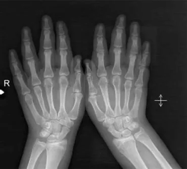
A 15-year-old patient was admitted with a diagnosis of drug-resistant epilepsy. According to the history, the patient showed normal development until 10 years of age, except for falling a little behind her peers in school performance. At this age, she began to experience sudden-onset drop attacks stimulated by sound. Her prenatal history was unremarkable, and she had a healthy older sister. The otherfive subsequent pregnancies of her mother resulted in spontaneous abor-tion. Her mother and father arefirst-degree cousins. Her dysmorphic features included orbital hypertelorism, downward-slanting palpebral features, aflat nasal bridge, a prominent forehead, anteverted nares, thickening of the ala nasi and nasal septum, and a long mouth with little everted lips. Her hands are broad and puffy, with broad, typically taper-ingfingers. In addition to her facial dysmorphism, the thinning of her distalfingers and a clumsy gait were remarkable findings from the physical examination (Fig. 1). She exhibited the skeletal deformity of kyphosis, and the X-ray findings of her hands revealed that their drumstick appearance was compatible with the disease (Fig. 2). She had moderate mental retardation. Because her SIDEs were misdiagnosed as atonic seizures, she was treated with several anti-epileptic drugs including valproic acid, levetiracetam, lamotrigine, primidone, carbamazepine, and clobazam. Since all of her interictal EEGs were normal despite the sudden drop attacks, the patient was admitted to our video monitoring unit for long-term monitoring. No pathological appearance was observed in the simultaneous EEGs, except for a muscular artifact during drop episodes. Her MR imaging revealed a normal appearance of the cerebellum and brain except for a mild dilatation of the lateral ventricles. Her genetic analysis of the RSK2 gene supported a diagnosis of Coffin–Lowry syndrome.
Sequence analysis was performed on all 22 of the exons and the exon–intron boundaries of RPS6KA3 by using the ABI 3130 capillary electrophoresis system. Heterozygote variation c.1973_1974insA (p.M659Dfs*55) was found on exon 21, generating a frameshift mutation beginning in p.Met659 and stopping at the stop codon at position 713. As this event constitutes a truncating mutation and frameshift variant, protein units after position 659 become defective. As seen inFig. 3, this variation causes damage to the functional protein kinase domain, resulting in a novel mutation. Based on our in silico evaluation of the functional effects by using the mutation
taster, Polyphen2, SIFT, and Mutation Assessor, this mutation seems to be the disease-causing entity.
All medications were discontinued following the diagnosis of SIDE. First, clonazepam was started because it is a treatment for this disease that is available in our country. While receiving a daily dose of clonaze-pam 40 mg (maximum) per day, the number of drop attacks decreased from two or three times a day to 5–6 times a week, depending on the stimuli. Interestingly, most of the drop attacks occurred during men-strual cycles. The reduction in the frequency of drop attacks and the co-ordination of the patient's movements and gait balance improved with clonazepam treatment. Although clonazepam reduced the frequency of drop attacks, the patient did not achieve a full recovery. For this reason, we began a secondary therapy of sodium oxybate andfluoxetine; how-ever, this treatment regimen did not affect the clinicalfindings of the patient.
3. Discussion
Coffin–Lowry syndrome is an X-linked inherited disease that is characterized by typical facial and radiological dysmorphism, mental retardation, short stature, pectus deformity, sensory–neuronal hearing loss, and mitral valve deformity. Multipoint linkage analysis of the many X-linked RFLP markers in 4 families was performed in 1988[8], and the causal gene was identified in 1996. These potential causes of CLS have been postulated as abnormalities in the MAPK/RSK signaling pathway[9]. The effects of RSK, which comprises 22 exons and encodes a protein spanning 740 amino acids, include proliferation, differentia-tion, cellular stress response, and apoptosis[10].
Coffin–Lowry syndrome is usually diagnosed based on clinical presentation and radiologicalfindings. However, as this diagnosis is difficult to make in very young children and females, a mutational anal-ysis of the RPS6KA3 gene is recommended[4]. Female carriers show a variable range of involvement in this disease, and individuals who are heterozygous are affected less[5]. Moreover, the present case exhibited a heterozygous variant of the disease. More than 100 different muta-tions have been identified up to 2007; still, 50% of clinically diagnosed patients have no mutations[6]. Highly heterogeneous loss-of-function mutations in the RPS6KA3 gene are responsible for CLS[4]. Both the widespread distribution of mutations and the rather large number of exons contribute to the difficulties in CLS. Because there is no Fig. 1. Photograph of the patient showing the thinning of the distalfingers.
Fig. 2. X-ray showing a drumstick appearance.
197 E.A. Arslan et al. / Epilepsy & Behavior Case Reports 2 (2014) 196–198
Downloaded for Anonymous User (n/a) at Istanbul Medipol University from ClinicalKey.com by Elsevier on March 26, 2020. For personal use only. No other uses without permission. Copyright ©2020. Elsevier Inc. All rights reserved.
relationship between the severity of disease and the identified muta-tions, genetic counseling is difficult[4].
‘Stimulus-induced drop episodes’ (SIDEs) have been proposed as the term for nonepileptic events in patients with CLS. Stimulus-induced drop episodes are usually characterized by a sudden loss of muscle tone induced after an unexpected tactile or auditory stimulus, the latter of which is more common[5]. Stimulus-induced drop episodes are clinically similar events as cataplexy. Clonazepam wasfirst used by Nakamura et al. in their successful treatment of the drop episodes of a 16-year-old female with Coffin–Lowry syndrome[8–11].
In our case, the patient did not achieve a full recovery with clonaze-pam; therefore, we started a secondary therapy of sodium oxybate and fluoxetine. Different treatment modalities have been applied in SIDEs. Despite our 6-month trial using sodium oxybate, no improvement in the SIDEs was noted. The effectiveness of sodium oxybate in cases exhibiting a sudden loss of muscle tone is considered an explanation of the mechanism seen in cataplexy rather than in hyperekplexia[7, 12,13]. Moreover, sodium oxybate is known to be effective in treating drop attacks, even though its exact mechanism remains undetermined
[14]. Some reports have indicated that instances of cataplexy, such as atonic falls, are observed at younger ages, whereas hyperekplexia is increasingly observed with advancing age[7]. Several case series have considered the use of valproic acid,fluoxetine, and clomipramine for the treatment of SIDEs. As valproic acid has previously been used with-out benefit, we added fluoxetine to the present therapy. Fluoxetine did not contribute to an improvement of our case.
4. Conclusion
Our patient performed all of her daily activities with the aid of her family prior to the treatment. After treatment with clonazepam, the family indicated that their child exhibited more balanced movements. Thus, we conclude that clonazepam is the most effective drug for the treatment of SIDEs. Moreover, published reports have shown that the severity and frequency of SIDEs increase with age.
Funding None.
Acknowledgments
We are very grateful to Dr. Jean Pierre Delaunoy for his aid in our ge-netic studies. We also thank the int.site“Picture:http://prosite.expasy. org/cgibin/prosite/ScanView.cgi?scanfile=443433327386.scan.gz” for displayingFig. 3and Dr. Dilek Yalnizoğlu for her helpful edits. Conflicts of interest
The authors declare that there are no conflicts of interest.
References
[1]Coffin GS, Siris E, Wegenkia LC. Mental retardation with osteocartilaginous anomalies. Am J Dis Child 1966;112:205–13.
[2]Lowry B, Miller JR, Fraser FC. A new dominant gene mental retardation syndrome. Am J Dis Child 1971;121:496–500.
[3]Temtamy SA, Miller JD, Hussels-Maumenee I. The Coffin–Lowry syndrome: an inherited faciodigital mental retardation syndrome. J Pediatr 1975;86:724–31.
[4]Pereira PM, Schneider A, Pannetier S, Heron D, Hanauer A. Coffin Lowry syndrome. Eur J Hum Genet 2010;18:627–33.
[5]Hahn JS, Hanauer A. Stimulus-induced drop episodes in Coffin–Lowry syndrome. Eur J Med Genet 2012;55:335–7.
[6]Micheli V, Sestini S, Parri V, Fichera M, Romano C, Ariani F, et al. RSK2 enzymatic assay as a second level diagnostic tool in Coffin Lowry syndrome. Clin Chim Acta 2007;384:35–40.
[7]Nelson GB, Hahn JS. Stimulus-induced drop episodes in Coffin Lowry syndrome. Pediatrics 2003;111:197–202.
[8]Hanuer A, Alembik Y, Gilgenkrantz S, Mujica P, Nivelon-Chevallier A, Pembrey ME, et al. Probable localisation of the Coffin–Lowry locus in Xp22.2-p22.1 by multipoint linkage analysis. Am J Med Genet 1988;30:523–30.
[9]Trivier E, De Cesare D, Jacquot S, Pannetier S, Zackai E, Young I, et al. Mutations in the kinase Rsk-2 associated with Coffin–Lowry syndrome. Nature 1996;384:567–70.
[10]Hanauer A, Young ID. Coffin–Lowry syndrome: clinical and molecular features. J Med Genet 2002;39:705–13.
[11]Nakamura M, Yamagata T, Momoi MY, Yamazaki T. Drop episodes in Coffin Lowry syndrome: exaggerated startle responses treated with clonazepam. Pediatr Neurol 1998;19:148e150.
[12]Yeomans JS, Frankland PW. The acoustic startle reflex neurons and connections. Brain Res Rev 1995;21:301–14.
[13]Guilleminault C, Gelb M. Clinical aspects and features of cataplexy. Adv Neurol 1995; 67:65–77.
[14]Havaligi N, Matadeen-Ali C, Khurana DS, Marks H, Kothare SV. Treatment of drop attacks in Coffin–Lowry syndrome with the use of sodium oxybate. Pediatr Neurol 2007;37:373–4.
Fig. 3. A truncating mutation and frameshift variant are shown. All protein units after position 659 are defective. This variation causes damage to the functional protein kinase domain. 198 E.A. Arslan et al. / Epilepsy & Behavior Case Reports 2 (2014) 196–198
Downloaded for Anonymous User (n/a) at Istanbul Medipol University from ClinicalKey.com by Elsevier on March 26, 2020. For personal use only. No other uses without permission. Copyright ©2020. Elsevier Inc. All rights reserved.