METASTASIS SUPPRESSOR GENES AND PROTEINS
IN NON-MELANOMA SKIN CANCERS
A THESIS
SUBMITTED TO THE DEPARTMENT OF MOLECULAR BIOLOGY AND GENETICS AND THE GRADUATE SCHOOL OF ENGINEERING AND SCIENCE
OF BILKENT UNIVERSITY
IN PARTIAL FULFILLMENT OF THE REQUIREMENTS FOR THE DEGREE OF DOCTOR OF PHILOSOPHY
By
Önder Bozdoğan
August, 2014
I certify that I have read this thesis and that in my opinion it is fully adequate, in scope and in quality, as a thesis for the degree of Doctor of Philosophy.
Assoc. Prof. Dr. Işık G. Yuluğ (Advisor) I certify that I have read this thesis and that in my opinion it is fully adequate, in scope and in quality, as a thesis for the degree of Doctor of Philosophy.
Prof. Dr. Hilal Özdağ
I certify that I have read this thesis and that in my opinion it is fully adequate, in scope and in quality, as a thesis for the degree of Doctor of Philosophy.
Assoc. Prof. Dr. Rengül Çetin-Atalay I certify that I have read this thesis and that in my opinion it is fully adequate, in scope and in quality, as a thesis for the degree of Doctor of Philosophy.
Assist. Prof. Dr. Özlen Konu I certify that I have read this thesis and that in my opinion it is fully adequate, in scope and in quality, as a thesis for the degree of Doctor of Philosophy.
Assist. Prof. Dr. Derya Beyza Sayın
Approved for the Graduate School of Engineering and Science:
Prof. Dr. Levent Onural
Director of the Graduate School of Engineering and Science
ABSTRACT
METASTASIS SUPPRESSOR GENES AND PROTEINS IN NON-MELANOMA SKIN CANCERS
Önder Bozdoğan
Ph.D in Molecular Biology and Genetics Supervisor: Assoc. Prof.Dr. Işık G. Yuluğ
August 2014
Skin cancers are the most common cancer in human population. They are practically
divided into two major group; melanoma and non-melanoma skin cancer (NMSC).
NMSC often refers to two common neoplasms; cutaneous squamous cell carcinoma (cSCC) and basal cell carcinoma (BCC). BCCs are slow growing, malignant,
significantly invasive but rarely metastasizing carcinomas. cSCCs are the malignant tumor of keratinocytes with significant squamous differentiation. In contrast to
BCCs, SCCs have significant metastatic capacity. Metastasis is a complex multistep
process and strictly positively or negatively controlled by tens of genes or proteins.
Besides supporting genes, a group of gene, called metastasis suppressor genes
(MSG), slow or inhibit metastasis without significantly affecting tumorigenicity.
The aim of this study was to find out distribution and importance of the seven
selected metastasis suppressor gene/proteins including NM23-H1, NDRG1,
E-cadherin, RHOGDI2 (ARHGDIB), CD82/KAI1, MKK4, and AKAP12 in NMSC.
Ninety six BCCs, 32 cSCCs, 6 in-situ SCCs, two cell lines (HaCaT, A-431) were included
for immunohistochemistry study. Eleven BCCs, 8 normal skin adjacent to the BCCs, 3
normal skin frozen tissue, and, two cell lines were inserted for qRT-PCR studies.
and 5 normal tissue samples by bisulfite sequencing method.
In immunohistochemistry study, NM23-H1 was protected in NMSC. Similarly,
relatively preserved cytoplasmic expressions of NDRG1 were also detected. AKAP12
and RHOGDI2 were decreased in both tumor groups. However, CD82/KAI1
downregulation was only detected in BCCs. E-Cadherin was relatively protected in
BCCs but significant lost was seen in cSCCs. Cytoplasmic positivity of MKK4 was
more pronounced in cSCC when compared to BCCs. Immunohistochemical study of
cell lines showed similar finding as in seen cSCC. In qRT-PCR study, we found
significant upregulation of NM23-H1 (1.4 fold; p=0.032) and downregulation of
AKAP12 (-1.2 fold; p=0.006) when BCC was compared to normal skin. NDRG1
showed significantly higher levels (2.2 fold, p=0.001) in BCC when compared to the
skin adjacent to the BCC. MKK4 (-2.1-fold, P=0.001), ARHGDIB (RHOGDI2) (-4.7-fold,
P=0.001), CD82/KAI1 (-2.4-fold, P=0.001) and AKAP12 (-9.7-fold, P=0.001) were
downregulated but NDRG1 (34.4-fold, p=0.001) was upregulated in A-431 cell line
when compared to HaCaT. CD82/KAI and MKK4 promoters were heavily
unmethylated in BCCs and normal skin.
In conclusion, we have demonstrated differential expression patterns for the seven
MSPs in NMSCs. In SCCs, the MSG expression signature is similar but not identical
to BCCs. The preserved levels of NM23-H1 and NDRG1 may contribute to the
non-metastatic features of NMSC.
Key Words: Metastasis suppressor gene, skin cancer, metastasis, NM23-H1, NDRG1, E-cadherin, RHOGDI2, CD82/KAI1, MKK4, AKAP12
ÖZET
MELANOM DIŞI DERİ KANSERLERİNDE METASTAZ BASKILAYICI GENLER VE PROTEİNLER
Önder Bozdoğan
Moleküler Biyoloji ve Genetik Doktorası Tez Yöneticisi: Doç.Dr. Işık G. Yuluğ
Ağustos 2014
Deri kanserleri insanlarda en sık görülen kanserlerdir. Pratik olarak melanoma ve
melanoma dışı deri kanserleri (MDDK) olmak üzere iki alt gruba ayrılabilir. MDDK sıklıkla bazal hücreli karsinom (BHK) ve deri kökenli skuamoz hücreli karsinomu
(dSHK) tanımlar. BHK’lar yavaş büyüyen, malign, invaziv ancak nadiren metastaz
yapan tümörlerdir. dSHK’lar ise belirgin skuamoz differansiyasyon gösteren
keratinositlerin malign neoplazileridir. BHK’lardan farklı olarak dSHK belirgin
metastaz kapasitesine sahiptirler. Metastaz, katı olarak pozitif veya negatif olarak
onlarca gen ve proteinle kontrol edilen karmaşık basamaklı bir sürectir. Metastazı
destekleyici genlerin yanı sıra metastaz baskılayıcı genler (MBG) adı verilen bir grup
gen tümorojeniteyi etkilemeden metastazı yavaşlatır veya durdurur.
Bu çalışmanın amacı NM23-H1, NDRG1, E-cadherin, RHOGDI2 (ARHGDIB),
CD82/KAI1, MKK4 ve AKAP12’nin dahil olduğu yedi seçilmiş metastaz baskılayıcı
genin/proteinin MDDK’ daki önemini araştırmaktır.
İmmunhistokimyasal çalışma için 96 BHK, 32 dSHK, 6 in-situ SHK, iki hücre hattı
(HaCaT, A-431) dahil edildi. 11 BHK, 8 tümör komşuluğunda normal deri, 3 normal
deri donuk dokuları ve hücre hatları qRT-PCR çalışmasına katıldı. Ayrıca 7 BHK ve 5
sekanslama yöntemiyle analiz edildi.
İmmunhistokimyasal çalışmada, MDDK’larda NM23-H1’in korunduğu izlendi.
Göreceli olarak sitoplazmik NDRG1 ekspresyonunun da korunduğu saptandı. Her iki
tümor grubunda da AKAP12 ve RHOGDI2 ekspresyonlarının azaldığı görüldü.
CD82/KAI düzeylerinin azalması sadece BHK’da saptandı. E-cadherin düzeyi BHK’da
göreceli olarak korunurken, belirgin düşme dSHK’da saptandı. MKK4 sitoplazmik
ekspresyonu dSHK’da BHK’a göre daha belirgindi. Hücre hatlarını
immunhistokimyasal çalışması dSHK’dakine benzer bulgular verdi. Kantitatif eş
zamanlı PCR çalışmasında BHK’da normal deri dokusuna göre NM23-H1 ‘de artış (
1,4 kat; p=0.032), AKAP12’de azalma (-1.2 kat; p=0.006) bulduk. NDRG1’de komşuluktaki deriye göre BHK’da artış (2.2 kat, p=0.001) saptandı. HaCaT hücre
hattına göre A-431’de MKK4 (-2.1 kat, P=0.001), ARHGDIB (RHOGDI2) (-4.7 kat,
P=0.001), CD82/KAI1 (-2.4 kat, P=0.001) ve AKAP12’de (-9.7 kat, P=0.001) azalma,
NDRG1’de ise (34.4 kat, p=0.001) artış bulundu. Promotor metilasyon
araştırmasında CD82/KAI1 ve MKK4 genlerinde metilasyon saptanmadı.
Sonuç olarak çalışılan yedi MBP/G ile MDDK’da farklı ekspresyon örüntüleri
saptadık. SHK’da MBG ekspresyonu BHK’a benzemekle birlikte, farklılıklar da
göstermektedir. NM23-H1 ve NDRG1 ekspresyonlarının korunması, MDDK’da
metastazın önlenmesinde katkısı olabilir.
Anahtar sözcükler: Metastaz baskılayıcı genler, deri kanseri, metastaz, NM23-H1, NDRG1, E-cadherin, RHOGDI2, CD82/KAI1, MKK4, AKAP12
ACKNOWLEDGEMENTS
I would like to gratefully and sincerely thank my academic supervisor Assoc. Prof.
Işık G.Yuluğ for her support, guidance, understanding and friendship during my PhD.
studies at Bilkent University. Her guidance and support helped me in all steps of my research and this thesis.
I would also like to thank Bala Gür Dedeoğlu for teaching me the first steps of
molecular biology techniques and her friendship during my first years at Bilkent
University.
I am indebted to Nilüfer Sayar and Gurbet Karahan, their help and contribution to
my studies.
I would also thank technical team of Pathology Department of Kırıkkale University
for their kind help.
I would like to express my very great appreciation to the academic and technical
team of Bilkent MBG department and I consider it an honor to work with MBG
family.
I wish to thank Dr. Aydın Yuluğ for formatting and editing this text.
I also thank my children, Umut and Ekin for their patience. Finally, and most
importantly, I would like to thank my wife Nazan for her endless support,
encouragement and patience in every step of my life and during this thesis study.
This work was supported grants (SBAG 108S184) from The Scientific and
TABLE OF CONTENTS
SIGNATURE PAGE……….. i
ABSTRACT……… ii
ÖZET……… iv
ACKNOWLEDGEMENTS………. vi
TABLE OF CONTENTS……….. vii
LIST OF TABLES……… ix
LIST OF FIGURES………. x
ABBREVIATIONS………. xii
CHAPTER 1………. 1
INTRODUCTION……….. 1
1.1. Skin Function and Histology………. 1
1.2. Skin Carcinomas……… 4
1.2.1. Basal Cell Carcinoma………. 5
1.2.1.1. Clinical Features………... 5
1.2.1.2. Etiology and pathogenesis……… 6
1.2.1.3. Histopathology………. 8
1.2.1.4. Aggressive-Non-aggressive Basal Cell Carcinoma………. 10
1.2.2. Squamous Cell Carcinoma………. 11
1.2.2.1. Clinical Features……….. 11
1.2.2.2. Etiology and Pathogenesis……… 13
1.2.2.3. Histopathology………. 14
1.3 Metastasis………. 17
1.3.1. Multistep Metastasizing Process………. 18
1.3.2. New Approaches………. 21
1.4. Metastasis Related Genes………. 22
1.4.1. Metastasis Suppressor Proteins/Genes Studied……… 24
1.4.1.1.N-Myc Downstream Regulated 1. (NDRG1)……… 24
1.4.1.2. Rho GDP Dissociation Inhibitor Beta (RHOGDI2, LY-GDI, D4-GDI, D4-GDI)………. 25
1.4.1.3. E-Cadherin……… 27
1.4.1.4. CD82/KAI1……… 28
1.4.1.5. Mitogen-Activated Protein Kinase Kinase 4 (MKK4,MEK4)……….. 29
1.4.1.6.Nucleoside Diphosphate Kinase 1 (NM23-H1)………. 30
1.4.1.7.A Kinase (PRKA) Anchor Protein 12(AKAP12, Gravin,AKAP250)………. 31
1.4.2.Other Metastasis Suppressor Proteins……….……. 31
1.5. OBJECTIVES AND RATIONALE... 32
1.5.1 Aim of the Study... 32
1.5.2 Rationale and Strategy... 32
CHAPTER 2………. 35
MATERIALS AND METHODS……… 35
2.1 Study Groups……… 35
2.1.1. Basal Cell Carcinoma Group………. 35
2.1.2 Squamous Cell Carcinoma Group……….. 36
2.1.3.Normal Skin Control Group……… 36
2.1.4. Quantitative Real-Time PCR Study Group………. 37
2.1.5. Clinicopathological Features……….. 37
2.1.6. Cell Lines……… 37
2.4.2. Amplification Efficiencies………. 42
2.4.3. qRT-PCR Studies……….. 43
2.4.4. qRT-PCR Data Analysis……… 44
2.5. Promoter DNA Methylation Analysis……… 45
2.5.1 Bisulfite Modification of DNA, Sequencing and Analysis………. 46
2.6. Statistical Analysis……….. 47
2.7. Ethical Issues and Support……… 47
CHAPTER 3………. 48
RESULTS……….. 48
3.1. Immunohistochemical Staining, HSCOREs and qRT-PCR Results……… 48
3.1.1. NM23-H1……….. 49 3.1.1.1. Immunohistochemical Analysis……… 49 3.1.1.2. HSCOREs……… 51 3.1.1.3. qPCR Results……….. 51 3.1.2. NDRG1……… 51 3.1.2.1. Immunohistochemical Analysis……….. 51 3.1.2.2. HSCOREs……….. 54 3.1.2.3. qPCR Results……….. 54 3.1.3. E-Cadherin……….…….. 56 3.1.3.1. Immunohistochemical Analysis……… 56 3.1.3.2. HSCOREs……… 57 3.1.3.3. qPCR Results……….. 59 3.1.4. RHOGDI2……… 59 3.1.4.1. Immunohistochemical Analysis……… 59 3.1.4.2. HSCORES……….. 60 3.1.4.3. qPCR Results……….. 61 3.1.5. MKK4……… 63 3.1.5.1. Immunohistochemical Analysis……… 63 3.1.5.2. HSCOREs……… 64 3.1.5.3. qPCR Results……….. 64 3.1.6. CD82/KAI1……… 67 3.1.6.1. Immunohistochemical Analysis……… 67 3.1.6.2. HSCOREs……… 67 3.1.6.3. qPCR Results……….. 67 3.1.7. AKAP12……….. 70 3.1.7.1. Immunohistochemical Analysis……… 70 3.1.7.2. HSCOREs……… 71 3.1.7.3. qPCR Results……….. 71 3.2. Correlation Analysis……….. 74
3.3. Bisulfite Sequencing Results……… 76
3.3.1. MKK4……… 76
3.3.2. CD82/KAI1……….. 77
CHAPTER 4………. 78
DISCUSSION AND CONCLUSION……….. 78
REFERENCES……….. 89
APPENDIX-A... 108
LIST OF TABLES
Table 1.1. Classification of Basal Cell Carcinoma. 10
Table 1.2. Classification of Squamous Cell Carcinoma 17
Table 1.3: Metastasis Suppressor Genes Studied 25
Table 1.4. Metastasis Suppressor Proteins described in the literature. 34
Table 2.1. Study groups. 35
Table 2.2. Primer antibodies used in this study. 42
Table 2.3. qRT-PCR primer sequences used in this study 42
Table 2.4. Amplification efficiencies of the used primer pairs. 43
Table 2.5. BSP primers 47
LIST OF FIGURES
Fig. 1. 1. Microanatomy of the normal skin. 2 Fig. 1.2. Simplified Hedgehog signal pathway. 7 Fig .1.3. Microscopic pictures of different types of basal cell carcinoma. 8 Fig. 1.4: Histopathologic appearances of the cutaneous squamous cell carcinoma 16 Fig. 1.5. Steps of classical metastasis process 20
Fig. 1.6. RhoGTPase pathway. 26
Fig. 1.7: MAP kinase pathway. 30
Fig. 1.8. The study design. 33
Fig. 2.1.The print screen of the simple Excel® macro. 41 Fig. 2.2. Amplification curve and melt curve graph. 45
Fig. 3.1. Summary of HSCORE Data. 48
Fig. 3.2. NM23-H1 immunohistochemistry. 50 Fig. 3.3. Boxplot graphs of NM23-H1. 52 Fig. 3.4. NDRG1 immunohistochemistry. 53 Fig. 3.5. Boxplot graphs of NDRG1. 55 Fig. 3.6. Amplification plots of NM23-H1 and NDRG1 genes. 56 Fig. 3.7. E-Cadherin immunohistochemistry. 57 Fig. 3.8. Boxplot graphs of E-Cadherin. 58 Fig. 3 .9. RHOGDI2 immunohistochemistry. 61 Fig. 3.10. Boxplot graphs of RHOGDI2. 62 Fig 3.11.Amplification plots of CDH1 (E-Cadherin) and ARHGDIB (RHOGDI2) genes 63 Fig. 3. 12. MKK4 immunohistochemistry. 65
Fig 3.13. Boxplot graphs of MKK4. 66
Fig 3.14. CD82/KAI1 immunohistochemistry. 68 Fig. 3.15. Boxplot graphs of CD82/KAI1. 69 Fig. 3.16. Amplification plots of MKK4 and CD82 genes 70
Fig. 3.17. AKAP12 Immunohistochemistry. 72 Fig. 3.18. :Boxplot graphs of AKAP12. 73 Fig. 3.19. Amplification plots of AKAP12 gene. 74 Fig 3.20.Schematic presentation of correlation in BCC study group. 75 Fig. 3.21. Schematic presentation of correlation in SCC study group. 76 Fig. 3.22 Bisulfite sequencing of MKK4 gene promoter. 77 Fig 3.23. Bisulfite sequencing of CD82 gene promoter. 77
ABBREVIATIONS
A-BCC Aggressive Basal Cell Carcinoma
AK Actinic Keratoses
AKAP12 A Kinase (PRKA) Anchor Protein 12, Gravin
bp Base Pairs
BCC Basal Cell Carcinoma
BCNS Basal Cell Nevus Syndrome
BSA Bovine Serum Albumin
cDNA Complementary Deoxyribonucleic Acid
cSCC Cutaneous Squamous Cell Carcinoma
-cyt Cytoplasmic Staining
DMEM Dulbecco’s Modified Eagle’s Medium
DMSO Dimethyl Sulfoxide
DNA Deoxyribonucleic Acid
dNTP Deoxyribonucleotide Triphosphate
EGFR Epidermal Growth Factor Receptor
EMT Epithelial–Mesenchymal Transition
FBS Fetal Bovine Serum
Fig. Figure
HPV Human Papilloma Virus
HSCORE Immunohistochemical Histological Score
IS-SCC In-situ Cutaneous Squamous Cell Carcinoma
KAI1 Kangai 1
kg Kilogram
m Meter
MEK4/ MKK4 Mitogen-Activated Protein Kinase Kinase 4
mg Milligram
min Minute
ml Milliliter
mRNA Messenger Ribonucleic Acid
MSG Metastasis Suppressor Gene
MSP Metastasis Suppressor Protein
μg Microgram
μl Microliter
μm Micrometer
N Normal Non-Lesional Skin
NE- BCC Normal Epidermis Adjacent To Basal Cell Carcinoma
NE-SCC Normal Epidermis Adjacent To Squamous Cell Carcinoma
NDRG1 N-Myc Downstream Regulated 1
NM23-H1 Nucleoside Diphosphate Kinase 1
NMSC Non-melanoma Skin Cancer
-nuc Nuclear Staining
Oligo(dT) Oligodeoxythymidylic Acid
PCR Polymerase Chain Reaction
PTCH Patched Homolog
PUVA Photochemotherapy
qRT-PCR Quantitative Real Time Reverse Transcription Polymerase Chain Reaction
RHOGDI2 Rho GDP Dissociation Inhibitor Beta
RNA Ribonucleic Acid
Rpm Revolutions Per Minute
RT PCR Reverse Transcription Polymerase Chain Reaction
Str. Stratum
SUFU Suppressor of fused
TAM Tumor-associated Macrophages
CHAPTER 1- INTRODUCTION
1.1. Skin, Function and Histology.
Skin is the largest organ of the human body covering the exterior of the whole
human body [1]. It weights approximately 3-5 kg and approaches 2 m2 in an adult
human [1, 2]. Main function of the skin is to provide a barrier for environment.
However, it has also important roles in thermoregulation, synthesizing important
products (vitamin D), cushioning the trauma and physiological and sociological
wellness [1, 3].
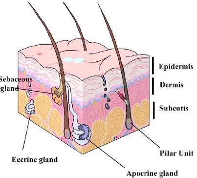
Skin is composed of three histologically and functionally different layers;
epidermis, dermis and subcutaneous tissue (Fig.1.1) [3]. Epidermis is a stratified
squamous epithelium and the main cell type is called keratinocyte. However, other
cells types, melanocytes, Langerhans cells, Merkel cells, and free nerve axons are
also found in the epidermis [3]. Histologically four well defined layers of epidermis
can be determined.
Basal cell layer (stratum basale)
Prickle cell or squamous layer (stratum spinosum) Granular cell layer (stratum granulosum)
Keratin or cornified layer (stratum corneum) [1].
Basal cell layer is composed of cuboidal or columnar cells with basophilic
cytoplasms [3]. This layer is often mitotically active and contains also melanocytes
and Merkel cells [1, 2]. The cells in the prickle layer are polygonal with wide
Langerhans cells are located at the mid and at the upper parts of this layer [3].
Granular layer is composed of 3-5 layer of flattened keratinocytes with basophilic
granular cytoplasms consists of keratohyaline protein [2]. Stratum corneum is the
uppermost layer of epidermis and composed of anucleated eosinophilic
keratinocytes [3]. An eosinophilic acellular keratinous layer (stratum lucidum) may
be recognized between str. granulosum and str. corneum in palm and soles [1].
Fig. 1. 1. Microanatomy of the normal skin. The figure is created by the author helping by the references 1-5
There are different types of skin adnexa or appendages, distribute in connective
tissue of the dermis or subcutis, include pilosebaceous unit and sweet glands [4].
Sweet glands in the human skin are generally divided into two major types; Eccrine
and apocrine glands [2]. Eccrine glands are simple coiled glands distributes in many areas of the skin and they are mainly responsible for the thermoregulation of the
products by decapitation; simply apical cytoplasms fell off into lumen [4]. Apocrine
glands are connected to pilosebaceous unit and open into the infundibulum of the
hair follicle [3, 4]. The main function of apocrine glands is not known in the human,
but they are responsible for production of the body scent and probably help sexual
attraction in other mammals [1].
Pilosebaceous unit includes hair, hair follicle, sebaceous gland and piloerector
muscle [3]. These units are distributed whole skin except palms and soles and a part
of genital skin [4]. The hair follicle divided into three different segments;
infundibulum, isthmus, and the inferior segment [3]. Infundibulum is an area
between opening of the follicle and sebaceous gland opening, and isthmus is
between sebaceous gland opening and piloerector muscle insertion [3]. The inferior segment includes papilla which is responsible for hair growth [4]. Sebaceous glands
are holocrine glands open to pilar follicle and empty their secretion. However, a
group of sebaceous glands opens directly to surface located at areola, eyelids and
vermilion border of lips [4].
Dermis mainly composed of connective tissue, blood vessels, nerves and skin
adnexa. Dermal connective tissue has significant amount of collagen and elastic
fibers which are responsible tensile strength of the skin [5]. Dermis can be divided
two different zones; papillary dermis and reticular dermis [3]. Papillary dermis is
below the dermoepidermal junction and composed of lose thin connective tissue
network of collagen I and III [3, 4]. The papillary dermis forms conic structures
from papillary dermis reticular dermis has more thick compact bundles of collagen
fibers basically composed of collagen I [3].
Subcutaneous tissue is located under the dermis and composed of mature fat
tissues which are divided into lobules with vascular connective tissue septa [1].
Subcutaneous tissue has important functions including thermo regulation,
insulation and cushioning the mechanical injuries [3].
1.2. Skin Carcinomas
Malignant skin tumors are the most common malign human neoplasms and an
important part of daily medical practice [6-9]. Because of their frequency and
increasing incidence, these neoplasms pose important medical, economical, and
social problems of healthcare services worldwide [6, 8, 10]. Despite established
detailed classification schemas for skin cancers, practically they are separated as
two different groups, melanoma and non-melanoma skin cancer (NMSC) [11].
Although there are other types of NMSC including skin adnexal tumors, soft tissue
tumors and lymphomas, this term commonly refers to two common neoplasms; cutaneous squamous cell carcinoma (cSCC) and basal cell carcinoma (BCC) [7]. BCCs
are more commonly seen and are at least 70% of diagnosed of NMSC [11]. The
incidence of NMSC changes due to geographic localization and race. The incidence is estimated more than 1000/100 000 person-per year in Australia, however it
incidence rate of 87.9 for BCC and 28.9 for SCC per 100 000 people [13]. Based on
the data of Turkish Health Minister Reports (2005), skin carcinoma is the third
common carcinoma and the incidence of is 18.91/100 000 person per year [14]. The
incidence in Turkey is probably higher when unregistered patients are taken into
account.
1.2.1. Basal Cell Carcinoma
1.2.1.1. Clinical Features
Basal cell carcinomas (BCCs) are slow growing, malignant, but rarely metastasizing
carcinomas and usually seen on sun exposed areas particularly head and neck of the elderly persons [15, 16]. In large published series, the mean age of the patients
is sixth or seventh decade. [17, 18] Although BCCSs are usually seen at elderly, the
age range is very wide; between second to ninth decade [17, 18]. Males are slightly
more affected than women [1, 17, 18]. Besides detected on sun exposed skin areas,
rarely BCCs may be seen on non-sun exposed area including vulva [19].
The clinical appearances of BCCs are closely related to histopathological subtype.
Clinically, the lesions may show nodular and/or ulcerative, diffuse, superficial
(multifocal) and pigmented appearances [1]. Nodular BCCs represent well defined
slow growing waxy nodules or papules sometimes with telangiectasias and
ulceration [20, 21]. Superficial BCCs are seen as an erythematous elevated plaque or
macule different color or hue from surrounding skin [21]. Superficial BCCs have a
plaque with ill–defined borders [20]. This type sometimes looks like a scar tissue
and clinical diagnosis may be difficult [21]. BCCs are usually asymptomatic but pain
may be rarely only symptom [16].
1.2.1.1. Etiology and Pathogenesis
The etiology of BCCs is shown to be related to multiple factors [22]. Ultra violet (UV)
radiation is a well known environmental factor contributes to the pathogenesis of
BCCs [8, 23]. UV radiation causes characteristic covalent bonds between adjacent
pyrimidines and generates cyclopyrimidine dimers (TT) and/or
pyrimidine-pyrimidine (6-4) adducts [8]. UVB is probably the major participant and more mutagenic than UVA [8, 22]. Besides UV radiation, a group of etiological factors are
described for BCCs including; Human papilloma virus (HPV), immunosuppression,
non-Hodgkin lymphoma, PUVA therapy, photosensitizing drugs, ionizing radiation,
occupational factors, arsenic, burns and scars [8, 22].
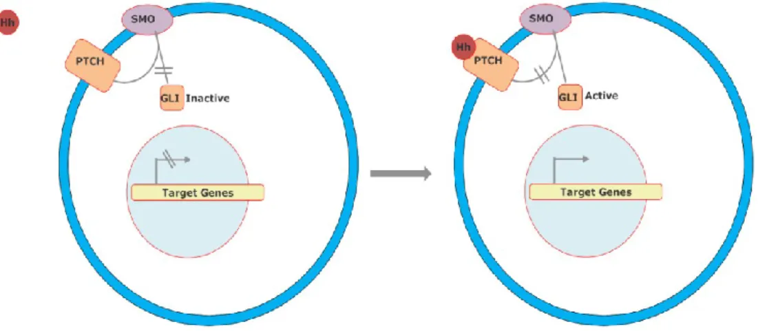
BCCs may be related to a group of familial inherited syndromes. One of well
known, Basal Cell Nevus Syndrome (BCNS), also named as Gorlin Syndrome or
Gorlin–Goltz Syndrome is characterized by multiple BCCs in early ages [24, 25]. Besides early and multiple onset of BCCs; keratocysts of jaw, palmoplantar pits,
skeletal anomalies, medulloblastomas, fibromas and calcification of falx cerebri may
be seen [26, 27]. Basal cell nevus syndrome (BCNS) is a relatively common
(PTCH1, PTCH2), signal transducer (smoothened), and transcription factors (Gli
proteins) [29]. This complex pathway is activated when ligands bind to PTCH
receptor. PTCH receptor releases bounded SMO to signal downstream. Eventually
Gli proteins act as a transcription factor for activating related genes (Fig. 1.2 ) [29].
The most common affected gene/protein in BCNS is PTCH1 (9q22.3) [28]. The others
are PTCH2 and SUFU in this pathway [30, 31]. PTCH genes act as a tumor suppressor
and have some important regressive roles in cell growth and differentiation [32].
The other responsible gene Suppressor of fused (SUFU) codes a negative regulator
of the Sonic Hedgehog pathway [33]. It has been showed that significant numbers
of sporadic BCCs share the same irregularities as seen in BCNS [34, 35]. After the molecular mechanism background of BCCs was established, the new therapy
strategies opened, such as SMO inhibitors [29].
Besides BCNS, the other syndromes related to BCCs are Rombo syndrome,
Bazex–Dupre–Christol syndrome, Multiple Hereditary Infundibulocystic Basal Cell
Carcinoma syndrome, and Xeroderma Pigmentosum [25, 36-38]. Furthermore, BCCs
are also an ancillary feature in other different cutaneous syndromes [27].
Fig. 1.2. Simplified Hedgehog signal pathway. Without ligand PTCH inhibits SMO. After ligand
binds to PTCH, It releases SMO and GLI activates. GLI translocates into the nucleus and induces target gene transcription. The figure is created by the author helping by the references 28, 29, 32.
1.2.1.3. Histopathology
Histopathologically, these tumors are classified into several distinct morphological
types but a significant percentage of mixed morphology BCCs may be seen in daily
practice [39]. Classically, BCCs are classified as superficial, nodular, infiltrative (with
or without sclerotic-morpheiform stroma), and micronodular subtypes (Table 1.1)
[15, 40]. Basosquamous cell carcinoma and metatypical BCCs are controversial
issues and it has been generally thought that these tumors are somewhere between
BCCs and SCCs [1, 40]. All subtypes are basically formed of small cells with scant
cytoplasm and hyperchromatic nucleus (Fig. 1.3) [40]. Besides the subtypes described above; there are also rare variants including divergent adnexal
Nodular BCCs are generally thought to be the most common subtype. Histologically, they are composed of different size basaloid nodules with peripheral
palisading (Fig. 1.3.A, B). There are also clefts between the stroma and the tumor
(Fig.1.3.B). The stroma often contains mucin and is stained blue-grey by H&E [1, 15, 40].
Superficial BCCs are more commonly seen on the trunk and the extremities than the other subtypes, however at least 40% of them are seen on the head and neck
area.[40] Histopathological examination reveals small multiple buds and nodules,
composed of small basaloid cells, which are attached to the atrophic epidermis (Fig. 1.3.C) [15, 40]. Superficial BCCs usually stay in the papillary dermis for a long time and usually do not invade the reticular dermis [15].
Infiltrative BCC consists of invasive cords and strands of basaloid cells with a different type of stroma. A group of infiltrative BCCs, that show significant collagen
deposition, is called morpheiform, sclerotic or fibrosing BCC (Fig. 1.3.D) [15].
Micronodular BCCs are a relatively new recognized variant of BCC [42]. Although tumor nodules are seen as in the nodular type, they are very small, approximately
near the size of hair bulb, and peripheral palisading is less obvious [40, 43]. There is
Table 1.1. Classification of Basal Cell carcinoma according to WHO (World Health Organization)
classification [44].
• Superficial basal cell carcinoma • Nodular (solid) basal cell carcinoma • Micronodular basal cell carcinoma • Infiltrating basal cell carcinoma • Fibroepithelial basal cell carcinoma
• Basal cell carcinoma with adnexal differentiation • Basosquamous carcinoma
• Keratotic basal cell carcinoma Other variants
1.1.1.4. Aggressive-Non-aggressive Basal Cell Carcinoma
BCCs have significant invasion capacity but rarely metastase [45]. The estimated
metastasis incidence is very low, between 0.0028% and 0.55% [45, 46]. However,
there is no adequate hypothesis to explain why this carcinoma cannot metastasize.
Since metastasis is so rare, the clinically important point of morbidity is the recurrence of the tumor. The recurrence rate is not easily estimated due to the
various factors including the surgical margin, the type of surgery (Mohs surgery or
classical excision), nonsurgical therapies, morphology and the subtype of BCCs. The recurrence rate of primer BCCs after surgical excision is estimated to be less than
5% [47]. Following Mohs micrographic surgery, the recurrence rates in the five-year
period are reported to be between zero and 6.5% for a primary tumor, and
From the clinicopathological point of view, BCCs may be practically separated
into two groups including high risk (aggressive) and low risk (non-aggressive)
[50-52]. Clinically possible aggressive features are large tumors (2 cm <); facial location,
especially the midline of the face, periocular area, nose, and ears; and neglected
tumors [15, 53]. Histopathological subtype of BCCs, perineural and vascular space
invasion, and positivity of the surgical margins are also important for recurrence of
the tumor [15, 54, 55] Infiltrative, micronodular and basosquamous types could be
classified as aggressive BCCs with a significant recurrence rate [15, 53]. Although
nodular and superficial BCCs are generally located in the low-risk group, finding the
exact surgical margin in surgical specimens may be difficult for superficial BCCs [15, 20].
1.2.2. Squamous Cell Carcinoma
1.2.2.1. Clinical Features
Cutaneous squamous cell carcinoma is the malignant tumor of keratinocytes with
significant squamous differentiation [11]. Squamous cell carcinoma (cSCC) is the
second most common skin cancer after BCC [56]. cSCCs are generally seen in the
elderly but they may also be detected in the younger age group [40]. Similar to
BCCs, cSCCs develop with several inherited conditions including Xeroderma
pigmentosa, Albinism, Dystrophic Epidermolysis Bullosa, Rothmund–Thomson
syndrome and Epidermodysplasia Verruciformis [1, 57]. In contrast to BCCs, SCCs have significant metastatic capacity [56]. The percentage of metastasis is described
between 1 and 9.9% in the literature [58, 59]. These varying rates are probably due
to data from different medical clinics or the inclusion of special locations such as the
lip, anal and vulval area into the case series [56]. It is well established that a group
of clinical and pathological features are associated with high risk [60, 61]. Thick
SCCs, localization, tumor size, tumor differentiation, histological subtypes,
perineural invasion, immunosuppression, DNA ploidy or aneuploidy, and high
proliferation antigen expression are thought to be important risk factors [56, 60].
There are two distinct clinicopathological types of precancerous or preinvasive
lesions; actinic keratoses and in-situ carcinoma (Bowen disease). Actinic keratoses (AKs) are common skin lesions, which are generally accepted as a precancerous
lesion for cSCCs [62]. AKs present as flesh-colored scaly macules and plaques, sometimes with hyperkeratosis. Erythematous and pigmented lesions may occur
[63]. The transformation rates of AK to cSCCs are reported as 0.075% and 20% per
one-year period [62, 64-66].
In-situ SCCs (IS-SCC) are seen in the skin as in other mucosal areas. Although
Bowen disease is often used as synonym of in situ SCCs, the usage of the last term is
more suitable [67]. IS-SCCs are generally seen on all skin areas but have a
predilection for sun-exposed areas of the head and neck, and the hands They are
often characterized as well-delineated, erythematous macules or plaques. They may
sometimes be pigmented, especially at genital areas [1, 68].
papules, nodules or plaques; and sometimes have a verrucous appearance [69, 70].
Advanced lesions may show significant hyperkeratosis, central ulceration, and
bleeding [69].
1.2.2.2. Etiology and Pathogenesis
cSCCs share nearly the same etiological factors as described for BCCs above[8].
Tobacco usage has been described as a risk factor for only SCC and not BCC, but this
is not supported with new epidemiological studies when lip SCCs are excluded [71,
72].
Although it is not clear that cSCCs shows similar multistep carcinogenesis as in
cervical carcinomas, there are important clues [73]. It is generally accepted that
tumor suppressor p53 inactivation mutation is probably the first step of the carcinogenesis [74, 75]. It has been reported that p53 mutations were detected
with high percentage (74%) in sun-exposed normal skin when compared to the
mutation rate (5%) in non-sun exposed skin [76]. p53 mutations are also detected
with high percentages in actinic keratoses and in cSCCs [74]. In cutaneous
carcinogenesis beside inactivation mutations, p53 levels may also be regulated by
activation or upregulation of several tyrosine kinases including EGFR [73, 77]. These
kinases down-regulate p53 by a c-JUN-dependent mechanism [73]. Similar to p53,
p14 and p16 (CDKN2A locus) genes are downregulated in cSCCs by mutation or
epigenetic mechanisms [78, 79]. There are also important clues for a role of the RAS
mutation rate is not high (21%) in human cSCCs [73, 80]. RAS activation probably
takes place by indirect mechanisms such as the EGFR-related pathway [81].
1.2.2.3. Histopathology
AKs are separated into various clinicopathological subtypes including Acantholytic,
Pigmented, Bowenoid, Atrophic, and Hypertrophic AKs [62]. All types of AKs except
Bowenoid AKs include atypical keratinocytes, mainly at the basal epidermal levels,
with orto, hyper and parakeratosis [82]. Bowenoid AKs show full thickness
keratinocytic atypia. However, they usually have less significant atypia and cellular
crowding than IS-SCCs [62, 68]. The acantholytic subtype represents discohesion of atypical keratinocytes at different levels of the epidermis [62].
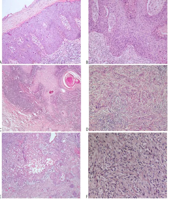
IS-SCC represents hyperkeratosis, parakeratosis, acanthosis, and full thickness atypical keratinocytes with mitoses, and loss of maturation and polarity (Fig.1.4.A,
B) [68, 69]. The basal epidermal layer is usually spared and they show more significant atypia than in Bowenoid actinic keratoses [68].
Invasive cSSCs can have various histopathology appearances due to the invasion
level, grade and subtype. Early invasive cSCCs are similar in morphology to AK or
IS-SCC with focal invasive areas. Well-differentiated cIS-SCCs consist of squamous nests
and islands with significant keratotic areas named “keratin pearls” (Fig. 1.4.C ) [62].
Besides classical cSCCs, several different morphological variants have been
described. However, most of cSCC variants demonstrate little significance for
prognosis and therapy (Table 1.2) [68].
Acantholytic squamous cell carcinoma is a rare variant of cSCCs, and
characterized by acantholysis and pseudoglandular appearance. Though
controversial, this subtype is usually considered as an aggressive variant of cSCCs
(Fig. 1.4. E) [83]. Verrucous squamous cell carcinoma is a low-grade specific variant of SCC most commonly seen skin, mucosal surfaces and genitalia. They consist of
verrucous architecture of well-differentiated squamous cells with little atypia [84]. Spindle cell cSCCs are composed of spindle or pleomorphic cells usually with no
keratinization, similar to spindle cell sarcomas, and immunohistochemistry may be needed to differentiate them (Fig. 1.4.F ) [85].
Adenosquamous carcinoma, as the name implies, consists of squamous and
adenocarcinoma areas in the same tumor.[88] It is often considered to be a
high-risk cSCC [86].
Besides the subtypes classified by WHO, other types of morphological variants
Fig 1.4. Histopathologic appearances of the cutaneous squamous cell carcinoma (cSCC). In-situ
SCCs show lost of maturation, significant atypia and mitoses (A, B). Classical well differentiated squamous cell carcinoma is composed of invasive squamous cell islands with keratin pearls (C).However moderately differentiated cSCC is more invasive and less differentiated (D). Acantholytic cSCC shows pseudoglandular features and acantholysis (E). Spindle cell tumor with no significant differentiated morphologic features. This tumor is immunohistochemically positive for cytokeratins (Spindle cell cSCC) (F). (A, C, D, E, x40; B, D, x100, F, x200)
Table 1.2. Classification of squamous cell carcinoma according to WHO (World Health
Organization) classification and as described by Cassarino et al.[44, 86, 87]
WHO Cassarino et al.
• Acantholytic squamous cell carcinoma
• Spindle-cell squamous cell
carcinoma
• Verrucous squamous cell carcinoma
• Pseudovascular squamous cell
carcinoma
• Adenosquamous carcinoma
Clear cell squamous cell carcinoma Acantholytic (adenoid) squamous cell
carcinoma
Signet ring cell squamous cell carcinoma
Papillary squamous cell carcinoma Pigmented squamous cell carcinoma Follicular squamous cell carcinoma Squamous cell carcinoma arising from
adnexal cysts
Squamoid eccrine ductal carcinoma Invasive Bowen’s disease
Malignant proliferating pilar tumor Desmoplastic squamous cell carcinoma Squamous cell carcinoma arising in
chronic conditions
Radiation-induced squamous cell carcinoma
Lymphoepithelioma-like carcinoma Squamous cell carcinoma arising in
actinic Keratosis
Tricholemmal carcinoma
Classically, cSCCs are graded by Broders’ system: However, this system is
complicated and not easily used. The classical textbook McKee's Pathology of the
Skin offers a simple three-tiered grading system: Well-differentiated, moderately
differentiated, and poorly differentiated squamous cell carcinoma. The fourth group
which includes anaplastic or indifferantiated carcinoma may be added [1]. This last
grading system is more commonly used in daily practice.
1.3. Metastasis
Metastasis is a complex multistep process briefly describes as spread of a disease
In the metastasis process, spread of tumor may occur through several pathways;
direct seeding of body cavities, lymphatic spread, and hematogenous spread [91].
Historically, the first accepted hypothesis about metastasis was emphasized by
Paget S. (1889). He described in this hypothesis that cancer cells (seed) migrate and
grow in a suitable biochemical and biological environment (soil) [92]. After Paget’s
description of “seed and soil hypothesis”, there was an extraordinary effort to
control metastasis in the basic and clinical science area [90, 92]. Today, we learn
that metastasis is a very complex and multistage process [93]. Furthermore this
process is very important in determining for prognosis of an oncology patient.
Despite better therapy options in controlling local cancer, investigators focus on systemic metastatic disease because of its fatal progress [89].
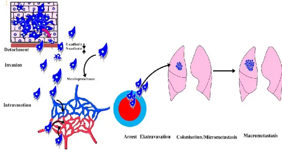
1.3.1. Multistep Metastasizing Process
The data from experimental and clinical studies support that metastasis is a
multistep process [89]. The tumor cells need a group of genetic and epigenetic
changes to regulate this complex multistep process (Fig. 1.5) [91]. The main stages
of metastasis are categorized briefly as:
- Detachment from main tumor. - Invasion
- Intravasation
- Metastatic colonization - Proliferation.
- Micrometastasis
- Macrometastasis [56, 94].
The first step in the metastatic cascade is detachment of tumor cells from
primary tumor mass. The tumor cells then invade the extracellular matrix, called the
invasion step [91]. The invasion and detachment steps need significant changes in tumor cell morphology and biology [90]. These steps are regulated by an important
and complex process called Epithelial–mesenchymal transition (EMT) [95]. EMT
was initially described for fetal development and wound healing but now it has
been thought that it is also a very important mechanism for tumor progress and spread [96]. It is well known that epithelial cells usually have polarized organization
and significant junctions to other cells and matrix proteins [90]. During the EMT
process, epithelial cancer cells lose their polarity and cell-cell adhesion and acquire mesenchymal characteristics which are required for detachment, invasion and
metastasis [96, 97]. In the invasion step, the cancer cells change their morphology,
gain spindle cell morphology, and look like fibroblasts [90].
There are important clues that the downregulation of E-Cadherin, an important
metastasis suppressor protein (MSP), by several pathways, triggers the EMT
process. Promoter methylation of the gene or by E-cadherin repressors including
SNAIL and SLUG are probably the reasons of E-cadherin downregulation at the early
steps of the tumor progression [98, 99]. Loss of E-cadherin expression provides a
Fig. 1.5. Steps of classical metastasis process.
Other adhesion molecules, such as NCAM, DCC, CEACAM1, Mel-CAM, are also down regulated in specific tumor types [90]. Despite downregulation of
E-cadherin and other adhesion molecules, there are important clues that another
cadherin, N-cadherin, is upregulated and positively controls the EMT process [100,
101]. Besides N-cadherin, vimentin is also upregulated and it is thought to be a
marker of EMT [96]. After the dissociation process, tumor cells release proteolitic
enzymes and also induce stromal cells for secreting [91]. At this point,
tumor-associated macrophages (TAM) and other inflammatory cells have important roles
for supporting the invasion step [102, 103]. One of the supportive roles of TAMs is
to secrete proteolitic enzymes [104]. These enzymes such as matrix
metalloproteinases may degrade the extracellular matrix and create a way through
[91]. Cell motility is generally realized by polymerization and depolymerization of
When malignant tumor cells approach the intravascular area, the circulating
surviving tumor cells may show a tendency for tropism to some tissues [105].
Though millions of tumor cells enter the bloodstream, only a very small fraction can
survive due to mechanical trauma and immune cells [106]. Organ tropism is well
defined for metastatic human tumors but the exact mechanism is not very clear
[107]. Probably intrinsic features of the tumor cells and the microenvironmental
factors of target tissue determine the organ specific metastasis [108]. When the
metastatic tumor cells reach the target tissue extravasation occurs. Two types of
tumor arrest can be described. In nonspecific arrest, tumor cells stick in the
capillaries because of their size. The other arrest type is specific to the interaction between tumor membrane protein (Selectins) and the target organ capillaries [106].
The tumor cells lose their mesenchymal characters and gain epithelial features
similar to the primary tumor (mesenchymal epithelial transmission). As a result, a
new colony is established [91, 97]. Colonization is regulated by close interactions of
tumor cells and the microenvironment [106].
1.3.1. New Approaches
Besides classical multistep sequential approach, some observation pointed out that
all metastasis is not differentiated and not similar to primary tumor [109]. It has
been postulated that there are two major types of metastasis (Brabletz); Type 1
plasticity metastasis, and Type 2 genetic type metastasis [97]. Type one metastasis
shows differentiated morphology. This type metastasis is probably regulated by
has undifferentiated phenotype and is usually related of fixed accumulated genetic
alternations [97, 109].
From classic point of view, metastasis is a late event in oncogenesis and tumor
cells need to acquire a group of genetic and epigenetic changes with time needed
for surviving and proliferating at distant size. This classical hypothesis is now called
linear model of metastasis [110]. However, there are also clues that cancer cells disseminate at very early stages of tumor progression even at precancerous lesions
and proliferate parallel to primary tumor. This fact is called parallel model of
metastasis [110, 111]. Probably both of the models are reliable [111].
1.4. Metastasis Related Genes.
The multistep and complex metastasis process is strictly positively and negatively controlled by tens of genes or proteins [56, 93, 112]. The genes and proteins
supporting metastasis are well known and have been studied extensively [112, 113].
According to Nguyen et al, the metastasis related genes are divided into three
groups; metastasis initiation, metastasis progression, and metastasis virulence
genes [113].
Metastasis initiation genes support and modulate basically invasion step [114]. EMT related genes TWiST1, SNAi1 and SluG, and other genes modulate invasion and
Metastasis progression genes code proteins for primer tumor growth and also modulate extravasation, survival and re-initiation and colonization at distant sites
[113]. PTGS2, EREG, MMP1, LOX, ANGPTL4, CCL5 may be given examples [115].
Metastasis virulence genes express at specific metastasis sites, e.g. bone, and help survival of the cancer cells at a specific microenvironment [113]. One special
gene coded parathyroid hormone-related protein (pTHRp) helps to establish
osteolytic metastasis in bone [115].
A group of proteins specifically inhibits metastasis is called as metastasis
suppressor proteins. Literally, a metastasis suppressor is a protein that acts to slow or prevent metastases from spreading in the body of an organism with cancer [116, 117]. However, these proteins are different from ones that act to suppress tumor
growth and they suppress development of metastasis without significantly affecting
tumorigenicity [117, 118].
Metastasis suppressor genes or proteins open a new approach and a study area
at metastasis research, and give hope to clinical therapy. NM23-H1 is first described
in 1988, and a prototype of MSGs [119, 120]. Nowadays, approximately, thirty
genes/proteins are described as MSG, however numbers are not exact and different
from one review to another [105, 121].
Pure MSGs would suppress metastases but have no effect on tumorigenicity
(proliferation) according to their definition [116, 121]. However, in the real world,
MSPs have also other important roles in cell functions.[122] MSG-coded proteins
have a diverse range of biomedical activities [121, 122]. They play various roles in
(MKK4), transcriptional regulation (BRMS1). MSGs also affect different metastasis
steps. They inhibit tumor cell motility and invasion, effects extravasation at the
secondary site or function at tumor dormancy [121].
1.4.1. Metastasis Suppressor Proteins/Genes Studied
In this study, we selected seven important genes/proteins which includes nearly all
steps of metastasis. These selected genes/proteins were summarized in table 1.3.
1.4.1.1. N-Myc Downstream Regulated 1. (NDRG1)
NDRG1, also called Cap43, is a member of NDRG family proteins which includes other proteins NDRG2, NDRG3, NDRG4 and it has been showed that this protein
reduces metastases in colon, breast and prostate neoplasms [123-126]. Although
absolute function of this protein is not well known, NDRG1 has various functions on
stress (hypoxic) response, nerve myelination, cell differentiation, interaction to
heavy metals, and hormones, recycling of E-cadherin, DNA damage response and
mast cell maturation [127-129]. Congenital NDRG1 mutation has been detected in
an autosomal recessive demyelinating polyneuropathy; Charcot-Marie-Tooth
disease type 4D (CMT4D) [130]. NDRG1 has also a role in mouse keratinocyte
differentiation [131, 132]. Though, the metastasis/tumor suppressor features of
mechanism of metastasis suppressor function by NDRG1 is not clear; however there
are some clues of interaction of NDRG1 with WNT signaling and E-cadherin
[137-139].
Table 1.3: Metastasis Suppressor Genes Studied
Gene
Abbrevation-Synonyms
Accesion Numbers
N-Myc Downstream Regulated 1 NDRG1, CAP43 DRG-1, RTP HGNC:7679, Entrez Gene: 10397, Ensembl: ENSG00000104419, UniProtKB: Q92597. NME/NM23 Nucleoside Diphosphate Kinase 1 nm23-H1, NM23-H1, NME1, NM23, NM23A, GAAD. HGNC: 7849, Entrez Gene: 4830, Ensembl: ENSG00000239672, UniProtKB: P15531
Rho GDP Dissociation Inhibitor (GDI) Beta
Rho GDI 2, ARHGDIB , GDID4, RhoGDI2 GDIA2, RAP1GN1, D4
HGNC: 679, Entrez Gene: 397, Ensembl: ENSG00000111348, UniProtKB: P52566.
Cadherin 1, Type 1, E-Cadherin (Epithelial) CDH1, CDHE, CAM 120/80, ECAD, LCAM, CD324. HGNC: 1748, Entrez Gene: 999, Ensembl: ENSG00000039068, UniProtKB: P12830.
CD82 Molecule KAI1, ST6, CD82, SAR2, IA4, TSPAN27, tetraspanin-27, Tspan-27. HGNC: 6210, Entrez Gene: 3732, Ensembl: ENSG00000085117, UniProtKB: P27701. Mitogen-Activated Protein Kinase Kinase 4 MKK4, MAP2K4, SERK1, JNKK1, PRKMK4, JNKK, MEK4, SAPKK-1, MAP2K4
HGNC: 6844, Entrez Gene: 6416,
Ensembl: ENSG00000065559, UniProtKB: P45985
A Kinase (PRKA) Anchor Protein 12
AKAP12, AKAP250, gravin, SSeCKS, AKAP-12
HGNC: 370, Entrez Gene: 9590, Ensembl: ENSG00000131016, UniProtKB: Q02952
1.4.1.2. Rho GDP Dissociation Inhibitor Beta (RHOGDI2, LY-GDI, D4‑GDI)
RHO family GTPases regulate several important cellular mechanism including
adhesion, migration and cell proliferation (Fig. 1.6) [140]. RHOGDIs are a small
cellular area is more complex, and besides RHOGTPase inhibitor, they are work as a
chaperons and transport RHOGTPases [140]. RHODGI family includes three proteins:
RHOGDI1, RHOGDI2, and RHOGDI3 [141]. RHOGDI1 is the well known and prototype
of the family and ubiquitously expressed in various human tissues [142]. However
the other member of the family, RHOGDI3, is expressed only a limited number of
the organs with low levels, including pancreas, brain, lung, testis, and kidney [140,
142]. Initially, RHOGDI2 is thought to be limited to hematopoietic cells, but now, its
expression has been shown in various tissues [143, 144]. The role of RHOGDI2 is
complex and type of tumor dependent [140]. RHOGDI2 acts as a metastasis
suppressor protein in bladder tumors and its expression is closely related to prognosis of the patient [144, 145]. Probably it acts as an MSP in the other types of
epithelial tumors [144, 146, 147]. Despite generally accepted as an MSP, it has been
shown that this protein has a more complex dual role in breast cancer [148-150].
Furthermore, RHOGDI2 supports invasion in pancreatic carcinoma cells [151].
1.4.1.3. E-Cadherin
Cadherins are big superfamily of proteins and classically separated as three groups,
classical cadherins (Type I), non-classical cadherins and protocadherins [152].
E-Cadherin is a member of the classical cadherins, a well-known member of cell-cell
adhesion proteins, and loss of its expression plays an important role in tumor
invasion and metastasis [152-155]. E-Cadherin is a transmembrane protein. The
extracellular part contains five elements that interact with other molecules on the
neighboring cells and the internal part of the molecule forms complexes with
β-catenin, gp-120 catenin and α-catenin.[156] Besides adhesion, E-cadherin also
functions as a negative regulator of the canonical WNT signaling pathway [154,
156].
E- Cadherin is an extensively investigated protein and has been studied in human
tumors [157-160]. The main role of E-cadherin in the metastasis cascade is at the
epithelial mesenchymal transmission (EMT) step. Loss of E-cadherin expression
triggers EMT and invasiveness of the carcinoma cells [161]. Downregulation of
E-cadherin is most commonly regulated by promoter methylation or transcription
repressors (e.g. SNAIL, SLUG, SIP1, ZEB1).[156] E-cadherin is also important in the differential diagnosis of breast cancer in daily practice; the downregulation or loss
of E-cadherin is a specific point in the diagnosis of lobular type breast carcinoma
1.4.1.4. CD82/KAI1
CD82/KAI1 protein, also called TSPAN27, is a member of big 4-span transmembrane
tetra-spantin superfamily (TMSF4) which has important roles in adhesion, motility
and also tumor progression [164-166]. In the human genome, 33 genes code
tetra-spantin proteins [165-166]. Main function of tetra-tetra-spantins is to organize other
transmembrane molecules including, growth receptors, integrins, and they form
tetraspantin-enriched microdomains (TEMs) on the cell surface [165-167].
CD82/KAI1 was first described experimentally in prostate carcinoma cell line AT6.1
as a MSG [168]. The importance of this protein was also demonstrated in breast cancer [169, 170]. However, CD82 expression is very complex in breast carcinoma.
CD82/KAI downregulation is mainly seen ER-positive breast cancer while CD82 is retained in ER-negative breast cancer [170, 171]. The prognostic significance and
reduced levels of CD82 have been shown in prostate and breast carcinoma,
squamous carcinoma of the penis, oral region and larynx, non-small cell lung
carcinoma, papillary carcinoma of the thyroid, gastric carcinoma, transitional cell
carcinoma, endometrial carcinoma, and cervical carcinoma [172-179].
CD82 is closely associated with EGFR, and its ectopic expression suppresses
EGFR-mediated cell migration.[180] New data have also shown that CD82/KAI1 is a
hypoxia target gene regulated by HIF1α [181].
CD82/KAI1 is suggested as a specific immunohistochemical marker for human
1.4.1.5. Mitogen-Activated Protein Kinase Kinase 4 (MKK4, MEK4)
MAP kinases (MAPK) are important intracellular enzymes which phosphorylate
effector proteins [183]. MAPKs are triggered by external or internal various stimuli
and convert the stimuli to different cellular responses including differentiation,
proliferation, survival and death (Fig. 1.7) [183]. MKK4 is an important component
and a key protein of the MAP kinase in stress activated protein kinase signaling
[184, 185]. MKK4 particularly activates and phosphorylates both Jun N-terminal
kinase (JNK) and p38 which have roles in tumor suppression [183, 185, 186].
Recently, it has been shown that the tumor suppressor function of the MKK4 may be related to inducing replicative senescence [187]. It has been shown that MKK4
inhibits metastases of prostatic and ovarian cancers experimentally in mice [188, 189]. Furthermore immunohistochemical expression of MKK4 is decreased in
prostate and ovarian tumors [188, 190]. Although tumor or metastasis suppressor
function of MKK4 is generally accepted, there are some clues MKK4 has a
pro-oncogenic roles at least a group of human tumors [185, 191, 192]. Some
experimental data which were come from breast cancer and pancreatic cell line
studies are shown pro-oncogenic role of MKK4 [193]. Furthermore, MKK4 were
Fig. 1.7: MAP kinase pathway.The figure is created by the author helping by the references[183] 1.4.1.6. Nucleoside Diphosphate Kinase 1 (NM23-H1)
NM23-H1 is the first described and prototype of MSPs [119]. NM23-H1 gene
encodes a nucleoside diphosphate kinase A (NDPK-A). Although, the metastasis
suppressor mechanism of NM23-H1 is not clear, its interaction with kinase
suppressors of RAS (KSR) and, as a result, alteration of the MAPK signaling pathway
is a probable mechanism [121]. It has also recently been suggested that it
suppresses metastasis by inhibiting the expression of EDG2 (lysophosphatidic acid
receptor) [194]. NM23-H1 has the 3’-5’ exonuclease activity, and it has been
postulated that this property is required for metastasis suppressor properties [195].
It has been shown that downregulation of NM23 is closely related to aggressive
1.4.1.7. A Kinase (PRKA) Anchor Protein 12 (AKAP12, Gravin, AKAP250)
A-kinase Anchoring Proteins (AKAPs) are a group of scaffold proteins which have
specific sites especially for protein kinase A and C, and also for phosphoprotein
phosphatases [201]. AKAP12, also called SSeCKS/Gravin, was first described as a
minor prognostic autoantigen in myestenia gravis [202, 203]. As similar to other
AKAPs its role is as a binding partner of protein kinase C (PKC) and A (PKA),
calmodulin, F-actin, cyclins, Src, and phospholipids.[204] Significant clinical and
experimental evidence has shown that AKAP12 is an important tumor and
metastasis suppressor [204]. AKAP12 expression is downregulated in various solid human cancers and leukemias [204, 205]. The downregulation AKAP12 is generally
controlled by epigenetic mechanism [206]. It has been shown that AKAP12 promoter methylation is widespread detected and significantly correlated with
Gleason score in human prostate carcinoma [207]. Similar to prostate carcinoma,
AKAP12 gene is significantly methylated and its expression is downregulated in
hepatocellular carcinoma [208]. Hypermethylation of AKAP12 promoter is also
documented in skin carcinoma, gastric carcinoma, and pancreatic cell lines
[209-211].
1.4.2. Other Metastasis Suppressor Proteins
Many proteins have some roles in negative regulation of metastasis. However, as a
definition, MSP should have no or minimal effect on tumor growth or proliferation
[121]. Nowadays, more than 30 proteins are generally accepted as MSP with more expected [212]. The list of well-known and generally expected MSPs is presented in
1.5. OBJECTIVES AND RATIONALE
1.5.1 Aim of the Study
As described above, metastasis is a complex multistep process and very important in determining for prognosis of an oncology patient. There has been an extraordinary
effort to prevent and retard metastasis in the basic and clinical science area.
Metastasis suppressor proteins give a hope that new therapy strategy for
metastasis will be found. Non-melanoma skin cancers differ from internal organ
cancers in that they have low metastatic rates and good prognosis. Thus, NMSCs are
interesting biological model for metastasis suppressor research. The main aim of
this study was to analyze differentially expressed genes and proteins which may
contribute to inhibit metastasis pathway in Non-Melanoma skin cancer. We also
established the association between these proteins and clinicopathological
parameters.
1.5.2 Rationale and Strategy
We collected fresh normal and pathological assessment of NMSC and paired
normal tissue samples. Parafin embedding biopsies were also selected from
archival specimens. Clinicopathological data for BCCs and cSCCs were collected by
expression profiles were analysed semi-quantitavely by immunohistochemistry
studies. The schema of the study design is demonstrated in Figure 1.8
Table1.4. Metastasis Suppressor Proteins described in the literature.
Metastasis Suppressor Proteins
Nonmetastatic 23 (Nm23) Kai1/Cd82
Mitogen Activated Protein Kinase Kinase
MKK4 MKK6 MKK7 P38
Rho Gdi-Dissociation Factor 2 (RHOGDI2)
N-Myc Downstream Regulated Gene 1 (NDRG1)
E-Cadherin
Src-Suppressed Protein Kinase C Substrate (SSECKS) (Akap12)
Breast Cancer Metastasis
Suppressor 1 (BRMS1) Gelsolin
Kiss1
Deleted In Liver Cancer 1 (DLC-1) Cd44
Cell Adhesion Molecule 1 (CADM1) Mdm2 Binding Protein (MTBP)
Caspase 8
Collapsin Response Mediator Protein 4 (CRMP4)
Deleted In Colorectal Cancer (DCC) Farnesoid X Receptor (FXR)
Growth Arrest-Specific 1 (Gas1) Leukemia Inhibitory Factor Receptor Alpha (LIFRA) Raf Kinase Inhibitory Protein (Rkip) Ribonucleotide Reductase Subunit M1
(RRM1) Stefin A
Lysine-Specific Demethylase 1 (LSD1) Hormonally Regulated Neu-Associated
Kinase (HUNK)
Tissue Inhibitor of Metalloproteases (TIMPS) Timp1 Timp2 Timp3 Timp4 Kruppel-Like Factor (Klf) 17 Caveolin-1
Ovarian Cancer G Protein-Coupled Receptor 1 (OGR1)
Lysine-Specific Demethylase 1 (LSD1) Lass2/Tmsg1
Metastamir and Non-Coding RNA *Adapted from references [121, 212-217]
![Fig. 1.7: MAP kinase pathway. The figure is created by the author helping by the references [183]](https://thumb-eu.123doks.com/thumbv2/9libnet/5876451.121189/44.892.172.750.103.501/fig-kinase-pathway-figure-created-author-helping-references.webp)