Gemcitabine Induced Radiation Recall Myositis:
Report of Two Cases
Gül ALCO1, Sefik IGDEM2, Maktav DINCER1, Coşkun TECIMER3,
Rana SENTURK4, Metin VURAL5, Sait OKKAN2
1Florence Nightingale Gayrettepe Hospital, Department of Radiation Oncology 2İstanbul Bilim University, Faculty of Medicine, Department of Radiation Oncology
3İstanbul Bilim University, Faculty of Medicine, Department of Medical Oncology 4Florence Nightingale Gayrettepe Hospital, Department of Radiology 5VKV Amerikan Hospital, Department of Radiology, Istanbul, TURKEY
ABSTRACT
Radiation recall phenomenon can appear as dermatitis, pseudocellulitis, panniculitis or myositis after the administra-tion of certain drugs and develop in the skin and soft tissue at previously irradiated sites. Several drugs are associat-ed with the radiation recall and gemcitabine is one of them. Gemcitabine-relatassociat-ed radiation recall preferentially involves internal organs and can also be observed at the fibromuscular tissue in the radiation portal. Herein, we report two cases with gemcitabine induced radiation myositis. One patient with non-small cell lung carcinoma, and the other one with pancreatic carcinoma developed myositis in the previously irradiated site after administration of gemcitabine based chemotherapy.
Key Words:Radiation recall, Gemcitabine, Myositis, Side effect
ÖZET Gemsitabine Bağlı Radyasyon “Recall” Myoziti: İki Olgu Sunumu
Radyasyon ‘recall’ fenomeni, ışınlama alanında radyoterapi sonrası uygulanan çeşitli ilaçlara bağlı olarak cilt ve yumuşak dokuda, dermatit, psödoselülit, pannikülit veya myozit olarak ortaya çıkabilen bir klinik tablodur. Gem-sitabin radyasyon ‘recall’ ile ilişkili bilinen ajanlardan biridir. GemGem-sitabine bağlı radyasyon ‘recall’ genellikle iç organlarda gözlense de radyoterapi alanı içindeki kas dokusunda da izlenir. Küçük hücreli dışı akciğer kanseri ve pankreas kanseri tanılarıyla radyoterapi uygulanan ve Gemsitabine bağlı radyasyon ‘recall’ myoziti gelişmiş iki olgu sunularak literatür eşliğinde tartışılmıştır.
Anahtar Kelimeler:Radyasyon recall, Gemsitabin, Myozit, Yan etki
ULUSLARARAS
ı H
EMATOLOJI-O
NKOLOJIDERGISI
C
ASER
EPORT/
INTRODUCTION
Radiation recall is a rare and infrequently reported adverse effect of radiotherapy (RT). Although skin has been the major site of radiation recall toxicity, the recall reaction may occur in previously irradi-ated sites such as mucous membranes in the upper respiratory tract, lung, muscles, and gastrointestinal tract. D’Angio et al.1have first described radiation recall
dermatitis in 1959 as an Actinomycin D potentiated X-ray effect. The recall is triggered by the admi-nistration of certain drugs and occur within days to years after the exposure to ionizing radiation.2,3
Gemcitabine (GEM), a nucleoside analogue with potent radiosensitizing activity, has been recently investigated with radiation in the treatment of panc-reatic cancer, non-small cell lung cancer (NSCLC) and other solid tumors. Most of the radiation recall reactions attributed to GEM affect internal organs, rather than dermis.4Almost all the radiation recall
myositis reactions reported in the literature have been related to GEM. Although a rare complicati-on, treating physicians must be aware of this poten-tial, and sometimes morbid side effect.
Herein, we present two cases with myositis consis-tent with a radiation recall reaction induced by GEM and report the clinical course, and treatment options in the light of the current literature.
CASE 1
A 42 year-old caucasian female presented to her surgeon with elevated levels of CA 19-9 which is incidentally found during a routine check-up. Ab-dominal MRI revealed a mass at the pancreatic tail. The patient underwent pancreatosplenectomy and lymph node dissection, and pathological examinati-on cexaminati-onfirmed the diagnosis of a moderately diffe-rentiated pancreatic ductal adenocarcinoma of 3.5 cm diameter in the corpus-cauda of the pancreas. Microscopically peripancreatic fat tissue invasion, perineural invasion and lymphovascular invasion were present with negative surgical margins, and none of five peripancreatic lymph nodes were in-volved. The patient was diagnosed with T2N0M0 carcinoma of the pancreas, and was referred to our department for postoperative RT. We treated the tu-mor bed and regional lymph nodes to a total dose of 45 Gy in 1.8 Gy fractions using conformally shaped four fields of 18 MV photons. The patient tolerated
the treatment well, no acute side effects of the gast-rointestinal tract, skin and soft tissue were observed during RT.
The patient received continuous 5-FU infusion con-comittantly with RT, followed by 4 cycles of adju-vant GEM 1250 mg/m2/week, 3 weeks in every
4-week-cycle. One month after the last cycle of GEM chemotherapy she developed a tender mass, pain and swelling of the abdominal wall muscles in the areas of the previous radiation fields. Abdominal MRI revealed diffuse edema and inflammation of the muscles of the anterior and right abdominal wall which was consistent with myositis (Figure 1). Her pain was moderate and scored 4-5 out of 10 with Visual Analogue Scale (VAS).5
The patient is diagnosed with radiation recall myo-sitis and treated with corticosteroids (CS) and non-steroid antienflammatory drugs (NSAID’s). Gaba-pentin was used to ease the neuropathic pain. Her pain and edema of the abdominal muscles regressed in a week and the clinical and radiologic findings disappeared in one month time, and medications were stopped. The patient is followed 38 months after the diagnosis without any evidence of disease.
CASE 2
A 67 year-old caucasian female presented with co-ughing and dyspnea. CT scan of the thorax revealed a mass at the left upper lobe and lingula which ca-used total atelectasis and bronchial obliteration wit-hout pathologic mediastinal lymph nodes. After fi-beroptic bronchoscopic biopsy she was diagnosed with NSCLC. PET-CT detected a FDG avid mass directly invading the mediastinum in the left upper lobe and ipsilateral N2 lymph nodes were also in-volved. She was diagnosed with stage IIIB NSCLC and received 62Gy curative RT with 3-D conformal technique to the FDG avid sites. No concurrent chemotherapy was administered during RT. The pa-tient tolerated RT well without any skin or soft tis-sue reaction. Following RT, GEM 1200 mg/m21-8
days and Carboplatin AUC 5.5 1 day / 30 days in 3 cycles was initiated. A dose reduction was done af-ter first cycle because of the intolerance of the pati-ent, and GEM dose was reduced to 800 mg/m2. The
patient complained of severe fatigue, muscle and joint pain, and chemotherapy was discontinued af-ter third cycle. Afaf-ter the completion of systemic therapy, CT scan of the thorax revealed partially
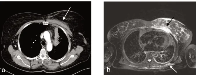
regression of the left upper mass, and total regres-sion of atelectasis. Two weeks after the last cycle of chemotherapy she presented with a painful swel-ling on her left breast and chest wall extending to her axilla. She described a burning pain and her left arm and shoulder movements were reduced beca-use of the pain. The pain was scored 7-8 with VAS. There were no erythema or pigmentation of the skin and the soft tissue on the irradiated areas were swollen. CT scan of the thorax was repeated and left sided pectoral and paraspinal muscles at the previously irradiated sites were found thickened from 1 cm to 3 cm (Figure 2). MRI showed a dra-matical muscle edema and soft tissue reaction at the left breast and subcutaneous soft tissue with hetero-genous contrast fixation, consistent with myositis. CS and NSAID’s, as well as opioids and antihista-minics were prescribed for recall reaction but they did not have any effect. After two months from the onset of the symptoms CS-induced diabetes emer-ged and the CS treatment was stopped. Although there was a spontaneous regression of pain, the VAS was still 5-6. Superoxide dismutase (SOD), pentoxifylline, Vitamin E 800 mg per day and sele-nium tablets were prescribed for the recovery of ra-diation injury. Gabapentin was tried to resolve ne-uropathic pain but again without any effect. Topical lidocaine 5% pomade was found effective in exa-cerbated pain attacks. Thorax CT and MRI were re-peated in every two months and revealed that tumor was under control but myositis findings were
stab-le. Four months after the first symptom of recall, the patient was able to handle the pain without anal-gesics and her pain level regressed to 3-4 according to VAS. There was no change in patient’s physical examination such as tenderness and swelling of the muscles except the pain level. She has had nine months of follow-up with recall myositis and she continued using SOD, pentoxifylline, Vitamin E and selenium. Her pain intensity regressed totally, but she still has difficulties in moving her left sho-ulder. Sixteen months after the diagnosis of NSCLC, she is still metastates-free and with radi-ologically stable local disease. Her broncoscopy was repeated because of the postobstructive pneumonia, and multipl broncoscopic biopsies were reported as inflammatory changes without malignant cells.
DISCUSSION
In the last half century, radiation recall reactions ha-ve been reported in the literature and most of the cases are examples of radiation recall dermatitis which mimic acute radiation reaction of the skin. In the last ten years, radiation recall reactions were se-en in the unconvse-entional areas such as cse-entral ner-vous system, gastrointestinal tract, lung and muscu-loskeletal systems.6,7
Gemcitabine is a newly developed drug which is very effective on solid tumors and has a potentiated radiosensitizing activity. Due to the increasing use of GEM, increasing numbers of recall phenomenon
Figure 1. MR scan of the abdomen showing muscle edema and thickening with contrast fixation (white arrows) in the radiation portal (a) 4 months after radiation, and (b) one months after symptomatic treatment with comp-lete regression
have been reported.4,6Seventy percent of GEM
in-duced radiation recall reactions effect internal or-gans and muscle tissue is effected more often.4We
performed a literature search using PubMed to find previously reported cases of GEM induced radiati-on recall myositis and found eleven cases.4,6, 8-15
Se-ven cases with NSCLC, and one case with bladder cancer were treated with palliative intent, 3 pancre-atic cases were treated with curative intent. In all these cases total radiation dose, dose per frac-tion and treatment technique seem to play an im-portant role in the severity of the myositis reacti-ons. Welsh et al.8treated the sacrum to a total dose
of 45 Gy in 2.5 Gy fractions in a patient with me-tastatic bladder cancer. To protect the neobladder, opposed two lateral fields were utilized. Therefore gluteal muscles, where the myositis developed, bi-laterally received a greater dose per fraction. Fo-garty et al.9treated the mediastinum of a lung
can-cer case palliatively and delivered 36 Gy in 12 frac-tions via ap-pa portals. Myositis involved the pos-terior chest wall. The symptoms of myositis settled on a tapering dose of oral CS and NSAID’s over a 6 week period and cessation of chemotherapy. The-re was however persistent subcutaneous fibrosis in the reaction area causing limitation of movement in the neck and right shoulder. Miura et al.10reported
two cases of myositis, where ap-pa portals were used and myositis was seen in the 50 Gy isodose li-ne. Our findings are consistent with those cases in terms of the severity of the reactions. We retrospec-tively contoured the muscles where recall reactions developed, and calculated doses to these structures
using dose volume histograms. The anterior abdo-minal wall muscles of our pancreatic case received a mean dose of 14.8 Gy (range, 1.6-20.6 Gy), whe-reas the right lateral abdominal wall muscle rece-ived a mean dose of 29 Gy (range, 6.6-35.3 Gy). When recall symptoms appeared, the patient comp-lained more pain at her right abdominal wall. The median dose to the abdominal muscles was low in this case and she recovered from myositis in one month time with simple symptomatic-analgesic medication. In contrast, in the other patient the tu-mor was located close to the chest wall. Therefore, pectoral muscle and dorsal wall received the full dose of the radiation. Mean pectoral and dorsal pa-raspinal muscle doses were 63 Gy (range, 34- 66.74 Gy) and 62.2 Gy (range, 31.2-66.2 Gy), respecti-vely. This patient had severe myositis and her symptoms slightly regressed despite CS, pentoxyp-hilline and antioxidant drugs.
These findings suggest that, in patients treated with two opposed fields the recall reactions were severe, necessitating long term medication, and sometimes leading to chronic morbidity, whereas in patients treated with multiple fields the severity of the reac-tions was mild, and symptomatic relief was achi-eved in a short period of time. Therefore using mul-tiple fields and conformal techniques must be enco-uraged, especially in patients prone to receive GEM. The relation between the recall and the dose of GEM has been investigated by Jeter et al.6They
re-ported of six cases and concluded that if GEM is used in doses of 600 mg/m2and higher, the
possibi-lity of radiation recall may also rise. All patients
Figure 2.CT scan (a) and MR scan (b) of the thorax showing marked thickening of the pectoral and dorsal paras-pinal muscles.
with recall myositis reported in the literature4,6,8-15
and our patients in the current study received 1000 mg/m2or more of GEM, supporting their findings.
Clinical course of the recall myositis differs from patient to patient and the unknown pathophysiolo-gic mechanism does not dictate a spesific therapy. Withdrawal of the offending agent is recommended first, rechallenge with the same chemotherapy agent is debatable. Many patients are treated with topical CS and NSAID’s.3,4,6In some cases with
se-vere recall reaction not responding to CS and/or NSAID’s, antioxidant drugs in a wide spectrum from tocopherol to SOD can be used.16
Pentoxifyl-line which improves blood flow in patients with cir-culation problems by decreasing the viscosity of the blood16 might be beneficial. For more aggressive
approaches with severe tissue breakdown hyperba-ric oxygen therapy may be considered.17 Early
physical therapy and rehabilitation are recommen-ded to reduce chances of developing long-term di-sability secondary to muscle fibrosis.18
In conclusion, GEM is the leading agent to cause radiation recall myositis. Treating physicians must be aware of this potential toxicity of GEM either gi-ven concomittantly or followed by radiation. Con-formal treatment techniques, using lower dose per fraction, and multiple fields are recommended to deliver lower doses to the soft tissue, even in the palliative cases treated with GEM. Muscle doses should be evaluated carefully. Recall reactions usu-ally regress with cessation of responsible agent or with symptomatic therapy. However, some cases do not respond to symptomatic therapy and limitation of movements may develop with severe impair-ment of quality of life.
REFERENCES
1. D’Angio GJ, Farber S, Maddock CL. Potentiati-on of x-ray effects by actinomycin-D. Radiology 73: 175-177, 1959.
2. Camidge R, Price A. Characterizing the pheno-menon of radiation recall dermatitis. Radiother Oncol 59: 237-245, 2001.
3. Azria D, Magné N, Zouhair A et al. Radiation recall: A well recognized but neglected pheno-menon. Can Treat Rev 31: 555-570, 2005. 4. Friedlander PA, Bansal R, Schwartz L et al.
Gemcitabine-related radiation recall preferenti-ally involves internal tissue and organs. Cancer 100: 1793-1799, 2004.
5. Wewers ME, Lowe NK. A critical review of vi-sual analogue scales in the measurement of clini-cal phenomena. Res Nurs Health 13: 227-236, 1990. 6. Jeter MD, Jänne PA, Brooks S et al. Gemcitabi-ne-induced radiation recall. Int J Radiat Oncol Biol Phys 53: 394-400, 2002.
7. Schwarte S, Wagner K, Kartens JH, Bremer M. Radiation recall pneumonitis induced by gemci-tabine. Strahlenther Onkol 183: 215-217, 2007. 8. Welsh JS, Torre TG, DeWeese TL, O’Reilly S.
Radi-ation myositis. Ann Oncol 10: 1105-1108, 1999. 9. Fogarty G, Ball D, Rischin D. Radiation recall
reaction following gemcitabine, Lung Cancer 33: 299-302, 2001.
10. Miura G, Matsumoto T, Tanaka N, et al. Two cases of radiation myositis probably induced by recall phenomenon. Nippon Igaku Hoshasen Gakkai Zasshi. 63: 420-422, 2003.
11. Horan G, Smith SL, Podd TJ. Gemcitabine-in-duced radiation necrosis of pectoralis major muscle. Clin Oncol 18: 85, 2006.
12. Squire S, Chan M, Feller E, et al. An unusual case of gemcitabine-induced radiation recall. Am J Clin Oncol 29: 636, 2006.
13. Ganem G, Solal-Celigny P, Joffroy A, et al. Radiation myositis: The possible role of gem-citabine. Ann Oncol 11:1615-1616, 2000. 14. Pinson PJ, Griep C, Sanders WH, Lelie B.
Myositis as a “radiatior-recall phenomenon” fol-lowing palliative chemotherapy with carbo-platin-gemcitabin for non-small cell polmonary carcinoma. Ned Tijdschr Geneeskd 150: 1891-1894, 2006.
15. Fakih MG. Gemcitabine-induced rectus abdominis radiation recall. J Pancreas 7: 306-310, 2006. 16. Moulder JE. Pharmacological intervention to
prevent or ameliorate chronic radiation injuries. Semin Radiat Oncol 13: 73-84, 2003.
17. Cohn GH, Hyperbaric oxygen therapy. Promot-ing healPromot-ing in difficult cases. Postgrad Med 79: 89-92, 1986.
18. Bentzen SM, Overgaard M, Thames HD. Frac-tionation sensitivity of a functional endpoint: Impaired shoulder movement after postmastec-tomy radiotherapy. Int J Radiat Oncol Biol Phys 17: 531-7, 1989.
Correspondence
Dr. Gül ALÇO
Florence Nightingale Gayrettepe Hastanesi Radyasyon Onkolojisi Bölümü
Gayrettepe 34340 İstanbul / TURKİYE Phone: (+90.212)336 55 70