Eastern Journal of Medicine 19 (2014) 28-32 Original Article
The relationship of mean platelet volume with
endogenous sex hormones and cardiovascular risk
parameters in postmenopausal women
Nilgün Güdücüa,*, Salih Serdar Kutayb, Güliz Sidara, Uzay Görmüşc, Zehra Neşe Kavakd, İlkkan Dündera
aDepartment of Obstetrics and Gynecology, İstanbul Bilim University, İstanbul, Turkey bDepartment of Cardiovascular Surgery, Marmara University, İstanbul, Turkey cDepartment of Biochemistry, İstanbul Bilim University, İstanbul, Turkey
dDepartment of Obstetrics and Gynecology, Marmara University, İstanbul, Turkey
Abstract. Evaluation of the relationship among mean platelet volume (MPV) and endogenous sex hormones and cardiovascular (CV) risk parameters.
We designed a retrospective study in postmenopausal women. Patient charts were reviewed for the results of mean platelet volume, hormonal and biochemical parameters.
MPV correlated only with white blood cell and platelet counts (r=-0.270, p=0.023 and r=-0.558, p=0.001 respectively). There was no statistically significant difference in MPV of patients with and without metabolic syndrome.
We could not detect any relationship between MPV and endogenous sex hormones and parameters indicating higher CV risk.
Key words: Endogenous sex hormones, postmenopausal women, mean platelet volume, cardiovascular disease 1. Introduction
Cardiovascular (CV) events increase in the postmenopausal period. Previous epidemiological studies suggested a risk reduction with hormone replacement therapy (HRT) in postmenopausal women (1), but the results of Women's Health Initiative (WHI) study showed an increased risk of CV events in the first 2 years of HRT and the risk of venous thrombosis also increased in women using combined HRT when compared to women using estrogen replacement therapy
*Correspondence: Nilgün GÜDÜCÜ, Assist. Prof., MD İstanbul Bilim University, Department of Obstetrics and Gynecology Kısıklı cad. No:106 Altunizade, 34692, İstanbul, Turkey Tel: +90 0533 6404010 Fax: +90 02163250104 E-mail: [email protected] Received: 10.07.2013 Accepted: 26.07.2013
(ERT) alone (2). Epidemiological studies also showed an increased risk of CV events related to increased platelet count (3) and mean platelet volume (MPV) (4). MPV is a marker used to calculate the volume of platelets and also to determine the functions of platelets. Larger platelets are more active and secrete more Thromboxane A2 which increases the risk of thrombosis (5). Estrogen and androgen receptors were detected on megakaryocytes and platelets (6). Autocrine estrogens were shown to promote change of megakaryocytes to platelets (7). Postmenopausal women with lower estradiol levels had lesser platelet activation when compared to premenopausal women (8). In addition, it was reported that HRT increased MPV (9) and platelet activity (10). These findings suggest an association between increased risk of CV events and platelet activity in the postmenopausal period. Does the hormonal differences of postmenopausal period lead to increased CV events by changing platelet activity? The aim of this study was to evaluate
N. Güdücü et al / MPV and endogenous hormones
the relationship among MPV and endogenous sex hormones and cardiovascular risk parameters in postmenopausal patients.
2. Materials and methods
Subjects for this retrospective study were recruited from the archives of our gynecology outpatient clinic. Charts of postmenopausal women attending to a previous study were searched and those with a documented complete blood count (CBC) analysis and sex steroids were included. Postmenopausal status was defined as no menses for more than twelve months in the presence of natural menopause or at the time of bilateral salpingoophorectomy in addition to a follicle stimulating hormone (FSH) level >30mIU/mL. We excluded the women who had
undergone hysterectomy without salpingoophorectomy and the women using hormone replacement therapy. Only postmenopausal women without any systemic diseases such as uncontrolled diabetes mellitus (fasting serum glucose>150mg/dL), previous thromboembolic diseases (such as coronary artery disease, stroke), autoimmune diseases, chronic
renal failure, hepatic diseases, malignancy, Cushing syndrome and congenital adrenal hyperplasia were included. Women using anticoagulant and antithrombotic drugs or those continuously using non-steroidal anti-inflammatory agents were excluded. The study protocol was in confirmation with the ethical guidelines of the Declaration of Helsinki.
Hip circumference (HC) and waist circumference (WC), weight, height and body mass index (BMI) of the subjects were obtained from patient charts. Levels of fasting blood glucose, insulin, total cholesterol (TC), High density lipoprotein (HDL), Low density lipoprotein (LDL), triglycerides (TG), Luteinizing hormone (LH), FSH, free testosterone (FT), estradiol (E2), sex-hormone binding globulin (SHBG), dehydroepiandrosterone sulfate (DHEAS) and C-reactive protein (CRP), hemoglobin, white blood cell count (WBC), hematocrite, platelet count, MPV were measured. Insulin resistance was determined by homeostasis model assessment (HOMA) of insulin resistance with the formula: HOMA-IR = fasting insulin (mU/mL) x fasting glucose (mg/dL)/405.
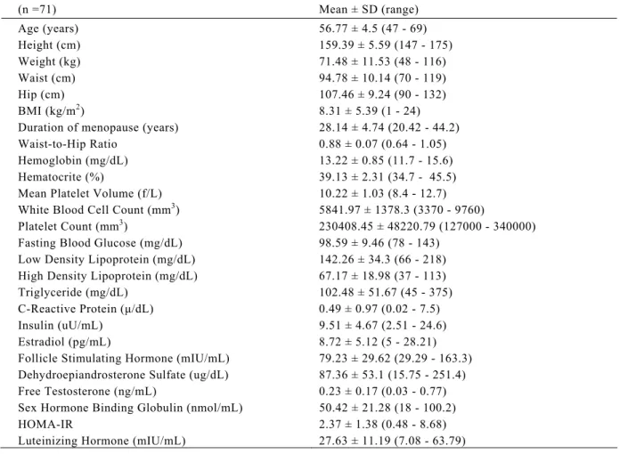
Table 1. Demographic and biochemical features of the patients
(n =71) Mean ± SD (range) Age (years) 56.77 ± 4.5 (47 - 69) Height (cm) 159.39 ± 5.59 (147 - 175) Weight (kg) 71.48 ± 11.53 (48 - 116) Waist (cm) 94.78 ± 10.14 (70 - 119) Hip (cm) 107.46 ± 9.24 (90 - 132) BMI (kg/m2) 8.31 ± 5.39 (1 - 24)
Duration of menopause (years) 28.14 ± 4.74 (20.42 - 44.2) Waist-to-Hip Ratio 0.88 ± 0.07 (0.64 - 1.05) Hemoglobin (mg/dL) 13.22 ± 0.85 (11.7 - 15.6) Hematocrite (%) 39.13 ± 2.31 (34.7 - 45.5) Mean Platelet Volume (f/L) 10.22 ± 1.03 (8.4 - 12.7) White Blood Cell Count (mm3) 5841.97 ± 1378.3 (3370 - 9760)
Platelet Count (mm3) 230408.45 ± 48220.79 (127000 - 340000)
Fasting Blood Glucose (mg/dL) 98.59 ± 9.46 (78 - 143) Low Density Lipoprotein (mg/dL) 142.26 ± 34.3 (66 - 218) High Density Lipoprotein (mg/dL) 67.17 ± 18.98 (37 - 113) Triglyceride (mg/dL) 102.48 ± 51.67 (45 - 375) C-Reactive Protein (μ/dL) 0.49 ± 0.97 (0.02 - 7.5) Insulin (uU/mL) 9.51 ± 4.67 (2.51 - 24.6) Estradiol (pg/mL) 8.72 ± 5.12 (5 - 28.21) Follicle Stimulating Hormone (mIU/mL) 79.23 ± 29.62 (29.29 - 163.3) Dehydroepiandrosterone Sulfate (ug/dL) 87.36 ± 53.1 (15.75 - 251.4) Free Testosterone (ng/mL) 0.23 ± 0.17 (0.03 - 0.77) Sex Hormone Binding Globulin (nmol/mL) 50.42 ± 21.28 (18 - 100.2)
HOMA-IR 2.37 ± 1.38 (0.48 - 8.68)
Eastern Journal of Medicine 19 (2014) 28-32 Original Article
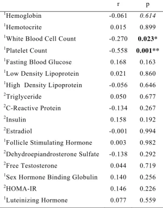
Table 2. Correlations among MPV and biochemical and hormonal parameters
r p
1Hemoglobin -0.061 0.614
1Hemotocrite 0.015 0.899
1White Blood Cell Count -0.270
0.023*
1Platelet Count -0.558 0.001** 1Fasting Blood Glucose 0.168 0.163 1Low Density Lipoprotein 0.021 0.860 1High Density Lipoprotein -0.056 0.646
2Triglyceride 0.050 0.677
2C-Reactive Protein -0.134 0.267
2Insulin 0.158 0.192
2Estradiol -0.001 0.994
1Follicle Stimulating Hormone 0.003 0.982 2Dehydroepiandrosterone Sulfate -0.138 0.292 2Free Testosterone 0.044 0.719 1Sex Hormone Binding Globulin 0.140 0.256
2HOMA-IR 0.146 0.226
1Luteinizing Hormone 0.077 0.559 1r= Pearson Correlation Coefficient *p<0.05
2r= Spearman Correlation Coefficient **p<0.01
To define metabolic syndrome (MS) we used The Third Report of the National Cholesterol Education Program (NCEP) expert panel on Detection, Evaluation and Treatment of High Blood Cholesterol in Adults (ATP-III). When ≥ 3 of the following criteria were present, patients were diagnosed with MS: 1. Systolic/Diastolic blood pressure ≥130/85mmHg or use of antihypertensive medications, 2. Waist circumference (WC) >88cm, 3. HDL-cholesterol < 50mg/dL, 4. Triglycerides ≥ 150mg/dL, 5. Fasting blood glucose ≥ 100mg/dL or use of hypoglycemic agents.
3. Results
There were 71 postmenopausal patients in the study group. Demographic and biochemical features of the patients were given in Table 1. Patients with MS (n=19) had a MPV of 10.17 ± 1.02 fL and patients without MS had a MPV of 10.23 ± 1.04 fL (p=0.818). MPV was not correlated with age (r=-0.071, p = 0.561), height (r = -0.044, p = 0.716), weight (r = 0.089, p=0.460), WHR (r = -0.036, p = 0.764), BMI (r=0.102, p=0.397) and duration of menopause (r=-0.156, p=0.194) in postmenopausal women.
The correlations among MPV, and biochemical and hormonal parameters were given in Table 2. MPV correlated with WBC (r=-0.270, p=0.023) and platelet count (r=-0.558, p=0.001). MPV correlated only with platelet count in patients with MS (r=-0.662, p=0.002).
4. Discussion
Mechanisms of arterial thrombosis and venous thrombosis are different. Arterial thrombosis develops by the adhesion of platelets to damaged endothelial surface, whereas venous thrombosis can develop without endothelial damage (11,12). The incidences of CV events are lower in premenopausal women when compared to men at the same age, but the rate increases after menopause. Platelet levels of men and women differ at every age group and decrease in both of them with aging (13). This oriented researchers to search the effects of hormones on platelet activity. Platelet aggregation changes in parallel to the changing estradiol levels of the menstrual cycle (14). Interruption of the natural menopause with HRT lead to an increase in platelet activity, microparticules originating from platelets increased (15). Another support to the relationship between platelets and hormones came from studies with selective estrogen receptor modulators, MPV increased in breast cancer patients using tamoxifen (16). These findings suggested sex hormones to have a role in CV events which might be related to platelet function instead of the coagulation cascade. To expect such an effect is logical because platelets contain both estrogenic and androgenic receptors (6). In this study we did not find a relationship between MPV, a marker of platelet activity and endogenous sex steroids and cardiovascular risk parameters and previous studies reported conflicting results about the effects of HRT on platelet functions (10-17). ERT was reported to increase MPV (9), both the estrogen and progesteron components of the oral contraceptives increased platelet aggregation (18). Our failure to detect a relationship might be due to different effects of endogenous and synthetic estrogen and progesteron on platelet functions, but in a previous study women with low levels of estradiol in the postmenopausal period had less platelet activation than premenopausal women (8).
In vitro studies showed human platelet aggregation to be enhanced by androgens (19). In vivo studies supported these findings, androgen therapy improved platelet counts in patients with myelodysplasia and thrombocytopenia (20) and
N. Güdücü et al / MPV and endogenous hormones
platelet production decreased with castration (21). DHEAS levels decrease with aging and CV events increase with aging. Women with polycystic ovary syndrome have a higher risk of CV events, they were reported to have a higher rate of MPV and their MPV correlated with DHEAS and FT levels (22). In an effort to decrease CV mortality, chronic DHEA supplementation was suggested, DHEA was expected to exert antiatherogenic effects, particularly in elderly subjects whom displayed low circulating levels of this hormone (23). In other reports physiologic doses of DHEAS prevented platelet activation and decreased risk of arterial thrombosis (24). When DHEAS was added to blood from postmenopausal women, platelet aggregation decreased (25). In our study neither DHEAS nor testosterone correlated with MPV. This suggests endogenous and synthetic hormones to exert different effects on platelets.
Limitations of this study might be the exclusion of postmenopausal women with systemic diseases, MPV might be higher in this group. The study group may be smaller to detect differences between the two groups.
In conclusion, we found no relationship among MPV and endogenous sex hormones and parameters indicating increased CV risk and we cannot suggest it as a marker of CV events in postmenopausal women.
References
1. Stampfer MJ, Willett WC, Colditz GA, et al. A prospective study of postmenopausal estrogen therapy and coronary artery disease. N Engl J Med 1985; 313: 1044-1049.
2. Rossouw JE, Anderson GL, Prentice RL, et al. Risks and benefits of estrogen plus progestin in healthy postmenopausal women: principal results from the Women’s Health Initiative randomized controlled trial. JAMA 2002; 288: 321-333.
3. Thaulow E, Erikssen J, Sandvik L, Stormorken H, Cohn PF. Blood platelet count and function are related to total and cardiovascular death in apparently healthy men. Circulation 1991; 84: 613-667.
4. Chu SG, Becker RC, Berger PB, et al. Mean platelet volume as a predictor of cardiovascular risk: a systematic review and metaanalysis. J Thromb Haemost 2010; 8: 148-156.
5. Martin JF, Trowbridge EA, Salmon G, Plumb J. The biological significance of platelet volume: its relationship to bleeding time, platelet thromboxane B2 production and megakaryocyte nuclear DNA concentration. Thromb Res 1983; 32: 443-460. 6. Khetwat G, Farday N, Nealen MI, et al. Human
megakaryocytes and platelets contain the estrogen receptor beta and androgen receptor (AR): testosterone regulates AR expression. Blood 2000; 95: 2289-2296.
7. Nagata Y, Yoshikawa J, Hashimoto A, et al. Proplatelet formation of megakaryocytes is triggered by autocrine estradiol. Genes Dev 2003; 17: 2864-2869.
8. Goodrow GJ, L’Hommedieu GD, Gannon B, Sites CK. Predictors of worsening insulin sensitivity in postmenopausal women. Am J Obstet Gynecol 2006; 194: 355-361.
9. Ranganath LR, Christofides J, Semple MJ. Increased mean platelet volume after oestrogen replacement therapy. Ann Clin Biochem 1996; 33: 555-560. 10. Thijs A, van Baal WM, van der Mooren MJ, et al.
Effects of hormone replacement therapy on blood platelets. Eur J Clin Invest 2002; 32: 613-618. 11. Libby P. Molecular bases of the acute coronary
syndromes. Circulation 1996; 91: 2844-2850. 12. Wu KK, Thiagarajan P. Role of endothelium in
thrombosis and hemostasis. Annu Rev Med 1996; 47: 315-331.
13. Santimone I, Di Castelnuovo A, De Curtis A, et al. White blood cell count, sex and age are major determinants of heterogeneity of platelet indices in an adult general population: results from the MOLI-SANI project Haematologica 2011; 96: 1180-1188. 14. Suzuki S, Matsuno K, Kondoh M. Primary
hemostasis during women's life cycle measured by Thrombostat 4000. Semin Thromb Hemost 1995; 21: 103-105.
15. Rank A, Nieuwland R, Nikolajek K, et al. Hormone replacement therapy leads to increased plasma levels of platelet derived microparticles in postmenopausal women Arch Gynecol Obstet 2012; 285: 1035-1041. 16. Karagöz B, Bilgi O, Alacacıoğlu A, et al. Mean
platelet volume increase after tamoxifen, but not after anastrazole in adjuvant therapy of breast cancer. Med Oncol 2009; 27:199-192.
17. Teede HJ, McGrath BP, Turner A, Majewski H. Effects of oral combined hormone replacement therapy on platelet aggregation in postmenopausal women. Clin Sci (Lond) 2001; 100: 207-213. 18. Poller L. Oral contraceptives, blood clotting and
thrombosis. Br Med Bull 1978; 34: 151-156. 19. Johnson M, Ramey E, Ramwell PW. Sex and age
differences in human platelet aggregation. Nature 1975; 253: 253-355.
20. Wattel E, Cambier N, Caulier MT, et al. Androgen therapy in myelodysplastic syndromes with thrombocytopenia: a report on 20 cases. Br J Haematol 1994; 87: 205.
21. Sullivan PS, Jackson CW, McDonald TP. Castration decreases thrombocytopoiesis and testosterone restores platelet production in castrated BALB/c mice: evidence that testosterone acts on a bipotential hematopoietic precursor cell. J Lab Clin Med 1995; 125: 326-333.
22. Kebapcilar L, Taner CE, Kebapcilar AG, Sari I. High mean platelet volume, low-grade systemic coagulation and fibrinolytic activation are associated with androgen and insulin levels in polycystic ovary syndrome. Arch Gynecol Obstet 2009; 280: 187-193. 23. Martina V, Benso A, Gigliardi VR, et al. Short-term
dehydroepiandrosterone treatment increases platelet cGMP production in elderly male subjects. Clin Endocrinol (Oxf) 2006; 64: 260-264.
24. Li S, Li X, Li J, et al. Experimental arterial thrombosis regulated by androgen and its receptor
Eastern Journal of Medicine 19 (2014) 28-32 Original Article
via modulation of platelet activation. Thromb Res 2007; 121: 127-134.
25. Munoz YC, Gomez GI, Moreno M, et al. Dehydroepiandrosterone prevents the aggregation of
platelets obtained from postmenopausal women with type 2 diabetes mellitus through the activation of the PKC/eNOS/NO pathway. Horm Metab Res 2012; 44: 625-631.