Original
Article
Introduction
The current guidelines recommend systematic medias-tinal lymph node sampling (MLNS) or systematic medi-astinal lymph node dissection (MLND) in patients with
resectable primary lung cancer for accurate staging and management.1) Mediastinal lymph node staging is also
reported to be a prognostic factor in case of metastatic lung cancers, therefore either MLNS or MLND are advised for patients undergoing metastasectomies for some certain types of malignancies.2) More than this, the dissection of
N1-level lymph nodes are considered to be highly import-ant due to (1) recurrence, (2) proper staging, and (3) for decision making of sleeve resection or pneumonectomy.
Anatomical pulmonary resections are performed either by thoracotomy or by minimally invasive approaches. Video-assisted thoracic surgery (VATS) and robotic- assisted thoracic surgery (RATS) are current surgical tech-niques in minimally invasive lung resections. Several authors compared thoracotomy to VATS in the dissection
Lymph Node Dissection in Surgery for Lung
Cancer: Comparison of Open vs. Video-Assisted
vs. Robotic-Assisted Approaches
Alper Toker, MD, Mehmet O˘guzhan Özyurtkan, MD, Özkan Demirhan, MD, Kemal Ayalp, MD, Erkan Kaba, MD, and Elena Uyumaz, MD
Purpose: We compared open, video-assisted and robotic-assisted thoracoscopic surgical techniques in the dissection of N1 and N2-level lymph nodes during surgery for lung cancer. Methods: This retrospective analysis is based on prospectively collected data of patients (excluding those with N2 or N3 diseases, and sleeve resections) undergoing mediastinal lymph node dissection via open (n = 96), video-assisted thoracoscopy (n = 68), and robotic-
assisted thoracoscopy (n = 106). The groups are compared according to the number of
lymph node stations dissected, the number of lymph nodes dissected, and the number of lymph nodes dissected by stations.
Results: Three techniques had similar results based on the number of the dissected N1 and N2-level lymph node stations. Robotic-assisted thoracoscopic surgery yielded signifi-cantly more lymph nodes in total (p = 0.0007), and in the number of dissected N1-level
nodes (p <0.0001). All techniques yielded similar number of mediastinal lymph nodes,
whereas robotic-assisted thoracic surgery (RATS) yielded more station #11 and #12 lymph nodes compared to the other groups.
Conclusions: In this study, robotic-assisted thoracoscopic surgery has been shown to dis-sect more lymph nodes at N1 level. However, taking the open approach as standard, we could claim that both currently robotic and video-assisted techniques may provide similar number of dissected N1 and N2-level lymph node stations.
Keywords: lymph node dissection, lung cancer, video-assisted thoracoscopy, robotic surgery,
thoracotomy
Department of Thoracic Surgery, Istanbul Bilim University Medical Faculty and Group Florence Nightingale Hospitals, Istanbul, Turkey
Received: March 29, 2016; Accepted: May 30, 2016
Corresponding author: Mehmet O˘guzhan Özyurtkan, MD. Depart-ment of Thoracic Surgery, Istanbul Bilim University and Group Florence Nightingale Hospital, Abide-i Hürriyet Cad. No: 166, ¸Si¸sli-Istanbul, Istanbul, Turkey
Email: [email protected]
©2016 The Editorial Committee of Annals of Thoracic and
Cardiovascular Surgery. All rights reserved.
of N1 and N2-level lymph nodes.3–10) Besides, there are
some reports comparing thoracotomy and RATS, and VATS and RATS.11–14) Based on these studies, the results
are controversial about the superiority of a “specific” sur-gical technique.
The above-mentioned studies, which are limited in number, compared only two modalities in the dissection of N1 and N2-level lymph nodes. Yet, there is not a specific study in the literature comparing open, video-assisted, and robotic approaches in the dissection of N1 and N2-level lymph nodes. This study was planned to compare these three modalities, taken the open approach as standard.
Materials and Methods
Patient selection
We performed a retrospective analysis on prospectively collected data of the patients with primary or metastatic lung cancers who underwent anatomic pulmonary resec-tions and mediastinal and N1-level lymph node dissection via open, video-assisted or robotic approaches, from October 2011 to March 2016. Among the patients, those who had pathologically proven N2 and/or N3 diseases, and who underwent sleeve resections were excluded. The remaining 270 patients were included in this study. This study received Institutional Review Board Approval, with individual patient consent being waived.
The patients were grouped into three according to the type of the operative approaches: 96 patients underwent thoracotomy (T), 68 patients underwent VATS, and 106 patients underwent RATS. The choice of the operative approach (open vs. minimally invasive) was at the discre-tion of the surgical team in the preoperative meetings. The decision to offer the approach, either VATS or RATS, was made according to the healthcare insurance systems, and the patients’ willingness.
Surgical technique
All operations were performed by the same surgeon. In all operations, double lumen intubation tube was placed. Thoracotomies were performed from either anterior mus-cle sparing or posterolateral thoracotomies. VATS opera-tions were performed either with 2 or 3 port techniques. RATS technique is described elsewhere,15) but generally
3 to 4 arm techniques were used including an access approach located at the 10th intercostal space posteriorly for upper lobes and anteriorly through the arm one (left side) or arm two (right side) as described by Veronesi and colleagues.12)
Mediastinal and N1-level lymph node dissection were performed by similar technique in all types of the approaches, systematic MLND was preferred. Basically, the surrounding fat tissue was resected together with the lymph nodes en bloc. Mediastinal lymph node stations that were typically dissected were 2R, 4R, 7, 8, 9 for right-sided procedures, and 5, 6, 7, 8, 9 for left-right-sided proce-dures. N1-level lymph nodes, station #10, #11 and #12 were dissected separately according to the flow during the surgery. Lymph nodes that were retrieved en bloc with the specimen or during the preresection mediastinoscopy were not added to the lymph node counting. Lymph node counting was done as previously described.16) Fissures
were divided by staplers or coagulation in all resections. Basically, for a segmentectomy operation level 11 and 12 nodes were dissected completely and sent for frozen sec-tion analysis. We defined #2 and #4 as superior mediasti-nal nodes, #5 and #6 as paraaortic nodes, #7 as subcarimediasti-nal nodes, and #8 and #9 as inferior mediastinal nodes.17)
N1-level lymph nodes were subdivided as hilar lymph nodes (#10) and level 11 and 12 lymph nodes.
Data collection
Patients demographics, operative reports, and final pathology reports were obtained from prospectively col-lected medical records. The type of the resection, the number of the lymph nodes and N1/N2 lymph node sta-tions were recorded. Three groups were compared accord-ing to (1) the number of lymph node stations dissected, (2) the number of lymph nodes dissected, and (3) the number of lymph nodes dissected by stations. To evalu-ate lymph nodes more specific to an ipsilevalu-ateral resec-tion, right and left-sided operations were investigated separately.
Statistical analysis
Excel software (Microsoft Corp, Seattle, WA) was used to analyze the data. The means and standard devia-tions of the continuous variables, and number and percent of categoric variables were given by using descriptive statistics. Categorical data were compared using the chi-squared test. To test significant differences between the groups, Shapiro-Wilk test was used to test the normality of the variables. One-way analysis of variance (ANOVA) followed by Tukey’s test was used for normally distrib-uted variables, and Kruskal-Wallis test was used for vari-ables that were not distrubuted normally. A p value of less than or equal to 0.05 was considered as statistically significant.
Results
Table 1 outlines the characteristisc of the patients in
each group. The groups were similar with respect to age, site of the disease, and the rate of primary lung cancer. The VATS and RATS group had more stage I lung cancer than the T group (53% and 60% vs. 27%, p <0.0001). While the mean pathological tumor size was similar in the VATS and RATS group, significantly larger lesions were resected in the T group (p <0.0001). Besides, more patients in the T group had pathologically proven N1 disease compared to the other groups (p = 0.02). According to the histology of the disease, significantly more patients had squamous
cell carcinoma in the T group compared to the others (p <0.001), while adenocarcinoma predominated in both VATS and RATS group compared to the T group (p = 0.0009). None of the patients in the VATS group underwent pneumo-nectomy, and the rate of pneumonectomy was only 2% in the RATS group. There were no significant differences between VATS and RATS group in terms of the type of the resection, but significantly more patients in the T group underwent pneumonectomy (18%, p <0.0001). All three groups had nearly equal rate of lobectomies, but segmentec-tomies were more common in the minimally invasive (VATS and RATS) groups compared to T group (p <0.0001). There were no significant differences between three groups in
Table 1 Characteristics of the patients
T VATS RATS p
N 96 68 106
Age (mean ± SD), years 60 ± 12 63 ± 8 62 ± 11 0.21 Male (n, %) 82 (85%) 47 (69%) 76 (72%) 0.02 Right-sided procedure (n, %) 50 (52%) 39 (57%) 64 (60%) 0.50 Diameter of the lesion (mean ± SD), cm 4.8 ± 3.0 3.5 ± 2.4 2.8 ± 1.9 <0.0001 N1 positivity (n, %) 13 (14%) 1 (1%) 9 (8%) 0.02 Number of primary lung cancer (n, %) 91 (95%) 62 (91%) 99 (93%) 0.76 Stage I lung cancer (n, %) 25 (27%) 33 (53%) 59 (60%) <0.0001 Histology (n, %)
Adenocarcinoma 25 (26%) 37 (54%) 45 (42%) 0.0009 Squamous cell carcinoma 54 (56%) 16 (24%) 34 (32%) <0.001 Carcinoid tumor 3 (3%) 4 (6%) 8 (8%) 0.39 Metastases 5 (5%) 6 (9%) 7 (7%) 0.66 Type of the resection (n, %)
Segmentectomy 3 (3%) 20 (29%) 34 (32%) <0.0001 Lobectomy 66 (69%) 45 (67%) 69 (65%) 0.86 Bilobectomy 10 (10%) 3 (4%) 1 (1%) 0.01 Pneumonectomy 17 (18%) 0 (0%) 2 (2%) <0.0001 VATS: video-assisted thoracic surgery; RATS: robotic-assisted thoracic surgery; SD: standard deviation
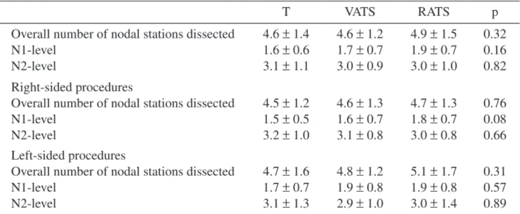
Table 2 Number of lymph node stations dissected
T VATS RATS p Overall number of nodal stations dissected 4.6 ± 1.4 4.6 ± 1.2 4.9 ± 1.5 0.32 N1-level 1.6 ± 0.6 1.7 ± 0.7 1.9 ± 0.7 0.16 N2-level 3.1 ± 1.1 3.0 ± 0.9 3.0 ± 1.0 0.82 Right-sided procedures
Overall number of nodal stations dissected 4.5 ± 1.2 4.6 ± 1.3 4.7 ± 1.3 0.76 N1-level 1.5 ± 0.5 1.6 ± 0.7 1.8 ± 0.7 0.08 N2-level 3.2 ± 1.0 3.1 ± 0.8 3.0 ± 0.8 0.66 Left-sided procedures
Overall number of nodal stations dissected 4.7 ± 1.6 4.8 ± 1.2 5.1 ± 1.7 0.31 N1-level 1.7 ± 0.7 1.9 ± 0.8 1.9 ± 0.8 0.57 N2-level 3.1 ± 1.3 2.9 ± 1.0 3.0 ± 1.4 0.89 Values are given in (mean ± SD). VATS: video-assisted thoracic surgery; RATS: robotic- assisted thoracic surgery; SD: standard deviation
terms of intraoperative complications like bleeding, hypo-tension or arrhythmia (p >0.05).
As demonstrated in Table 2, three different approaches had similar results based on the number of the dissected N1 and N2-level lymph node stations, regardless of the site of the operation. Although more lymph node stattions have been dissected, both from the left and the right, via RATS, the results were not significant (p >0.05).
Table 3 outlines the details of the number of lymph
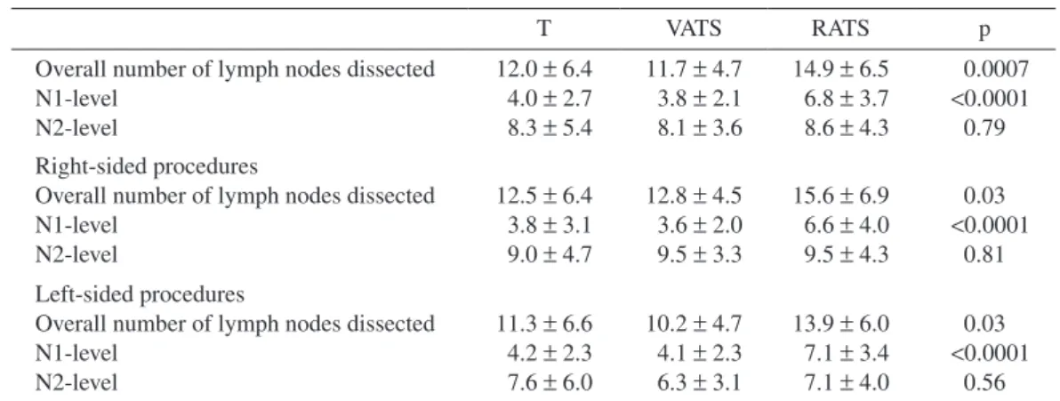
nodes dissected, both during the right-sided and the left-sided resections. RATS yielded significantly more lymph nodes in total, compared to the T and VATS groups (p = 0.0007). There was also significant difference in the num-ber of N1-level nodes dissected in the RATS group com-pared to the other groups (p <0.0001). Regarding the operation site, the total number and number of N1-level lymph nodes dissected in the RATS group were signifi-cantly higher than the other approaches (p <0.05). All the groups were same with regard to the number of overall
and N2-level lymph nodes dissected, either from the left or the right.
Overall, the mean numbers of the N1-level lymph nodes dissected during segmentectomy procedure in three different approaches were 5.0 ± 3.6, 3.4 ± 1.9, and 6.0 ± 3.0, respectively (p = 0.19). Significantly more N1-level lymph nodes were dissected during lobectomy and bilo-bectomy procedures in the RATS group (4.1 ± 2.8, 4.0 ± 2.2, and 7.3 ± 4.0, respectively with p <0.05).
When examining specific mediastinal nodal stations for all patients, there was not a significant difference in the number of nodes dissected for stations #2, #4, #7, #8 and #9 for right-sided, and #5, #6, #7, #8 and #9 for left-sided operations (Table 4). Table 5 demonstrated the number of lymph nodes dissected by N1-level stations. All three approaches resulted in similar numbers of hilar (#10) lymph nodes, but RATS yielded significantly more level 11 and 12 lymph nodes, compared to the other two approaches (p <0.0001).
Table 3 Number of lymph nodes dissected
T VATS RATS p
Overall number of lymph nodes dissected 12.0 ± 6.4 11.7 ± 4.7 14.9 ± 6.5 0.0007 N1-level 4.0 ± 2.7 3.8 ± 2.1 6.8 ± 3.7 <0.0001 N2-level 8.3 ± 5.4 8.1 ± 3.6 8.6 ± 4.3 0.79 Right-sided procedures
Overall number of lymph nodes dissected 12.5 ± 6.4 12.8 ± 4.5 15.6 ± 6.9 0.03 N1-level 3.8 ± 3.1 3.6 ± 2.0 6.6 ± 4.0 <0.0001 N2-level 9.0 ± 4.7 9.5 ± 3.3 9.5 ± 4.3 0.81 Left-sided procedures
Overall number of lymph nodes dissected 11.3 ± 6.6 10.2 ± 4.7 13.9 ± 6.0 0.03 N1-level 4.2 ± 2.3 4.1 ± 2.3 7.1 ± 3.4 <0.0001 N2-level 7.6 ± 6.0 6.3 ± 3.1 7.1 ± 4.0 0.56 Values are given in (mean ± SD). VATS: video-assisted thoracic surgery; RATS: robotic-assisted thoracic surgery; SD: standard deviation
Table 4 Number of lymph nodes dissected by mediastinal stations
T VATS RATS p Right-sided procedures
Superior mediastinal lymph nodes dissected 5.0 ± 3.6 5.6 ± 2.6 5.2 ± 3.1 0.64 Subcarinal lymph nodes dissected 3.5 ± 2.3 3.7 ± 2.0 3.8 ± 2.3 0.75 Inferior mediastinal lymph nodes dissected 0.6 ± 1.0 0.3 ± 0.5 0.5 ± 1.0 0.18 Left-sided procedures
Paraaortic lymph nodes dissected 2.7 ± 2.6 3.1 ± 1.6 2.8 ± 2.1 0.71 Subcarinal lymph nodes dissected 3.4 ± 3.3 2.4 ± 1.8 2.7 ± 1.8 0.24 Inferior mediastinal lymph nodes dissected 1.2 ± 1.4 0.8 ± 1.2 1.3 ± 1.8 0.39 Values are given in (mean ± SD). VATS: video-assisted thoracic surgery; RATS: robotic-assisted thoracic surgery; SD: standard deviation
Discussion
The excision and pathological assesment of mediasti-nal and N1-level lymph nodes in lung cancer resections is important in the prognosis and treatment. Besides non- invasive techniques, several surgical approaches includ-ing open or minimally invasive techniques, have been performed to obtain the oncologic effectivity based on the completeness of lymph node dissection.18)
Systematic MLND is considered to be the single most effective technique to decide and evaluate the extention of the nodal disease. Excision of occult mediastinal nodal dis-ease may be diagnosed with a proper MLND. The effectiv-ity of a particular technique, especially a new one, should be able to provide the similar results at least as the common and standard one (which is thoracotomy in this situation). In this study, we aimed to analyze our capability at lymph node dissection with the robotic and videothoracoscopic surgical techniques.
According to the literature, there exist several studies comparing T and VATS, T and RATS, and VATS and RATS approaches separately, based on the N1 and N2-level lymph node dissection. This study differs from the pre-vious studies, because we investigated three different approaches (T, VATS, and RATS) at the same time, and to our knowledge, this is the first study comparing these three modalities.
Several studies on patients with stage I lung cancer, demonstrated that lymph node assessment can be perfor-med as effectively by the VATS as by the T approach.3,4,6)
In a multicenter study, D’Amico et al.5) compared 199
patients undergoing VATS with 189 patients undergoing T, and found similar results. Unlike the previous studies concerning of patients with N0 diseases, the latter one included also patients with N2 and N3 diseases. In our study, we excluded patients with pathologically proven N2 or N3 diseases, and included only patients with N0 and N1 diseases.
Three recent studies compared the completeness of the lymph node dissection in patients with clinical N0 primary
lung cancers, undergoing lobectomy via VATS or T. Both Denlinger et al.,9) and Merritt et al.10) investigated the
patients with only stage I lung cancers, therefore Ramos et al.8) also included patients with stage II lung cancers in
their study. All three studies demonstrated that more lymph nodes (total and N2-level, but not N1-level) could be dis-sected with open approach. The authors concluded that thoracotomy was superior in lymph node dissection, because of the long learning curve of the VATS proce-dure.9,10) It was claimed that in VATS, more experienced
surgeon could perform a proper mediastinal lymph node dissection. Sato et al.19) mentioned that it is easier, by using
thoracotomy, to create efficient retraction of organs and sufficient working place to dissect lymph nodes located in deep groove between the superior vena cava and trachea, or between the two main bronchi and the esophagus.
However, in a multicenter study of 5620 patients under-going lobectomy with lymphadenectomy (2703 VATS vs. 2917 T), Wang et al.7) concluded that it was possible to
dissect more lymph nodes using VATS, compared to T. The authors claimed that the thoracoscopic vision pro-vides better surgical exposure and has a visual zoom effect to magnify the surgical field, and this helps to identify and expose clearly the hilar structures and mediastinal lymph node stations.
One of the benefits of robotic pulmonary resection is the superior and stable vision allowing the surgeon to perform an extensive lymphadenectomy.12) Although the number of
lymph nodes removed tended to increase with increasing experience, there were no significant differences between RATS and T in terms of lymph nodes removed even in the early experience.11,12) Then it could be speculated that, in
robotic MLND, even in the early experience, similar num-ber of lymph nodes could be dissected. This superiority, provided by the capability of robotic instruments may help to solve the experience problem faced in VATS, as described above.9,10) Louie et al.13) and Lee et al.14) also
com-pared RATS and VATS, and concluded that both approaches were similar in lymph node dissection. However we believe that the lymph node dissection in robotic surgery requires
Table 5 Number of lymph nodes dissected by N1-level stations
T VATS RATS p #10 dissected 1.3 ± 1.0 1.0 ± 0.9 1.2 ± 1.1 0.37 #11 dissected 2.4 ± 2.2 2.2 ± 1.8 4.1 ± 2.6 <0.0001 #12 dissected 0.4 ± 0.6 0.7 ± 0.9 1.6 ± 1.5 <0.0001 Sum of #11 and #12 dissected 2.8 ± 2.3 2.9 ± 1.9 5.7 ± 3.1 <0.0001 Values are given in (mean ± SD). VATS: video-assisted thoracic surgery; RATS: robotic- assisted thoracic surgery; SD: standard deviation
a sharp dissection unlike that in VATS, which is generally performed with blunt dissection.
Our team has been performing VATS lobectomy since 2006, and the surgeons have developed their capabilities of MLND with VATS. In our study, MLND capability developed on an old technique, which is VATS, was com-pared to the most recent one, which is robotic. It is inter-esting that the most recent technique provided the higher number of lymph node dissection of N1-level, but similar number of nodes were dissected at N2-level.
Our study demonstrated that, compared to T and VATS, RATS can yield more N1-level lymph nodes, in both from the right or left-sided procedures (p <0.05). As mentioned above, RATS provided the surgeon greater confidence and capability in dissecting N1 lymph nodes adjacent to the pulmonary artery.13) We believe that, there may be two
explanations for such a higher number of N1-level lymph node dissection in RATS. As a first explanation, we spec-ulate that, as the console surgeon and table surgeon are different, the console surgeon tries to provide a clear area around artery and bronchus for the table surgeon, who is going to staple these dangerous structures, and so, resects each lymph node around arteries and branches to be divided to provide a safer area. Contrary to this, in VATS and thoracotomy, the dissecting and stapling surgeons are the same persons, thus depending on the personal experi-ence, the surgeon may not need to dissect all lymph nodes to provide a clear area around artery and bronchus. The second explanation may be the greater capabilitiy of the instruments used in RATS. Robotic instruments provide sharp surgical techniques and prevent the violation of the capsule of the lymph node, and by this way the number of nodes may increase.
We demonstrated that hilar (#10) lymph node dissec-tion yielded similar results in three approaches. On the contrary, significantly more level 11 and 12 lymph nodes could be obtained by RATS, which increased the overall sum of the N1-level lymph nodes. The significant increase in the rate of segmentectomy procedures in the RATS group, was not correlated with the increased number of dissected N1-level lymph nodes.
Very few studies investigated lymph node dissection by specific nodal stations, up to now. Ramos et al.8) found
that using T, surgeon could dissect more N1-level lymph nodes on the left-sided operations compared to VATS, but did not give an explanation for this finding. Denlinger et al.9) also demonstrated that, compared to VATS, T was
advantageous significantly in the dissection of station #7, and insignificantly in that of station #5 and #6. They
concluded that it was challenging for VATS to dissect paraaortic lymph node region in lower lobectomy cases, since the visualization was at times difficult. We believe that all techniques are safe. We also believe that the stan-dard technique should be the open technique. As long as a new technique provides similar number of lymph nodes with the classical open technique, then the innovative technique may be an acceptable alternative for the MLND.
In 2013, Palade et al.6) published their study compairing
VATS and open mediastinal lymphadenectomy. They clas-sified N1 and N2-level lymph nodes into different zones, and made a detailed comparison between two approaches. They reported similar results with respect to the number of lymph nodes in each zone. They stated that although the visualization of the anatomical structures with the optic system was excellent, the subcarinal station posed the problem of a sufficient lung retraction and a clear exposure of the depth to dissect along the contralateral mainstem bronchus compared to open technique.
This study has several limitations. Being retrospective in nature is the first limitation, although the data were collected prospectively. Histology, stage and size of the tumors are different which may possibly affect the num-ber of the dissected lymph nodes. The main limitation of our study is that the types of the operations were not uni-form in three groups. But the abovementioned limitations have shown to be also valid for similar studies we have referenced, comparing the different modalities in the dis-section of the N1 and N2-level lymph nodes.
Conclusion
This study analyzes prospectively collected data on N1 and N2-level lymph node dissection in patients with lung cancer, which compares three different approaches (T, VATS, and RATS). This study is the updated form of our previous study including 170 patients (T = 60 patients, VATS = 44 patients, and RATS = 66 patients), and pre-sented before.20) In conclusion, our study shows that all
three methods can provide similar numbers of N1 and N2-level lymph node stations, and there exist no signifi-cant difference in the number of lymph nodes dissected from specific nodal stations. But, using RATS, signifi-cantly more N1-level lymph node could be dissected. Considering this is the results of an early experience of RATS technique, we can claim that RATS may have a potential in the future of lung cancer surgery, if the choice will be a minimally invasive surgery. However, we still believe that this difference should not be overrated, and
should only be considered as a sign of similar capabilities of RATS technique compared to VATS and thoracotomy techniques in the current thoracic surgery practise.
Disclosure Statement
Alper Toker and other co-authors have no cinflict of interest.
References
1) Lardinois D, De Leyn P, Van Schil P, et al. ESTS guide-lines for intraoperative lymph node staging in non-small cell lung cancer. Eur J Cardiothorac Surg 2006;
30: 787-92.
2) Shiono S, Matsutani N, Okumura S, et al. The prog-nostic impact of lymph-node dissection on lobectomy for pulmonary metastasis. Eur J Cardiothorac Surg 2015; 48: 616-21; discussion 621.
3) Sugi K, Kaneda Y, Esato K. Video-assisted thoraco-scopic lobectomy achieves a satisfactory long-term prognosis in patients with clinical stage IA lung cancer. World J Surg 2000; 24: 27-30; discussion 30-1. 4) Scott WJ, Allen MS, Darling G, et al. Video-assisted
thoracic surgery versus open lobectomy for lung can-cer: a secondary analysis of data from the American College of Surgeons Oncology Group Z0030 random-ized clinical trial. J Thorac Cardiovasc Surg 2010; 139: 976-83.
5) D’Amico TA, Niland J, Mamet R, et al. Efficacy of mediastinal lymph node dissection during lobectomy for lung cancer by thoracoscopy and thoracotomy. Ann Thorac Surg 2011; 92: 226-31; discussion 231-2. 6) Palade E, Passlick B, Osei-Agyemang T, et al. Video-
assisted vs open mediastinal lymphadenectomy for Stage I non-small-cell lung cancer: results of a prospec-tive randomized trial. Eur J Cardiothorac Surg 2013;
44: 244-9; discussion 249.
7) Wang W, Yin W, Shao W, et al. Comparative study of systematic thoracoscopic lymphadenectomy and con-ventional thoracotomy in resectable non-small cell lung cancer. J Thorac Dis 2014; 6: 45-51.
8) Ramos R, Girard P, Masuet C, et al. Mediastinal lymph node dissection in early-stage non-small cell lung cancer: totally thoracoscopic vs thoracotomy. Eur J Cardiothorac Surg 2012; 41: 1342-8; discussion 1348.
9) Denlinger CE, Fernandez F, Meyers BF, et al. Lymph node evaluation in video-assisted thoracoscopic lobec-tomy versus lobeclobec-tomy by thoracolobec-tomy. Ann Thorac Surg 2010; 89: 1730-5; discussion 1736.
10) Merritt RE, Hoang CD, Shrager JB. Lymph node eval-uation achieved by open lobectomy compared with tho-racoscopic lobectomy for N0 lung cancer. Ann Thorac Surg 2013; 96: 1171-7.
11) Cerfolio RJ, Bryant AS, Skylizard L, et al. Initial con-secutive experience of completely portal robotic pulmo-nary resection with 4 arms. J Thorac Cardiovasc Surg 2011; 142: 740-6.
12) Veronesi G, Galetta D, Maisonneuve P, et al. Four-arm robotic lobectomy for the treatment of early-stage lung cancer. J Thorac Cardiovasc Surg 2010; 140: 19-25. 13) Louie BE, Farivar AS, Aye RW, et al. Early experience
with robotic lung resection results in similar operative outcomes and morbidity when compared with matched video-assisted thoracoscopic surgery cases. Ann Thorac Surg 2012; 93: 1598-604; discussion 1604-5.
14) Lee BE, Korst RJ, Kletsman E, et al. Transitioning from video-assisted thoracic surgical lobectomy to robotics for lung cancer: are there outcomes advantages? J Thorac Cardiovasc Surg 2014; 147: 724-9.
15) Toker A, Özyurtkan MO, Kaba E, et al. Robotic anatomic lung resections: the initial experience and description of learning in 102 cases. Surg Endosc 2016; 30: 676-83. 16) Toker A, Tanju S, Ziyade S, et al. Alternative
paratra-cheal lymph node dissection in left-sided hilar lung cancer patients: comparing the number of lymph nodes dissected to the number of lymph nodes dissected in right-sided mediastinal dissections. Eur J Cardiothorac Surg 2011; 39: 974-80.
17) Watanabe A, Koyanagi T, Obama T, et al. Assessment of node dissection for clinical stage I primary lung cancer by VATS. Eur J Cardiothorac Surg 2005; 27: 745-52. 18) Goldstraw P, Crowley J, Chansky K, et al. The IASLC
Lung Cancer Staging Project: proposals for the revi-sion of the TNM stage groupings in the forthcoming (seventh) edition of the TNM Classification of malig-nant tumours. J Thorac Oncol 2007; 2: 706-14.
19) Sato Y, Tezuka Y, Kanai Y, et al. Novel retractor for lymph node dissection by video-assisted thoracic sur-gery. Ann Thorac Surg 2008; 86: 1036-7.
20) Toker A, Özyurtkan MO, Demirhan Ö, et al. Lymph node dissection in surgery for lung cancer: comparison of open vs. video-assisted vs, robotic approaches. 23rd European conference on General thoracic surgery abstract book, 2015, p. 206.