Uric Acid and Colorectal Cancer
Asian Pac J Cancer Prev, 18 (2), 485-490
Introduction
Colorectal cancer is the most common malignancy of the gastrointestinal tract (Ciombor et al., 2015). Unfortunately, despite of rapid developments in the interventional radiology, surgery, regional and systemic therapy colorectal cancer is still the third most common cause of cancer-related deaths in the United States (Ciombor et al., 2015; Compton et al., 2000). Moreover after the adjuvant treatment of stage III cancer, metastatic disease develops more than half of these patients (Ciombor et al., 2015). In addition, despite the increase of overall survival with the more frequent use of the current targeting agents such as bevacizumab, cetuximab, panitimumab, and regorafenib in the metastatic disease, 5-years survival rate is still significantly below the hope expected (Ciombor et al., 2015).
In actual studies, prognostic and predictive features of different biomarkers, which produce a wide array of molecules such as preoperative carcinoembryonic antigen (CEA), carbohydrate antigen 19-9 (CA19-9), N-RAS, K-RAS, BRAF and microRNA, was to investigate in colorectal cancer patients (Ciombor et al., 2015; Loupakis
Abstract
Purpose: In this pilot study we investigated whether serum levels of uric acid, the final breakdown product of
purine metabolism, might influence cancer-related events in stage II and III colorectal cancer patients. Material and
methods: In this cross-sectional retrospective study, a total of 150 stage II and III colorectal adenocarcinoma patients
with no exclusion criteria were included. Serum uric acid levels of the patients measured at diagnosis and demographic, clinical, laboratory analyses were performed and histologically features recorded. Patients were stratified into quartiles according to serum uric acid level with different cut-off values. Results: The mean serum uric acid level of all patients was determined o be 6.97±2.87 md/dL (range, 3.1-12.4). Median follow-up time was 98 (range 9-98) months and the proportion of patients who did not relapse was 22%. It was determined that majority of the patients who had serum uric acid levels over 8.37 mg/dLhad stage IIIB lesions, were smokers, were among those who lost weight, with metastasis to the liver and with significantly leukocyte and platelet counts. Conclusions: Serum uric acid levels measured at the time of diagnosis may be associated with cancer-related events so we can conclude a prognostic value of increase in colorectal cancer patients.
Keywords: Uric acid- colorectal cancer- prognosis
RESEARCH ARTICLE
Hyperuricemia at The Time Of Diagnosis is a Factor for Poor
Prognosis in Patients With Stage II and III Colorectal Cancer
(Uric Acid and Colorectal Cancer)
Ahmet Ozan Cetin
1, Muhyettin Omar
1, Serkan Calp
1, Hasan Tunca
1, Nevin
Yimaz
2, Burak Ozseker
2, Ozgur Tanriverdi
3*
et al., 2015).
Still the most important prognostic indicator in colorectal cancer TNM staging which was mainly based on three components including tumor penetration into the bowel wall (T), presence of nodal involvement (N), and metastasis (M) (Compton et al., 2000). In clinical practice while adjuvant treatment is recommended for stage III disease, stage II is recommended only in the presence of poor prognostic factors which are T4 stage, lymph node sampling of less than 12, clinical intestinal obstruction or perforation, poor histologic grade, and lymphovascular and perineural invasion (Ciombor et al., 2015; Compton et al., 2000; Loupakis et al., 2015). Uric acid is a last product in circle of nucleotide metabolism, which indicates process of nucleic acids catabolism along with free purine nucleotides, hypoxanthine and xanthine. With our previous knowledge, uric acid is most important molecule because of the antioxidant feature (Ames et al. 1981). However, the relationship between serum UA and cancer is not still clear as well as studies on this topic are very limited in the English literature (Fini et al., 2012). Conversely the theoretical explanation that increased serum UA levels might be protective against cancer due 1Mugla Sitki Kocman University Faculty of Medicine, Department of Internal Medicine, 2Division of Gastroentero-Hepatology, 3Division of Medical Oncology, Mugla Sitki Kocman University Faculty of Medicine, Department of Internal Medicine, Mugla. *For Correspondence: [email protected]
to its antioxidant properties, some studies reported that hyperuricemia increases cancer prevalence in mankind as well as rises up mortality among cancer patients (Ames et al. 1981; Fini et al., 2012; Tanriverdi et al., 2014). Despite these findings, a study of the literature on the relation between increased serum UA and colorectal cancer prognosis has not been reached.
In this study, we aimed to investigated whether the serum UA level’s effects of cancer-related events in stage II and III colorectal cancer patients.
Materials and Methods
In this cross-sectional study, the medical records of 198 patients with colorectal adenocarcinoma, whom diagnosis proven as histologically between 2006 and 2015 were searched retrospectively, and a total of 150 stage II and III patients with no exclusion criteria were included in the study.
Date of diagnosis, age, gender, smoking habit, weight loss within the past three months, disease stage, histological type of tumor, tumor diameter, area(s) of metastasis at the first recurrence, neoadjuvant and adjuvant treatment options of all patients were recorded.
Patients with history of diabetes mellitus, metabolic syndrome, hypertension, rheumatologic diseases, hematological malignancies, alcoholism, gout, previously chemotherapy, and/or radiotherapy applications; patients who had acute coronary syndrome or cerebrovascular diseases within the past 6 months; patients on medication for hyperuricemia and hypertension; and patients whose serum UA level or other study related data were not available in the records were excluded from the study.
Serum UA levels of the patients that measured at the time of diagnosis were recorded, and the patients were stratified into quartiles according to serum UA levels.
Blood samples for laboratory analyses were drawn following an 8- to 12-h fasting. Measurements of serum UA and lactate dehydrogenase (LDH) levels were done using the “Abbott/Aeroset system (™)” device. Hemoglobin level (Hb;), leukocyte (Leu;) and platelet counts were the other variables of the study measured by ABX-PENTRA 120 DX® Hematology Analyzer (ABX Diagnostics, France). In addition, preoperative CEA and CA19-9 were recorded.
The data are expressed as the mean ± standard deviation or the median and interquartile range (25- 75%). Statistical methods such as the Kolmogorov-Smirnov test, the independent Student’s t test, the Mann-Whitney U test, the Chi-square test, Fisher’s test, the Kruskal-Wallis test were used for analyses of data. Additionally, univariate and multivariate analyses, multiple logistic regression method for identify independent risk factors, and Kaplan–Meier curves for disease-free survival analyses were used. Statistical significance level was set as P<0.05.
Results
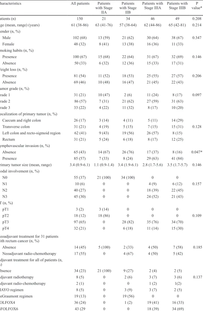
Mean serum uric acid level of all patients was determined as 6.97±2.87 mg/dL (range, 3.1-12.4). Among the patients, the majority was males (68%), smokers
(67%), patients who had lost more than 10 kilos in the past 3 months (54%), tumor grade II (57%), tumors located in the left colon and recto-sigmoid (41%), presence of lymphovascular invasion (57%), pT3 (65%), and pN0 (37%) (Table 1).
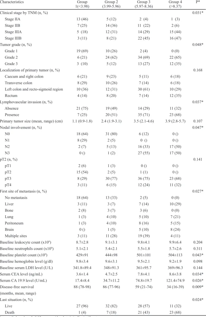
Median follow-up time was 98 (range 9-98) months and the proportion of patients who did not relapse was 22%. The first area of metastasis was multiple (25 %) among metastatic patients. Based on the analyses, patients were stratified according to quartiles of serum uric acid with cutoff values defined as <3.98 mg/dL (lowest quartile, Group 1), 3.99-5.96 mg/dL (Group 2), 5.97-8.36 mg/dL (Group 3), and >8.37mg/dL (highest quartile, Group 4) (Table 2).
It was determined that increased serum uric acid level was significantly positively correlated with stage IIA and stage IIIB (r=0.598, p=0.029), N3 disease, (r=0.618, p=0.024), grade III tumor (r=0.631, p=0.029), presence of weight loss (r=0.539, p=0.037), presence of lymphovascular invasion (r=0.621, p=0.031), recurrence with liver metastases (r=0.648, p=0.018), baseline platelet counts (r=0.496, p=0.042), baseline CEA levels (r=0.509, p=0.036), baseline CA19-9 levels (r=0.645, p=0.028), and a shorter disease-free survival rates (r=0.651, p=0.018).
Patients with serum uric acid levels over 8.37 mg/dL had a worst prognosis, illustrated as short progression-free survival compared to other groups (Table 2).
The univariate analysis conducted with models which prepared for disease-free survival yielded that serum uric acid level (Odds ratio (OR) with 95% Confidential Interval (CI) 3.41 (1.45-3.56), p=0.025), stage IIA and stage IIIB (2.45 (1.54-3.42), p=0.034 and 1.86 (1.14-3.17), p=0.032, respectively), grade III tumor (3.24 (2.18-4.34), p=0.027), presence of lymphovascular invasion (2.84 (1.91-3.76), p=0.036), N disease (3.11 (1.69-3.28), p=0.039), baseline platelet (2.41 (2.04-6.32), p=0.024), and leukocyte counts (2.48 (1.97-3.84), p=0.035) had prognostic value. Multivariate analysis showed that serum uric acid level (2.86 (1.14-5.94), p=0.023) and presence of Lymphovascular invasion (3.18 (2.09-4.93), p=0.024) were independent prognostic factors.
Discussion
In this cross-sectional study we investigated serum UA levels that measured at the time of diagnosis whether the effects of cancer-related events in total 150 stage II and III colorectal cancer patients. We determined that the patients included in our study who had serum UA levels over 8.37 mg/dL, metastases have developed in a shorter time and first metastatic area was liver.
Increased serum UA level was first supposed to have a possible protective effect against developing of cancer among mankind by Ames et al., (1981). This hypothesis was based on the knowledges that lipid peroxidation is inhibited and the free oxygen radicals are cleared through xanthine oxidoreductase when UA is in levels with high concentrations. (Tanriverdi et al., 2014). However, in some recent studies, antioxidants have been reported to act as prooxidants in certain cases (Giavonnucci, 2007; Petersson et al., 1983; Petersson et al., 1984; Shin et al.,
Uric Acid and Colorectal Cancer
Characteristics All patients Patients
with Stage IIA Patients with Stage IIB Patients with
Stage IIIA Patients with Stage IIIB value*P
Patients (n) 150 21 34 46 49 0.208
Age (mean, range) (years) 61 (38-86) 63 (41-76) 57 (38-64) 62 (44-86) 65 (42-81) 0.214 Gender (n, %) Male 102 (68) 13 (59) 21 (62) 30 (64) 38 (67) 0.347 Female 48 (32) 8 (41) 13 (38) 16 (36) 11 (33) Smoking habits (n, %) Presence 100 (67) 15 (68) 22 (64) 31 (67) 32 (69) 0.146 Absence 50 (33) 6 (32) 12 (36) 15 (33) 17 (31) Weight loss (n, %) Presence 81 (54) 11 (52) 18 (53) 25 (55) 27 (57) 0.206 Absence 69 (46) 10 (48) 16 (47) 21 (45) 22 (43) Tumor grade (n, %) Grade 1 31 (21) 10 (47) 2 (6) 11 (24) 8 (17) 0.097 Grade 2 86 (57) 7 (31) 21 (62) 27 (59) 31 (63) Grade 3 33 (22) 4 (22) 11 (32) 8 (17) 10 (20)
Localization of primary tumor (n, %)
Caecum and right colon 26 (17) 3 (14) 4 (11) 5 (11) 14 (29)
Transverse colon 31 (21) 4 (19) 5 (15) 7 (15) 15 (31) 0.128
Left colon and recto-sigmoid region 62 (41) 9 (43) 19 (56) 26 (57) 8 (15)
Rectum 31 (21) 5 (24) 6 (18) 8 (17) 12 (25)
Lymphovascular invasion (n, %)
Absence 65 (43) 14 (67) 26 (76) 17 (37) 8 (16) 0.047*
Presence 85 (57) 7 (33) 8 (24) 29 (63) 41 (84)
Primary tumor size (mean, range) 3.4 (0.9-6.1) 1.1 (0.9-1.4) 3.4 (1.9-6.1) 2.8 (1.7-5.6) 3.5 (1.7-5.7) 0.146 Nodal involvement (n, %) N0 55 (37) 21 (100) 34 (100) 0 0 N1 10 (6) 0 0 4 (9) 6 (12) 0.157 N2 40 (27) 0 0 18 (39) 22 (45) N3 45 (30) 0 0 24 (52) 21 (43) pT (n, %) pT1 3 (2) 3 (14) 0 0 0 pT2 18 (12) 18 (86) 0 0 0 0.109 pT3 97 (65) 0 28 (82) 35 (76) 34 (70) pT4 32 (21) 0 6 (18) 11 (14) 15 (30)
Neoadjuvant treatment for 31 patients with rectum cancer (n, %)
Absence 14 (45) 5 (100) 2 (33) 4 (50) 7 (58) 0.185
Neoadjuvant radio-chemotherapy 17 (55) 0 4 (67) 4 (50) 5 (42)
Adjuvant treatment for all of patients (n, %) Absence 34 (23) 21 (100) 9 (27) 2 (4) 2 (5) Adjuvant radiotherapy 8 (5) 0 2 (6) 3 (7) 3 (6) 0.137 Adjuvant radio-chemotherapy 2 (1) 0 0 1 (2) 1(2) MAYO regimen 8 (5) 0 3 (9) 3 (7) 2 (5) DeGraumont regimen 19 (13) 0 19 (56) 0 0 FOLFOX4 36 (24) 0 1 (2) 19 (41) 16 (33) mFOLFOX6 43 (29 0 0 18 (39) 34 (69)
Table 1. The Baseline Demographical, Histological, Clinical, and Laboratories Characteristics of All Patients in This Study
Characteristics All patients Patients with
Stage IIA Patients with Stage IIB Patients with Stage IIIA Patients with Stage IIIB value*P
Recurrence rate (%) 78 14 68 91 100 0.014* First site of metastasis (n, %) No metastasis 33 (22) 18 (86) 11 (32) 4 (8) 0 (0) Liver 23 (15) 0 3 (9) 9 (20) 11 (23) Bone 8 (6) 0 2 (6) 3 (6) 3 (6) 0.037* Lung 17 (11) 0 2 (6) 8 (18) 7 (14) Peritoneum 18 (12) 3 (14) 6 (18) 7 (15) 2 (4) Brain 14 (9) 0 1 (2) 8 (18) 5 (10) Multiple sites 37 (25) 0 9 (27) 7 (15) 21 (43) Baseline leukocyte count (x109) 9.7±7.2 6.4±4.3 6.7±4.2 8.1±3.9 9.2±6.8 0.034* Baseline neutrophils count (x109) 5.8±2.1 4.4±1.3 4.7±1.6 5.4±1.9 5.6±2.3 0.041* Baseline platelet count (x109) 499 ± 112 424±98 446±94 465±97 498±114 0.036* Baseline hemoglobin level (g/dl) 9.8±2.8 9.7±2.1 9.5±2.6 9.6±2.9 9.4±2.7 0.264
Baseline serum uric
acid level (mg/dL) 9.67±4.87 5.64±1.78 6.44±2.46 7.98±2.59 9.46±5.21 0.024* Baseline serum LDH
level (U/L) 368.4±98.5 345.2±94.4 358.3±91.4 354.6±94.3 367.8±98.9 0.064
Serum CEA level
(ng/mL) 11.4±9.7 1.94±0.58 1.96±0.89 5.6±4.3 11.2±6.8 0.042* Serum CA 19.9 level (U/mL) 112.4±84.7 21.6±6.8 24.9±7.4 48.9±24.9 119.25±86.2 0.038* Follow-up period (months) (mean, range) 61 (18-65) Estimated
disease-free survival rates (95% CI; number still at risk)
86 (32-92) 79 (24-82) 57 (18-76) 38 (16-47) 0.036*
1-year rates 89 (51-100; 150) 100 (100-100; 21) 100 (100-100; 34) 95 (86-100; 46) 75 (51-100; 49)
3-year rates 64 (34-100; 98) 95 (93-100; 20) 89 (81-100; 32) 74 (69-100; 30) 42 (34-100; 16) 0.015* 5-year rates 48 (0-100; 33) 86 (81-100; 18) 71 (68-100; 11) 59 (54-100; N/A
Last situation (n, %)
Live 98 (65) 20 (95) 31 (91) 24 (52) 23 (47)
Death 52 (35) 1 (5) 3 (9) 22 (48) 26 (53) 0.032*
*A two tailed p value of <0.05 was considered statistically significant
Table 1. Continued
Characteristics Group 1 (<3.98) Group 2 (3.99-5.96) Group 3 (5.97-8.36) Group 4 (>8.37) P*
Patients (n) 28 39 49 34 0.114
Age (mean, range) (years) 64 (41-78) 52 (38-64) 61 (38-83) 66 (43-86) 0.218
Gender (n, %) 0.094 Male 19 (68) 21 (54) 36 (74) 26 (77) Female 9 (32) 18 (46) 13 (26) 8 (23) Smoking habits (n, %) 0.116 Presence 14 (50) 23 (60) 36 (73) 27 (79) Absence 14 (50) 16 (40) 13 (27) 7 (21)
Table 2. Comparison of Characteristics of Demographical, Histological, and Clinical and Analyses of Survival According to the Divided the Level of Serum Uric Acid
Uric Acid and Colorectal Cancer
Characteristics Group
1(<3.98) (3.99-5.96)Group 2 (5.97-8.36)Group 3 Group 4(>8.37) P*
Clinical stage by TNM (n, %) 0.031* Stage IIA 13 (46) 5 (12) 2 (4) 1 (3) Stage IIB 7 (25) 14 (36) 11 (22) 2 (6) Stage IIIA 5 (18) 12 (31) 14 (29) 15 (44) Stage IIIB 3 (11) 8 (21) 22 (45) 16 (47) Tumor grade (n, %) 0.048* Grade 1 19 (69) 10 (26) 2 (4) 0 (0) Grade 2 6 (21) 24 (62) 34 (69) 22 (65) Grade 3 3 (10) 5 (12) 13 (27) 12 (35)
Localization of primary tumor (n, %) 0.168
Caecum and right colon 6 (21) 9 (23) 5 (11) 6 (18)
Transverse colon 8 (29) 10 (26) 7 (14) 6 (18)
Left colon and recto-sigmoid region 10 (36) 12 (31) 30 (61) 10 (29)
Rectum 4 (14) 8 (20) 7 (14) 12 (35)
Lymphovascular invasion (n, %) 0.037*
Absence 21 (75) 19 (49) 14 (29) 11 (32)
Presence 7 (25) 20 (51) 35 (71) 23 (68)
Primary tumor size (mean, range) (cm) 1.1 (0.9-1.8) 2.4 (1.9-3.1) 3.5 (2.1-4.6) 3.9 (2.8-5.7) 0.107
Nodal involvement (n, %) 0.047* N0 18 (64) 31 (80) 6 (12) 0 () N1 8 (29) 2 (5) 0 () 0 () N2 2 (7) 5 (13) 16 (33) 17 (50) N3 0 () 1 (2) 27 (55) 17 (50) pT2 (n, %) 0.141 pT1 2 (6) 1 (3) 0 () 0 () pT2 15 (54) 2 (5) 1 (1) 0 () pT3 8 (29) 30 (77) 36 (75) 23 (68) pT4 3 (11) 6 (15) 12 (24) 11 (32)
First site of metastasis (n, %) 0.027*
No metastasis 18 (64) 13 (33) 2 (5) 0 (0) Liver 3 (11) 3 (7) 7 (14) 10 (29) Bone 2 (8) 3 (7) 3 (6) 0 (0) Lung 1 (3) 4 (10) 5 (10) 7 (21) Peritoneum 1 (3) 4 (10) 8 (16) 5 (15) Brain 0 () 1 (5) 5 (10) 8 (24) Multiple sites 3 (11) 11 (28) 19 (39) 4 (11)
Baseline leukocyte count (x109) 8.7±2.8 9.1±3.1 9.8±4.1 9.9±6.4 0.204
Baseline neutrophils count (x109) 5.1±2.1 5.4±2.1 5.5±1.8 5.7±2.6 0.311
Baseline platelet count (x109) 429±91 444±98 501±101 504±113 0.043*
Baseline hemoglobin level (g/dl) 9.8±3.4 9.6±3.1 9.5±2.1 9.2±1.9 0.098
Baseline serum LDH level (U/L) 341.8±89.4 348±91.3 361±95.7 369±96.3 0.144
Serum CEA level (ng/mL) 3.6±1.4 4.7±2.5 7.8±4.1 8.6±3.8 0.034*
Serum CA 19.9 level (U/mL) 17.4±8.4 34.7±11.2 74.8±19.7 121.4±74.9 0.026*
Disease-free survival 88 (78-98) 86 (77-96) 59 (21-74) 34 (16-39) 0.009*
(months, mean, range)
Last situation (n, %) 0.024*
Live 27 (96) 32 (82) 28 (57) 11 (32)
Death 1 (4) 7 (18) 21 (43) 23 (68)
Table 2. Continued
Conflicts of interest None
Preliminary results of this study were presented as a poster in 16th ESMO World Congress on Gastrointestinal Cancer, in Barcelona, 2014 (Ann Oncol 2014; 25 (suppl 2): ii14-ii104. 10.1093/annonc/mdu165).
References
Abdel-Salam OME, Youness ER, Hafez HF (2011). The antioxidant status of the plasma in patients with breast cancer undergoing chemotherapy. Open J Mol Integr Physiol, 1, 29-35.
Ames BN, Catheart R, Schwiers E, Hochstein P (1981). Uric acid provides an antioxidant defense in humans against oxidant- and radical-caused aging and cancer: a hypothesis.
Proc Natl Acad Sci U S A, 78, 6858-62.
Bozkir A, Simsek D, Gungor A, Torun M (1999). Ascorbic acid and uric acid levels in lung cancer patients. J Clin Pharm
Ther, 24, 43-7.
Ciombor KK, Wu C, Goldberg RM (2015). Recent therapeutic advances in the treatment of colorectal cancer. Annual
Reviews, 66, 83-95.
Compton CC, Fielding LP, Burgart LJ, et al (2000). Prognostic factors in colorectal cancer. college of American pathologists consensus statement 1999. Arch Pathol Lab Med, 124, 979-94.
Fini MA, Elias A, Johnson RJ, Wright RM (2012). Contribution of uric acid to cancer risk, recurrence, and mortality. Clin
Transl Med Clinical and Translational Medicine, 1, 16-30.
Giavonnucci E (2007). Metabolic syndrome, hyperinsulinemia, and colon cancer: a review. Am J Clin Nutr, 83, 836-2 Loupakis F, Yang D, Yau L, et al (2015). Primary tumor
localization as a prognostic factor in metastatic colorectal cancer. JNCI J Natl Cancer Inst, 107, dju427.
Lawol AO, Kolude B, Adeyemi BF (2012). Serum uric acid levels in oral cancer patients seen at tertiary institution in Nigeria. Ann Ibd Pg Med, 10, 9-12.
Petersson B, Trell E (1983). Raised serum urate concentration as risk factor for premature mortality in middle-age men: relation to death from cancer. Br Med J (Clin Res Ed), 287, 7-9
Petersson B, Trell E, Henningsen NC, Hood B (1984). Risk factors for premature death in middle-age men. Br Med J
(Clin Res Ed), 288, 1264-8.
Rose DP, Hoffner SM, Baillargeon J (2007). Adiposity, the metabolic syndrome, and breast cancer in African-American and white American women. Endocr Rev, 28, 763-7. Shin HS, Lee HR, Lee DG, et al (2006). Uric acid as a prognostic
factor for survival time: a prospective cohort study of terminally ill cancer patients. J Pain Symptom Manage,
31, 493-501.
Strasak AM, Rapp K, Hilbe W, Oberaigner W (2007). The role of serum uric acid as an antioxidant protecting against cancer: prospective study in more than 28,000 older Austrian women. Ann Oncol, 18, 1893-7.
Tanriverdi O, Cokmert S, Oktay E, et al (2014). Prognostic significance of the baseline serum uric acid level in non-small cell lung cancer patients treated with first-line chemotherapy: a study of the Turkish Descriptive Oncological Researches Group. Med Oncol, 31, 217.
2006). Especially in studies conducted on cardiovascular diseases, uric acid has been shown to act as an antioxidant in early stages of atherosclerosis but may also contribute to the pro-inflammatory effects in later stages (Ames et al., 1981; Rose et al., 2007; Tanriverdi et al., 2014). When all these results are considered, the role of uric acid in cancer biology can be assumed to be more complicated than that can be explained with its common antioxidant features.
The association between serum UA level and cancer has not been clarified yet and studies on this matter are highly limited (Ames et al., 1981). Several studies have demonstrated that premature cancer mortality rates increased significantly in both genders when urate level, a dominant monosodium form of uric acid in physiological pH, were high (Ames et al., 1981; Rose et al., 2007; Tanriverdi et al., 2014). In other studies analyzing all types of cancer, serum uric acid level was demonstrated to have a significant epidemiologic association with age, gender, and increased mortality (Ames et al., 1981; Rose et al., 2007; Tanriverdi et al., 2014). Moreover, this association may also be assumed to be affected by conditions related to increased serum UA levels such as diabetes, cardiovascular diseases, metabolic syndrome, gout, obesity, insulin resistance, alcoholism, and diet (Ames et al., 1981; Rose et al., 2007; Tanriverdi et al., 2014). Many studies have proven that obesity and metabolic syndrome, co-morbidities associated with serum UA level, are significant prognostic factors especially for breast cancer and that they are associated with reduced survival and increased mortality rate (Ames et al., 1981; Petersson et al., 1984). In this study, we excluded such conditions by defining them among the exclusion criteria.
Prior studies have shown that reduced or absent XOR in tumor cells was significantly associated with poor clinical prognosis and reduced survival in breast, stomach, colorectal, ovarian, and NSCLC cancers (Abdel-Salam et al., 2011; Ames et al., 1981; Bozkir et al., 1999; Lawol et al., 2012; Strasak et al., 2007; Rose et al., 2007; Tanriverdi et al., 2014). As a result of these studies, XOR activity was reported to be reduced in highly aggressive tumor cells and that this condition is associated with both carcinogenesis and metastasis as much as it is with the unexpected impacts on the cell differentiation (Ames et al., 1981; Tanriverdi et al., 2014).
In conclusion, the association between serum UA level and poor prognosis and increased mortality among cancer patients is not clear and a study about colorectal cancer has not been demonstrated. However several of the prior studies reported that the significant increase in serum UA levels in some cancer patients may be associated with the malignant process itself and that the high mortality rate is associated with aggressive cancers where increased nucleic acid turnover is observed (Ames et al., 1981; Rose et al., 2007; Tanriverdi et al., 2014).
The relatively small sample size and the analysis of overall survival has not been made because of not enough data for statistical analysis of all groups in our study may cause limitations. Even so we can conclude that in stage IIIA/ IIIB colorectal cancer patients, who have high serum UA levels, may develop early metastases independent of all the variables.