Access this article online
Quick Response Code:
Website: www.njcponline.com
DOI: 10.4103/1119-3077.187311
How to cite this article: Ekingen E, Yilmaz M, Yildiz M, Atescelik M, Goktekin MC, Gurger M, Alatas OD, Basturk M, Ilhan N. Utilization of glial fibrillary acidic protein and galectin-3 in the diagnosis of cerebral infarction patients with normal cranial tomography. Niger J Clin Pract 2017;20:433-7.
This is an open access article distributed under the terms of the Creative Commons Attribution-Non Commercial-Share Alike 3.0 License, which allows others to remix, tweak, and build upon the work non-commercially, as long as the author is credited and the new creations are licensed under the identical terms.
For reprints contact: [email protected] Acceptance Date: 09-06-2016
Objective: It was aimed to determine whether levels of glial fibrillary acidic protein (GFAP) and Galectin-3 contribute to the diagnosis of cerebral infarction in clinically suspected ischemic stroke patients with normal computerized cranial tomography (CCT) in the emergency department. Materials and Methods: In this study, patients above the age of 18 years who presented to emergency department of Firat University between December 2011-November 2012 and were diagnosed with cerebral infarction were included. Exclusion criteria were as follows: symptom onset exceeding 24 hours, trauma, pregnancy, acute myocardial infarction, acute pulmonary embolism, chronic renal insufficiency and steroid therapy. Results: A total of 90 participants, forty patients with ischemic infarction who were diagnosed by CCT and clinical findings (Normal CCT in 17 patients and CCT with an area of infarction in 23 patients) and fifty healthy controls, were included in this study. Compared with the control group, levels of Galectin-3 and GFAP were found to be significantly increased in patients with ischemic infarction (P <0.001 and P = 0.01, respectively). It was found that levels of Galectin-3 and GFAP were significantly increased in ischemic stroke patients with normal CCT compared to the control group (P = 0.04 and P = 0.025, respectively). In ROC curve analysis, we detected %70.59 sensitivity and 70% specificity (AUC = 0.684, P = 0.0213, 95% CI: 0,558-0.792) with a cutoff value of 33.24 ng/ml for GFAP and 76.47% sensitivity and 68% specificity (AUC = 0.734, P = 0.0048, 95% CI: 0.611-0.834) with a cutoff value of 0.84 ng/ml for Galectin-3. No correlation was found between National Institutes of Health Stroke Scale (NIHSS) scores and Galectin-3 and GFAP (r = 0.251, P = 0.118 and r = 0.164, P = 0.311, respectively). Conclusion: The levels of Galectin-3 and GFAP were increased in acute ischemic stroke patients.
Utilization of Glial Fibrillary Acidic Protein and Galectin-3 in the
Diagnosis of Cerebral Infarction Patients with Normal Cranial
Tomography
Address for correspondence: Dr. M Yilmaz, Department of Emergency Medicine, School of Medicine, Firat University, 23200 Elazig, Turkey. E-mail: [email protected]
to the central nervous system, and thus, an increase in GFAP expression is observed.[5] Increased levels of
GFAP have been demonstrated in studies on ischemic stroke patients, trauma patients with accompanying brain damage, and discrimination of ischemic stroke and intracerebral hemorrhage.[1,6-10] GFAP has been reported to
increase to its maximum levels in 2–4 days in ischemic stroke patients.[1,11] However, there are also studies
indicating increased levels of GFAP within first 6 h.[6,8]
I
ntroductIonS
troke, included in cerebrovascular diseases, is one of the most important causes of mortality and disability. To determine prognosis and clarify the pathophysiology, biomarker studies are carried out in the diagnosis of cerebrovascular diseases.[1-3]Glial fibrillary acidic protein (GFAP) and galectin-3 are among these biomarkers. GFAP, abundant in the cytoplasm of fibrous astrocytes in the brain, is a 50-kDa intracellular protein and is an intermediate filament that is essentially expressed by astrocytes. It is also found in oligodendroglia, ependymal cells, and Schwann cells.[1,4]
Glial cells initiate a cellular response in case of an injury
E Ekingen, M Yilmaz, M Yildiz, M Atescelik, MC Goktekin, M Gurger, OD Alatas1, M Basturk2, N Ilhan3
A
bstr
A
ct
Keywords: Galectin-3, glial fibrillary acidic protein, ischemic stroke
Department of Emergency Medicine, Batman State Hospital, Batman, 1Department of Emergency Medicine, Mugla Sitki Kocman University, Mugla, 2Department of Emergency Medicine, Kanuni Sultan Suleyman Training and Research Hospital, Istanbul, 3Department of Biochemistry, School of Medicine, Firat University, Elazig, Turkey
Galectin-3 is approximately 30-kDa and is a member of the beta-galactoside-binding lectin family.[12] Galectin-3,
which is found on the cell surface and in the cytoplasm, has been reported to be expressed by basophils, mast cells, eosinophils, neutrophils, and peripheral blood monocytes that differentiate into macrophages.[13]
Galectin-3 expression actively regulates cell proliferation, cell differentiation, and activation of viral proteins during inflammation.[14-16] Galectin-3 levels and
galectin-3 positive cells have been reported to increase in atherosclerotic lesions that are rich in foam cells. Galectin-3 positive cells were close to a lipid core, or to the areas with fibrosis, hemorrhage, or thrombosis in these atherosclerotic lesions.[17] Furthermore, the levels
of galectin-3 have been reported to increase in ischemic brain damage, and it was proposed that it might play a role in the postischemic tissue remodeling by taking part in angiogenesis and neurogenesis.[18-20]
Computerized cranial tomography (CCT) remains the mainstay of imaging modality for diagnosis in the emergency department. Detection of pathological changes is possible typically after 6–18 h although ischemia can be visible within the first 2 h. Diffusion-weighted magnetic resonance imaging (MRI) is more sensitive than CT and is frequently used for the diagnosis of ischemic stroke.[21-23]
In this study, it was aimed to determine whether levels of GFAP and galectin-3 contribute to the diagnosis of cerebral infarction in clinically-suspected ischemic stroke patients with normal CCT in the emergency department.
mAterIAls And methods
A total of ninety participants, forty patients who presented to Emergency Department of Firat University between December 2011 and November 2012 and fulfilled the WHO cerebral infarction criteria and fifty healthy controls, were included in this study. Patients with determined ischemic stroke were divided into two groups, namely ischemic stroke patients with a normal CCT (n = 17) and ischemic stroke patients with areas of infarction on CCT (n = 23). The healthy control group included healthy volunteers. Local Ethics Committee approval was obtained before the study was initiated. In addition, written consent was obtained from all participants enrolled in the study.
Inclusion criteria were as follows: Being diagnosed as having cerebral infarction, 18 years of age and over, acute onset, having persistent complaints, admission within 24 h, nontrauma patients, and patients who agreed to participate in the study. Patients with trauma, pregnancy, acute myocardial infarction, acute pulmonary embolism, chronic renal failure, and patients on steroid
therapy were excluded from the study.
Patient data and National Institutes of Health Stroke Scale (NIHSS)[24] scores were recorded in the standard
data form. NIHSS is a tool used to examine neurological function in patients with ischemic stroke and gives an idea about the long-term prognosis. The NIHSS is subdivided into five major areas including the level of consciousness, visual assessment, motor functions, sensory function and neglect, and cerebellar functions. The individual scores from each item are summed to calculate a patient’s total NIHSS score. The maximum possible score is 42, with the minimum score being a 0.[25,26]
Five milliliter blood samples were collected from the antecubital veins for serum GFAP and galectin-3 level measurements. Serum samples were allowed to clot 10–15 min and the sera were collected following centrifugation for 10 min at 1000 × g. The obtained sera were stored at -70°C for later use after being placed in Eppendorf tubes for each patient.
Measurement of serum galectin-3 levels
Serum galectin-3 levels were analyzed via Human Galectin-3 Platinum Elisa Kit (Catalog No: BMS279/2, Bender MedSystems GmbH, Vienna, Austria) in accordance with the analysis procedure specified in the catalog. Absorbance was read spectrophotometrically via ELx800™ Absorbance Microplate Reader at 450 nm. BioTek. ELx50™ Microplate Strip Washer was used as an automatic microplate washer.
Measurement of serum glial fibrillary acidic protein levels
Serum GFAP levels were analyzed via Human Glial Fibrillary Acidic Protein Elisa Kit (Catalog No: E90068hu, Uscn Life Science Inc., Wuhan, China) in accordance with the analysis procedure specified in the catalog. Absorbance was read spectrophotometrically via ELx800™ Absorbance Microplate Reader at 450 nm. BioTek™ ELx50™ Microplate Strip Washer was used as an automatic microplate washer.
Statistical analysis
Statistical analysis was performed using SPSS (21.0 software, Chicago, IL, USA). Numerical data, qualitative data, and nonnormally distributed data were expressed as mean ± standard deviation, percentage, and median (inter quantifier ratio, 25–75%), respectively. Kolmogorov– Smirnov and the Shapiro–Wilk normality tests were used to determine the distribution of continuous variables. Mann–Whitney U-test was used in determining the relationship between two independent, nonnormally distributed groups. Receiver operating characteristic (ROC) curve analysis was used to assess the utilization
Compared with the control group, significantly increased levels of galectin-3 and GFAP were detected in cerebral infarction patients (P < 0.001 and P < 0.001, respectively). In addition, no significant difference was detected between clinically-suspected ischemic stroke patients with normal CCT and patients with areas of infarction on CCT in terms of galectin-3 and GFAP (P = 0.366 and P = 0.924, respectively).
The levels of galectin-3 and GFAP were significantly increased in clinically-suspected ischemic stroke patients with normal CCT compared to the control group (P = 0.04 and P = 0.025, respectively). A ROC curve analysis was performed to assess the usefulness of galectin-3 and GFAP levels in assisting the diagnosis of ischemic stroke in clinically-suspected ischemic stroke patients with normal CCT. In ROC curve analysis, we detected 70.59% sensitivity and 70% specificity (AUC = 0.684, P = 0.0213, 95% CI: 0.558–0.792) with a cutoff value of 33.24 ng/ml for GFAP, and 76.47% sensitivity and 68% specificity (AUC = 0.734, P = 0.0048, 95% CI: 0.611– 0.834) with a cutoff value of 0.84 ng/ml for galectin-3 were detected [Figure 1].
No correlation was found between NIHHS, and galectin-3 and GFAP (r = 0.251, P = 0.118 and r = 0.164, P = 0.311, respectively) [Table 3].
of levels of GFAP and Galectin-3 in the diagnosis of ischemic stroke. ROC curve analysis results were expressed as specificity (%), sensitivity (%), area under the curve (AUC), P value, and 95% confidence interval (CI). Spearman’s correlation test was used to determine the correlation of scales and scores that consist of ordinal data. P < 0.05 was considered statistically significant.
results
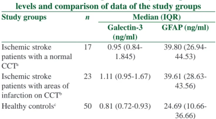
In this study, data of 40 patients diagnosed with ischemic infarction (17 patients with a normal CCT and 23 patients with areas of infarction on CCT) and 50 healthy individuals were analyzed. General characteristics of the patients with ischemic stroke were given in Table 1. Galectin-3 and GFAP levels and comparisons were given in Table 2.
Figure 1: Receiver operating characteristic curve of serum glial fibrillary
acidic protein and serum galectin-3
Table 1: Demographic and clinical characteristics of patients with ischemic stroke
Ischemic stroke patients (n=40)
Gender female (%) 21 (52.5)
Age median (mean±SD) 70.72±9.8 Admission time (Median)
(IQR) 180 (90-300) History of CVD, n (%) 8 (20) History of HT, n (%) 25 (62.5) History of DM, n (%) 6 (15) History of IHD, n (%) 16 (40) History of hyperlipidemia, n (%) 22 (55) Atrial fibrillation, n (%) 4 (10) Smoking, n (%) 3 (7.5)
NIHSS (Median) (IQR) 3 (1.25-4)
CVD=Cerebrovascular disease; HT=Hypertension; DM=Diabetes mellitus; IHD=İschemic heart disease; NIHSS=National Institutes of Health Stroke Scale; SD=Standard deviation; IQR=Inter quantifier ratio
Table 2: Galectin-3 and glial fibrillary acidic protein levels and comparison of data of the study groups
Study groups n Median (IQR)
Galectin-3 (ng/ml)
GFAP (ng/ml)
Ischemic stroke patients with a normal CCTa 17 0.95 (0.84-1.845) 39.80 (26.94-44.53) Ischemic stroke
patients with areas of infarction on CCTb
23 1.11 (0.95-1.67) 39.61 (28.63-43.56) Healthy controlsc 50 0.81 (0.72-0.93) 24.69
(10.66-36.66) For galectin-3; P=0.366 (comparison of Groups a and b) and P<0.001 (comparison of Groups a and c); For GFAP; P=0.924 (comparison of Groups a and b) and P<0.001 (comparison of Groups a and c). GFAP=Glial fibrillary acidic protein; IQR=Interquantifier ratio; CCT=Computerized cranial tomography
Table 3: Correlation table of National Institutes of Health Stroke Scale score with galectin-3 and glial
fibrillary acidic protein
NIHSS Galectin-3 GFAP
Sperman's correlation coefficient (r)
0.251 0.164
P 0.118 0.311
GFAP=Glial fibrillary acidic protein; NIHSS=National Institutes of Health Stroke Scale
to increase galectin-3 levels during inflammation. This information shows that galectin-3 is affected seriously from physiological and pathological events and that it is involved in inflammatory responses.[28] In an
experimental animal study by Yang et al.[16] on induced
ischemic brain damage in mice, they reported increased levels of galectin-3 and suggested that it might play a role in the postischemic tissue remodeling by taking part in angiogenesis and neurogenesis. There are not enough clinical studies in the literature regarding galectin-3 levels in ischemic stroke patients. We detected that galectin-3 levels could be used to support the diagnosis of ischemic stroke, especially during acute phase, where patients have normal CCT with no visible ischemic lesions.
conclusIon
The levels of galectin-3 and GFAP were increased in acute ischemic stroke patients. Furthermore, the levels of galectin-3 and GFAP were increased in the early stages of acute ischemic stroke patients that CCT was normal.
Study limitations
Limitations of our study are as follows: (1) The volumes of the ischemic area on CCT were not calculated (2) the affected cranial artery was not identified (3) absence of diffusion MRI of patients with clinically-suspected infarction but with normal CCT (4) lower numbers of clinically-suspected ischemic stroke patients with normal CCT (5) control group including healthy individuals (without chronic illness). For the use of inflammatory markers, it might be more appropriate if the patient and the control groups included individuals with similar clinical characteristics. Repetitive measurements of biomarkers at certain hours would provide guidance regarding biomarker levels in response to treatment and how long inflammation lasts in future studies.
Acknowledgment
We thank to the Firat University Scientific Research Unit for funding this project (Project Grant No: TF1204).
Financial support and sponsorship
Firat University Scientific Research Unit for Funding tis project (Protect Grant No: TF 1204).
Conflicts of interest
There are no conflicts of interest.
references
1. Herrmann M, Vos P, Wunderlich MT, de Bruijn CH, Lamers KJ. Release of glial tissue-specific proteins after acute stroke: A comparative analysis of serum concentrations of protein S-100B and glial fibrillary acidic protein. Stroke 2000;31:2670-7. 2. Ma A, Pan X, Xing Y, Wu M, Wang Y, Ma C. Elevation of serum
CXCL16 level correlates well with atherosclerotic ischemic stroke. Arch Med Sci 2014;10:47-52.
dIscussIon
Cerebral infarction is the pathological damage of cerebral blood vessels, and many inflammatory substances are involved in this pathological process.[27] In this study, it
was aimed to detect the utilization of levels of GFAP and galectin-3, which are involved in this pathological process, in the diagnosis, and no significant difference was detected between clinically-suspected ischemic stroke patients with normal CCT and patients with areas of infarction on CCT in terms of galectin-3 and GFAP. However, significantly higher levels of galectin-3 and GFAP were detected in patients with cerebral infarction compared with the healthy control group. The onset of cellular response of glial cells in case of central nervous system damage is known to cause increased expression of GFAP.[5] Therefore, studies for determining the level
of GFAP in cardiovascular disease (CVD) have been conducted. In a study by Kamchatov et al.[7] on biomarker
levels, also including GFAP, in 42 patients with cerebral infarction, statistically significant increase in GFAP levels was shown in patients with acute ischemic CVD compared to the control group. Similarly, in a study by Herrmann et
al.[1] on 32 patients with acute stroke, they reported that
there is an increase in measured GFAP levels after acute stroke and that it can be used in the detection of minor lacunar infarcts that are difficult to diagnose with CCT, especially in the first period of the disease. With these results, it was considered that biomarkers, such as GFAP, could be utilized in discrimination of ischemic stroke and normal patients, especially in the first few hours where CCT is normal and no hemorrhage can be detected. GFAP has been reported to increase to its maximum levels in 2–4 days in ischemic stroke patients.[1,11]
However, in a study by Foerch et al.[8] on 42 patients with
intracerebral hemorrhage and 93 patients with ischemic stroke, they found an increase in GFAP levels within the first 6 h and argued that GFAP levels could be used as a marker in the diagnosis of intracerebral hemorrhage in the early stages. In our study, time of admission to the emergency department after disease onset was also approximately 3 h, and significantly increased levels of GFAP were detected. Similarly, in a study by Dvorak et
al.[6] on 18 patients with intracerebral hemorrhage and
45 patients with ischemic stroke, they reported higher levels of GFAP, especially in hemorrhagic CVD patients compared to ischemic stroke patients within the first 2–6 h and suggested that distinction of hemorrhagic and ischemic CVD could be done within the first 6 h.
Galectin-3, thought to be a molecule involved in inflammatory response, plays an active role in cell proliferation, differentiation, and activation of viral proteins.[14-16] Tumor necrosis factor-α has been shown
15. Hsu DK Hammes SR, Kuwabara I I, Greene WC, Liu FT. Human T lymphotropic virus-I infection of human T lymphocytes induces expression of the beta-galactoside-binding lectin, galectin-3. Am J Pathol 1996;148:1661-70.
16. Yang RY, Hsu DK, Liu FT. Expression of galectin-3 modulates T-cell growth and apoptosis. Proc Natl Acad Sci U S A 1996;93:6737-42.
17. Nachtigal M, Al-Assaad Z, Mayer EP, Kim K, Monsigny M. Galectin-3 expression in human atherosclerotic lesions. Am J Pathol 1998;152:1199-208.
18. de Boer RA, Yu L, van Veldhuisen DJ. Galectin-3 in cardiac remodeling and heart failure. Curr Heart Fail Rep 2010;7:1-8. 19. Lalancette-Hébert M, Swarup V, Beaulieu JM, Bohacek I,
Abdelhamid E, Weng YC, et al. Galectin-3 is required for resident microglia activation and proliferation in response to ischemic injury. J Neurosci 2012;32:10383-95.
20. Doverhag C, Hedtjärn M, Poirier F, Mallard C, Hagberg H, Karlsson A, et al. Galectin-3 contributes to neonatal hypoxic-ischemic brain injury. Neurobiol Dis 2010;38:36-46.
21. Ceylan A, Erdem AB, Büyükcam F, Çavus UY. Epidemiological study of the patients diagnosed as ischemic stroke in the emergency department. J Acad Emerg Med
2014;13:10-22. Schaefer PW, Grant PE, Gonzalez RG. Diffusion-weighted MR imaging of the brain. Radiology 2000;217:331-45.
23. Jauch EC, Saver JL, Adams HP, Bruno A, Connors JJ, Demaerschalk BM, et al. Guidelines for the early management of patients with acute ischemic stroke: A guideline for healthcare professionals from the American Heart Association/American Stroke Association. Stroke 2013;44:870-947.
24. Adams HP, Davis PH, Leira EC, Chang KC, Bendixen BH, Clarke WR, et al. Baseline NIH Stroke Scale score strongly predicts outcome after stroke: A report of the Trial of Org 10172 in Acute Stroke Treatment (TOAST). Neurology 1999;53:126-31. 25. Brott T, Adams HP, Olinger CP, Marler JR, Barsan WG, Biller
J, et al. Measurements of acute cerebral infarction: A clinical examination scale. Stroke 1989;20:864-70.
26. Wunderlich MT, Ebert AD, Kratz T, Goertler M, Jost S, Herrmann M. Early neurobehavioral outcome after stroke is related to release of neurobiochemical markers of brain damage. Stroke 1999;30:1190-5.
27. Hargrove J, Nguyen HB. Bench-to-bedside review: Outcome predictions for critically ill patients in the emergency department. Crit Care 2005;9:376-83.
28. Jeng KC, Frigeri LG, Liu FT. An endogenous lectin, galectin-3 (epsilon BP/Mac-2), potentiates IL-1 production by human monocytes. Immunol Lett 1994;42:113-6.
3. Pawelczyk M, Chmielewski H, Kaczorowska B, Przybyla M, Baj Z. The influence of statin therapy on platelet activity markers in hyperlipidemic patients after ischemic stroke. Arch Med Sci 2015;11:115-21.
4. Gao S, Zheng Y, Cai Q, Wu X, Yao W, Wang J. Different methods for inducing adipose-derived stem cells to differentiate into Schwann-like cells. Arch Med Sci 2015;11:886-92.
5. Gomi H, Yokoyama T, Fujimoto K, Ikeda T, Katoh A, Itoh T, et al. Mice devoid of the glial fibrillary acidic protein develop normally and are susceptible to scrapie prions. Neuron 1995;14:29-41.
6. Dvorak F, Haberer I, Sitzer M, Foerch C. Characterisation of the diagnostic window of serum glial fibrillary acidic protein for the differentiation of intracerebral haemorrhage and ischaemic stroke. Cerebrovasc Dis 2009;27:37-41.
7. Kamchatov PR, Ruleva N, Dugin SF, Buriachkovskaia LI, Chugunov AV, Mikhailova NA, et al. Neurospecific proteins and autoantibodies in serum of patients with acute ischemic stroke. Zh Nevrol Psikhiatr Im S S Korsakova 2009;109: 5 Suppl 269-72.
8. Foerch C, Curdt I, Yan B, Dvorak F, Hermans M, Berkefeld J, et al. Serum glial fibrillary acidic protein as a biomarker for intracerebral haemorrhage in patients with acute stroke. J Neurol Neurosurg Psychiatry 2006;77:181-4.
9. Pelinka LE, Kroepfl A, Schmidhammer R, Krenn M, Buchinger W, Redl H, et al. Glial fibrillary acidic protein in serum after traumatic brain injury and multiple trauma. J Trauma Acute Care Surg 2004;57:1006-12.
10. Vos PE, Lamers KJ, Hendriks JC, van Haaren M, Beems T, Zimmerman C, et al. Glial and neuronal proteins in serum predict outcome after severe traumatic brain injury. Neurology 2004;62:1303-10.
11. Niebrój-Dobosz I, Rafalowska J, Lukasiuk M, Pfeffer A, Mossakowski MJ. Immunochemical analysis of some proteins in cerebrospinal fluid and serum of patients with ischemic strokes. Folia Neuropathol 1994;32:129-37.
12. Baydas G, Nedzvetskii VS, Tuzcu M, Yasar A, Kirichenko SV. Increase of glial fibrillary acidic protein and S-100B in hippocampus and cortex of diabetic rats: Effects of Vitamin E. Eur J Pharmacol 2003;462:67-71.
13. Sano H, Hsu DK, Yu L, Apgar JR, Kuwabara I, Yamanaka T, et al. Human galectin-3 is a novel chemoattractant for monocytes and macrophages. J Immunol 2000;165:2156-64.
14. Sano H, Hsu DK, Apgar JR, Yu L, Sharma BB, Kuwabara I, et al. Critical role of galectin-3 in phagocytosis by macrophages. J Clin Invest 2003;112:389-97.