Immunohistochemical investigation of the damage to and repair of
myelin, and astrocyte activity in small ruminants resulting from with
natural meningoencephalitic listeriosis
Zafer ÖZYILDIZ
1, Güngör Çağdaş DİNÇEL
*2, Osman Safa TERZİ
3, Şule Yurdagül ÖZSOY
4,
Oğuz KUL
51Mehmet Akif Ersoy University, Faculty of Veterinary Medicine, Department of Pathology, Burdur; 2Aksaray University, Laboratory and Veterinary Science, Eskil Vocational High School, Aksaray; 3Ankara University, Faculty of Veterinary Medicine, Department of Internal Medicine, Ankara; 4Mustafa Kemal University, Faculty of Veterinary Medicine, Department of Pathology, Hatay; 5Kırıkkale
University, Faculty of Veterinary Medicine, Department of Pathology, Kırıkkale, Turkey.
Summary: Meningoencephalitic listeriosis is a significant bacterial disease in various species. Common characteristics include microabscesses in the brainstem, midbrain and cerebellum. Many aspects of damage to the central nervous system caused by the disease remain obscure. We investigated central nervous system damage by assessing the presence of biomarkers such as galactocerebroside (GAL-C), glial fibrilary acidic protein (GFAP) and myelin basic protein (MBP) in brain tissues of sheep and goats of various ages, which naturally infected with listeriosis. We examined sections of pons, medulla oblongata, rostral colliculus, cerebellum, thalamus and cornu ammonis and found increased MBP (P<0.01), GAL-C (P<0.01) and GFAP (P<0.01) expression. This study showed that myelin damage in meningoencephalitic listeriosis was an important neuropathological finding. The most prominent finding of this study is the beginning of the regeneration as soon as myelin damage occurs.
Keywords: Astrocytes, GAL-C, GFAP, Listeria monocytogenes, MBP.
Küçük ruminantlardaki doğal meningoensefalitik listerioziste miyelin hasar ve onarımı ile astrosit
aktivitesinin immunohistokimyasal olarak incelenmesi
Özet: Meningoencephalitic listeriosis çeşitli türlerde önemli bir bakteriyel hastalıktır. Hastalığın karakteristik özelliği beyin sapı, orta beyin ve beyincikte görülen mikroapselerdir. Hastalık sebebiyle merkezi sinir sisteminde meydana gelen hasarlar birçok açıdan hala belirsizliğini korumaktadır. Bu çalışmada, listeriozis ile doğal enfekte değişik yaşlardaki küçük ruminantların merkezi sinir sistemlerinde görülen hasarın varlığı, galaktoserebrosid (GAL-C), glial fibrilary asidik protein (GFAP) ve miyelin temel protein (MBP) gibi biyolojik belirteçlerle araştırıldı. Pons, medulla oblongata, colliculus rostralis, cerebellum, thalamus ve cornu ammonis gibi bölümler incelendi ve beyin hasarını gösteren MBP (P<0.01) ve GFAP (P<0.01) ile birlikte GAL-C (P<0.01) ekspresyonlarının da arttığı görüldü. Bu çalışma, meningoensefalitik listeriyozisdeki miyelin hasarının önemli bir nöropatolojik bulgu olduğunu gösterdi. Araştırmanın en belirgin bulgusu, miyelin hasarı meydana gelirken rejenerasyonun da başladığının gösterilmesidir.
Anahtar sözcükler: Astrosit, GAL-C, GFAP, Listeria monocytogenes, MBP.
Introduction
Listeriosis is a zoonotic bacterial infection caused by Listeria monocytogenes. The disease is characterized by abortion, mastitis, meningoencephalitis and septicemia. Except for the meningoencephalitic form, clinical findings are nonspecific (2, 21). Although microabcesses, glial nodules and perivascular mononuclear cell infiltration in the brainstem are characteristic of listeriosis, a definitive diagnosis can be made by identification of the infective agent using immunopathology and electron microscopy or by isolating bacteria from the tissue (13, 21). Use of an immunoperoxidase staining technique has been reported to enable rapid and sensitive diagnosis of both cerebral
infection and the abortive septicemic and subclinical forms of the disease (12, 13, 21).
Astrocyte and oligodendrocyte activity are observed during central nervous system repair. This repair involves an increased number of astrocytes and neighboring destroyed neurons, which is also known as astrocytosis. During astrocytosis, marked increase is observed in astrocytes that have star-shaped extensions and swollen cytoplasm. Damaged areas are surrounded by astrocytes, which form a type of scar tissue. During the repairment period, immunohistochemical staining of glial fibrillar acidic protein (GFAP) in the astrocytes is strong (9, 19). Oligodendrocytes adjacent to healthy neurons are believed
to connect these neurons to blood vessels.
Oligodendrocytes also are responsible for the
characteristic appearance of satellitosis as these cells wrap themselves around degenerated neurons and participate in the repair of damaged myelin sheaths. When oligodendrocytes become active, fibrillar structures develop. And galactocerebrocide (GAL-C) can be detected by immunostaining (19). Destruction of myelin sheaths is indicated by the presence of myelin basic
protein (MBP), which is detectable by
immunohistochemical staining (9, 19).
We investigated central nervous system of small ruminants which naturally infected by L. monocytogenes and exhibited neurological signs. Bacteria were detected using anti-L. monocytogenes specific antibody visualized using a streptavidin-biotin peroxidase method. In addition, astrocytes, oligodendroglia and myelin antigens were visualized using GFAP, GAL-C and MBP primary antibodies, respectively.
The aim of this study is to investigate the relationship between destruction of myelin sheaths and neurological signs in animals naturally infected by Listeria monocytogenes and to demonstrate whether there is a
correlation between neuropathology seen in
meningoencephalitic listeriosis and damaged myelin sheaths repairment period. The activity of the astrocytes were also assessed in terms of its GFAP expressions in infected animals and healthy control groups.
Materials and Methods
Study material: Paraffin blocks of brain tissue diagnosed with meningoencephalitis from 11 sheep (2 months to 3 years old) and one goat (1-year-old), obtained from the archive of the Department of Pathology, Faculty of Veterinary Medicine, University of Kırıkkale were used. These samples had been collected between 2003 and 2009. Five samples with no brain lesions constituted the control group. Tissues from the pons, medulla oblongata, rostral colliculus, cerebellum, thalamus and cornu ammonis were selected for study. Also, one slide from a case diagnosed as canine distemper was used as a positive control, which was characterized by severe demyelination of the cerebellum. Listeria cases were numbered from L1 to L12 according to the date they were obtained; details are given in Table 1.
Histopathology: Sections 5 µm thick were cut from the blocks and all slides were stained with both hematoxylin and eosin (H&E) and Kluver-Barrera (luxol fast blue) methods (15) and mounted with cover slips. Sections of tissue blocks were evaluated in three groups: brainstem (pons, medulla oblongata, rostral colliculus), midbrain (thalamus, cornu ammonis,) and cerebellum. Three randomly selected fields were assessed for each slide using a light microscope at 20X. Gliosis, microabscesses, neuronal degeneration, perivascular mononuclear cell infiltrates and meningitis were assessed in each section and assigned a numerical score. The scoring system is described in Table 2 and the results are summarized in Table 3.
Table 1. Numbers of animals with neural symptoms suspected to be due to meningoencephalytic listeriosis. Data were collected 2003–2009.
Tablo 1. Meningoensefalitik listeriozis ile ilişkili nörolojik semptom gösteren hayvan sayıları. Veriler 2003-2009 yılları arasında toplandı.
Case no. Year Species and age Clinical signs Province of origin
Control 1 Control 2 Control 3 Control 4 Control 5 L1 2009 2009 2009 2009 2009 2003 Lamb, 1 year Goat, 8 months Lamb, 6 months Goat, 1 year Lamb, 3 months Lamb, 3 months No No No No No Tremor excitation Kırıkkale Kırıkkale Ankara Ankara Kırıkkale Kastamonu
L2 2003 Lamb, 5 months Tremor excitation Kastamonu
L3 2004 Sheep, 3 years Death after neural signs Balıkesir
L4 2004 Sheep, 2 years Bolu
L5 2004 Lamb, 4 months Torticollis circling Ankara
L6 2004 Lamb, 5 months Torticollis circling Ankara
L7 2004 Lamb, 2 months - Kırıkkale
L8 2004 Goat, 1 year - Kırıkkale
L9 2005 Sheep - Kırıkkale
L10 2005 Lamb - Kırıkkale
L11 2006 Sheep, 3 years Animal was dead Kırşehir
Table 2. Scoring criteria. Tablo 2. Skorlama kriterleri.
Scor Scoring system
Microabscess Gliosis ND and N PVMI MNG
4 ≥ 3 foci included 20 and more neutrophils ≥ 3 foci ≥ 10 necrotic neurons ≥ 5 foci More severe 3 2 foci included 20 ≥ neutrophils 2 foci 6–9 necotic neurons 3–4 foci Severe 2 1 focus included 20 ≥ neutrophils 1 focus 1–5 necrotic neurons 2 foci Mid 1 1 foci included 10–20 neutrophils Mild glial proliferation Neuronal shrinkage and
chromatolysis
1 focus Mild
0 No lesions No lesions No lesions No lesions No lesions
ND: neuronal degeneration; N: necrosis; PVMI: perivascular mononuclear cell infiltration; MNG: meningitis. ND: nöronal dejenerasyon; N: nekrozis; PVMI: perivasküler mononükleer hücre infiltrasyonu; MNG: meningitis.
Table 3. Histopathological lesion scores. Tablo 3. Histopatolojik lezyon skorları.
Case no. Microabcess Gliosis
Neuronal degeneration Perivascular cell infiltration Meningitis TLS BS MB C BS MB C BS MB C BS MB C BS MB C L1 4 4 2 4 4 1 4 4 2 4 4 2 3 3 3 48 L2 4 3 2 4 3 2 3 4 3 3 3 3 3 3 3 46 L3 3 2 0 1 2 2 1 2 1 1 1 3 1 4 3 27 L4 4 0 1 3 2 1 4 3 2 4 4 2 1 3 2 36 L5 3 2 2 3 2 1 4 2 2 3 3 1 2 1 1 32 L6 1 2 3 3 3 4 1 4 4 3 1 1 2 2 1 35 L7 3 4 0 4 4 1 4 4 2 4 4 2 2 2 2 42 L8 2 3 3 4 2 1 4 2 1 4 3 2 3 2 2 38 L9 2 1 3 3 4 2 4 4 1 2 1 3 4 2 1 37 L10 0 0 1 2 0 2 2 1 3 1 1 1 4 2 4 24 L11 0 1 1 2 3 2 2 3 1 1 4 0 4 2 1 27 L12 1 1 0 2 2 1 2 2 1 2 2 0 1 1 0 18 Total 21 18 10 26 22 13 26 25 16 24 26 15 22 22 20
BS: brainstem; MB: midbrain; C: cerebellum; TLS: total lesion score H&E stained sections. BS: beyin sapı; MB: ortabeyin; C:beyincik; TLS: H&E boyalı kesitlerde toplam lezyon skoru.
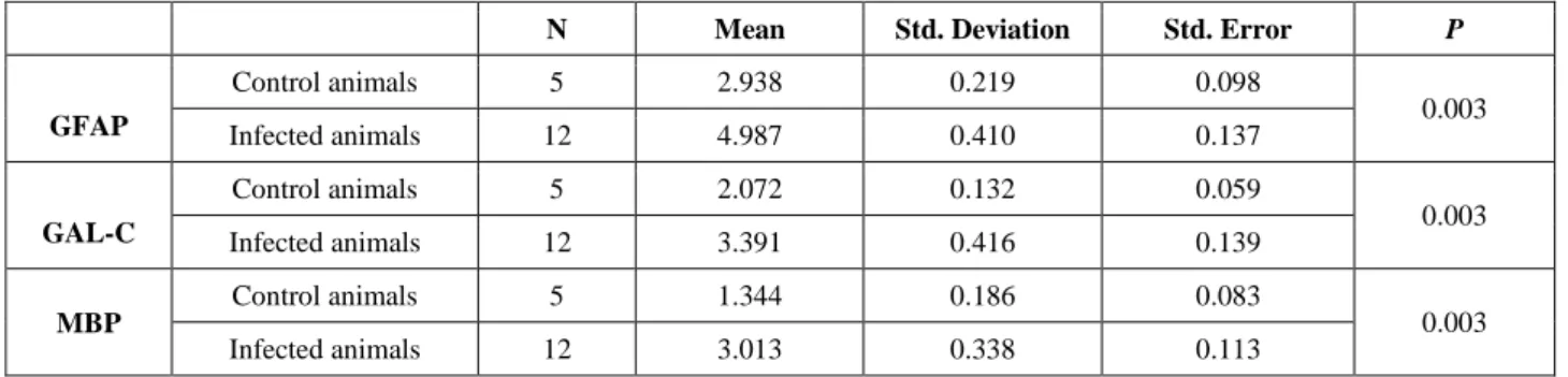
Table 4. Immunoperoxidase test results and statistical data. Tablo 4. İmmünoperoksidaz test sonuçları ve istatiksel veriler.
N Mean Std. Deviation Std. Error P
GFAP Control animals 5 2.938 0.219 0.098 0.003 Infected animals 12 4.987 0.410 0.137 GAL-C Control animals 5 2.072 0.132 0.059 0.003 Infected animals 12 3.391 0.416 0.139 MBP Control animals 5 1.344 0.186 0.083 0.003 Infected animals 12 3.013 0.338 0.113
Histopathology: Sections 5 µm thick were cut from the blocks and all slides were stained with both hematoxylin and eosin (H&E) and Kluver-Barrera (luxol fast blue) methods (15) and mounted with cover slips. Sections of tissue blocks were evaluated in three groups: brainstem (pons, medulla oblongata, rostral colliculus), midbrain (thalamus, cornu ammonis,) and cerebellum. Three randomly selected fields were assessed for each slide using a light microscope at 20X. Gliosis, microabscesses, neuronal degeneration, perivascular mononuclear cell infiltrates and meningitis were assessed in each section and assigned a numerical score. The scoring system is described in Table 2 and the results are summarized in Table 3.
The total lesion score, which indicated the severity of the lesions, was obtained by adding the numerical scores along the rows from the left to right for each animal in Table 3. In this way, the most severe cases could be identified, which allowed us to relate the effects of factors such as age and species to prognosis of the disease. A score ≥ 30 was considered as severe while lower scores were considered as mild.
Affected brain areas with lesions were counted by adding the numerical scores in each column from up to down for each animal in Table 3. We determined that more severe brain lesion in which region. A score ≥ 20 was considered as severe; lower scores were considered as mild.
Immunohistochemistry:
Detection of Listeria: Immunohistochemistry was performed on sections of brain stem, midbrain and cerebellum to determine the distribution of Listeria specific antigens. All steps were carried out following the procedure described by Dinçel and Kul (5). Sections were incubated with rabbit-anti-Listeria monocytogenes sera diluted at 1:500 Antibody Diluent (S0809; DAKO, Denmark) for 60 min at room temperature. We used a commercial streptavidin-biotin complex peroxidase (ABC-P) kit according to the supplier’s protocol (LSAB2 Kits, Universal, K0675 HRP, Rabbit/Mouse, DAKO) for visualization of all immunohistochemical staining. 3-amino-9-ethylcarbazole was used as the chromogen with counterstaining by Mayer's hematoxylin. The same procedure was applied to the tissue sections of the control group animals.
Glial fibrillary acidic protein (GFAP),
galactocerebroside (GAL-C) and myelin basic protein (MBP) expressions: Serial tissue sections of experimental and control groups also were stained with the ABC-P method using anti-GFAP (1:100 dilution), anti-MBP (1:100 dilution) and anti-GAL-C (1:500 dilution) specific commercial antibodies. A case diagnosed as canine distemper, which is characterized by severe demyelination of the cerebellum, was used as a positive control for
GFAP, MBP and GAL-C staining. Negative controls consisted of sections incubated with normal goat serum instead of the primary antibody prior to performing GFAP, MBP, GAL-C immunostaining.
The density of immunopositive staining was measured using a computerized image system composed of a Leica CCD camera DFC420 (Leica Microsystems Imaging Solutions, Ltd., Cambridge, UK), connected to a Leica DM4000 B microscope (Leica Microsystems Imaging Solutions, Ltd.) was used according to the procedure described by Dincel and Kul 2015 (6). Five representative fields were selected under high-power view and consecutive pictures were captured by the Leica QWin Plus v3 software by a 20X objective (Leica Microsystems Imaging Solutions) at a setting identical to the image system for analyzing. For examining the staining of each antibody, we used the same setting for all slides. Integrated optical density of all positive stainings of GFAP, GAL-C and MBP in each photograph were measured. For the quantification of the mean GFAP-, GAL-C- and MBP- positive area/total area were measured and calculated by Leica Qwin Plus on the pictures. All images were collected under the same lighting conditions. Data were statistically described in terms of mean and standard deviation (mean±SD) for area %. After calculating the proportion (% pixels) of stained area to the whole field, the mean (in % pixels) staining area for each slide was determined. Statistical analysis of GFAP, GAL-C and MBP immunohistochemical results were compared between groups using Mann Whitney U test. The data were presented as mean±SD.
Results
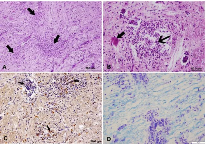
Histopathological findings: The histopathology of the brainstem, midbrain and the meninges were investigated using the H&E stained sections. Typical multifocal microabscesses (Figures 1A, B) and perivascular mononuclear cell infiltration were detected near the brainstem. In addition, meninges covering the brainstem exhibited acute inflammatory cell infiltration with a predominance of neutrophils (Figure 1B). Meninges were thickened and inflammatory cells were observed migrating from vessel walls to meninges in
lesioned areas. Perivascular mononuclear cells
(predominantly lymphocytes) and microabscesses were observed adjacent to themes aquaductus mesencephali. Microglial proliferation (gliosis) was evident around the microabscesses. Astrocyte proliferation, neuronal degeneration and satellitosis (Figure 1B) were detected in lesioned regions including brain stem, midbrain and cerebellum gray matter. Some cases (L5, L6, L9) exhibited no lesions around listeriosis-specific
microabscess, no perivascular mononuclear cell
excluded from our study. Lesion scores are summarized in Table 3. The brainstem, cerebellum and midbrain were the parts of the brain affected most adversely. Cases L1−L8 were considered severe and the remainder were considered mild based on criteria described in the Methods section. Age or species predisposition were not detected among cases (Table 3). We observed that the most affected area was the brain stem (9 cases).
Luxol fast blue findings (LFB): Listeria
monocytogenes-infected animals showed significantly blanched areas of demyelination, while high intensity staining was evident in the healthy-control group. LFB staining revealed significant demyelination in the white matter of the cerebellum and brainstem in the Listeria monocytogenes-infected animals (Figure 1D).
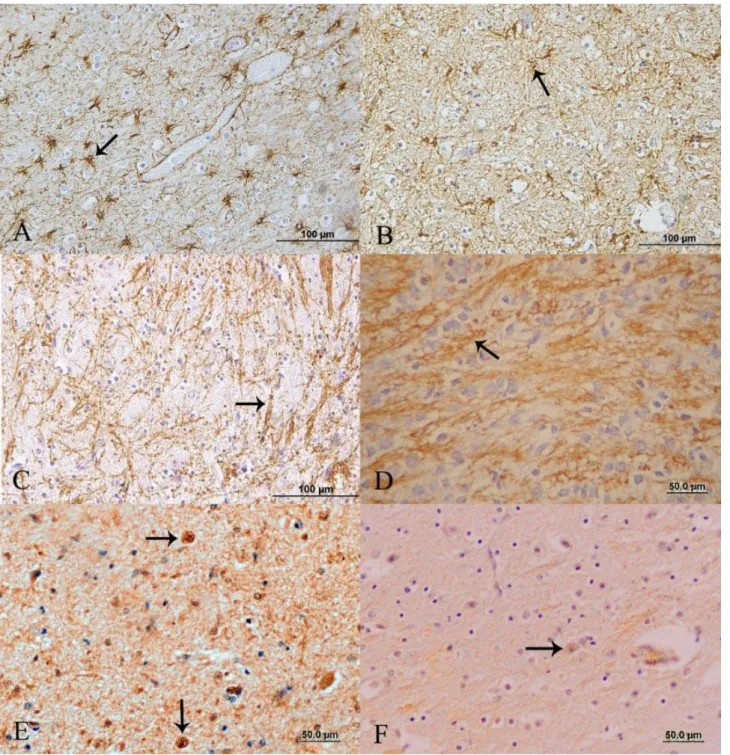
Immunohistochemical findings: Listeria spp. were confirmed by streptavidin-biotin peroxidase methods in selected tissue sections. We observed immunopositive Listeria spp. in microabscesses and perivascular mononuclear cell infiltrations in brain parenchyma and
meninges (Figure 1C). We examined reactive astrocytes (gemistocytic astrocytes) in both gray and white matter, and these showed increased anti-GFAP immunopositive areas compared to control group (P<0.01, 4,987-0,41) (Figure 2A). The control group tissue sections exhibited fewer stained organisms than the listeriosis group (Figure 2B). We also observed intensely positive MBP staining as disorganized linear or particulate entities, especially in damaged areas (P<0.01, 3,013-0,338) (Figure 2C). In control group sections, stained material appeared as regular cords or rows (Figure 2D). Strong GAL-C staining was observed in oligodendrocyte cytoplasm surrounding degenerated neurons, demyelinized areas and perivascular regions (P<0.01, 3.391-0,416) (Figure 2E). By contrast, weak GAL-C staining was observed in the control group sections (Figure 2F).
Statistical analysis of the data on MBP, GAL-C and
GFAP expression in the brain, measured by
immunostaining in all the groups, are listed in Table 4.
Figure 1. A) Sheep, case no. L1. Miliary microabcesses (arrows) in the pons. H&E. Bar=200μm. B) Sheep, case no. L1. A microabcess in the pons. Degenerated neuron with eosinophilic cytoplasm (thick arrow); thin arrow indicates focus of microabcess. H&E, Bar=50μm C) Sheep, case no. L4. Listeria positive areas in cells within a microabcess (arrow) in the thalamus. Streptavidin-biotin peroxidase method, Bar=50μm. D) Sheep, case no. L7. Degenerated myelin sheaths surrounding a microabcess in the medulla. Kluver-Barrera method, Bar=50μm.
Şekil 1. A) Koyun olgu no L1. Pons’ta miliar mikroapseler (oklar). H&E, Bar=200μm B) Koyun olgu no L1. Pons’ta mikroapse. Eozinofilik sitoplazmalı dejeneratif nöron (kalın ok); ince ok mikroapseyi göstermektedir. H&E, Bar=50μm. C) Koyun olgu no L4. Thalamus’ta mikroapse içinde listeria pozitif alanlar (ok). Streptavidin-biotin peroksidaz metodu, Bar=50μm. D) Koyun olgu no L7. Medulla’da mikroabseyi çevreleyen dejenere miyelin. Kluver-Barrera metodu, Bar=50μm.
Figure 2. A) Sheep, case no. L2. Intense GFAP staining of the pons showing numerous stellate cells (arrow). Streptavidin-biotin peroxidase method, Bar=100μm. B) Sheep, control no 1. Weak GFAP staining of the pons in a control group animal (arrow). Streptavidin-biotin peroxidase method, Bar=100μm. C) Sheep, case no. L7. Disorganized MBP stained areas among inflammatory cell infiltration demonstrating myelin cluster residues in the rostral colliculus (arrow). Streptavidin-biotin peroxidase method, Bar=100μm. D) Sheep, control no 3. MBP stained cords or rows in the rostral colliculus of a control animal (arrow). Streptavidin-biotin peroxidase method, Bar=50μm. E) Sheep, case no. L10. Strong GAL-C staining of oligodendrocytes (arrows) in the thalamus. Streptavidin-biotin peroxidase method, Bar=50μm. F) Sheep, control no 5. Oligodendrocyte (arrow) from the thalamus of a control animal unstained for GAL-C. Streptavidin peroxidase method, Bar=50μm.
Şekil 2. A) Koyun, olgu no L2. Pons’ta çok sayıdaki stellata hücrelerinde yoğun GFAP pozitif boyanmalar (ok). Streptavidin-biotin peroksidaz metodu, Bar=100μm. B) Koyun, kontrol no 1. Kontrol grubu hayvanlarda pons’taki zayıf GFAP boyanmaları (ok). Streptavidin-biotin peroksidaz metodu, Bar=100μm C) Koyun, olgu no L7. Rostral kollikulusta görülen infiltratif hücrelerdeki organize olmayan MBP boyanmaları (ok). Streptavidin-biotin peroksidaz metodu, Bar=100μm. D) Koyun, kontrol no 3. Kollikulusta kontrol grubu hayvanlara ait MBP boyanmaları (ok). Streptavidin-biotin peroksidaz metodu, Bar=50μm. E) Koyun, olgu no L10. Thalamus’ta oligodendrositlerde güçlü GAL-C boyanmaları (oklar). Streptavidin peroksidaz metodu, Bar=50μm. F) Koyun, kontrol no 5. Kontrol grubu hayvanların thalamus’undaki oligodengrositlerde negatif GAL-C boyanmaları (oklar). Streptavidin peroksidaz metodu, Bar=50μm.
Discussion and Conclusion
Listeriosis is a common zoonotic bacterial disease in domestic cattle, goats and sheep (1). Adult goats and sheep are particularly sensitive to this disease (3), but outbreaks also have been reported in lambs (16, 20). Previously, the axonal degenerations caused by Listeria monocytogenes are described (10). However, there is an in vitro study in which MBP expressions are examined (18). However, limited studies have investigated the particular mechanisms of myelin degeneration and regeneration in meningoencephalitic listeriosis (11). In this study, it was demonstrated that in meningoencephalitic listeriosis observable regenerations were detected as soon as myelin degeneration occurred. In addition to these findings, it has been shown that myelin damage in meningoencephalitic listeriosis was an important part of neuropathological evaluations.
Listeria is a widely distributed flagellated bacterium that is resistant to many environmental conditions. In addition, Listeria can be spread by feces, nasal and vaginal discharges, and tears (9). Consequently, Listeria is observed most commonly in intensive animal farming. Compero et al. (2002) (3) reported that morbidity is greater in sheep than cattle owing to poorer housing and feeding conditions, and greater stress. Small ruminants in Turkey commonly are raised and fed in fields with negligible use of silage. Accordingly, the animals investigated in this study were also fed with a similar diet. We suspect that infection may have occurred due to contaminated feed or water.
How Listeria reaches brain tissue remains a matter of debate. Some claim that this occurs through the trigeminal nerve from the nasal or oral mucosa or from the conjunctiva (1, 3, 7). Others suggest that Listeria reach the central nervous system as a result of septicemia. Listeria can cross the placental barrier, which is somewhat similar to the blood-brain barrier (4, 14). Our total lesion score data indicate that the most severe lesions were in the brainstem and midbrain. Neuronal degeneration and gliosis in those regions suggest that Listeria migrate through neurons and that neuronal degeneration evokes inflammation. Therefore, inflammatory infiltrates in the form of perivascular mononuclear cell infiltration are located in the brainstem and midbrain following neuronal degeneration.
Nevertheless, it is still unclear how Listeria reaches the meninges and it requires further investigation. Ondorff et al. (17) reported that Listeria reached the meninges owing to septicemia, but did not explain how the organisms crossed the blood-brain barrier. Dramsi et al. (8) reported that Listeria can survive in phagocytic cells and can spread to the meninges from brain parenchyma through the cerebrospinal fluid, blood and lymph circulations. Concomitant to the previous study we
observed Listeria immunostaining within phagocytic cells in the meninges.
Astrocytes participate in repairing damaged parts of the brain. The presence of reactive astrocytes can be detected by GFAP expression in brain tissue (9, 19). We found that GFAP expression was increased more in severe cases than in mild cases of listeriosis in the experimental group. We demonstrated that the expression of the GFAP is directly related to the severity of the lesions of Listeria. We also found that more MBP is present in Listeria infected animals than in healthy animals, which indicates that myelin damage plays an important role in listeriosis; myelin damage is also greater in more severe infections. The most striking finding of this study was that there was a positive correlation between GAL-C and MBP expressions. This positive correlation showed that together with degenerations of the myelin, regeneration was observed at the same time. However, according to the LFB findings, the regeneration of myelin from the spindle cannot be fully achieved. It is evident from these findings that the destructive effects of the agent are more intense than the regenerative process of metabolism. Therefore, a complete myelin regeneration should not be expected during the infectious period.
There are limited studies on demyelination in listeriosis (11). The pathogenesis of myelin damage is still not fully understood. In this study, the existence of myelin damage has been revealed. However, it is interesting that MBP expressions are increased. In addition, myelin damage was revealed with LFB. On the other hand, myelin did not disappear and presence of MBP expressions in microenvironment. This study shows that myelin damage from listeriosis is out of the ordinary situation. There is myelin damage in this disease but myelin is also present in the environment. The same increase in GAL-C expressions confirms this theory. The same intensity increase of GAL-C and MBP expressions that are in harmony with each other is a significant finding in listeriosis, in which myelin damage is observed. Demonstrating a significant increase in GAL-C and MBP expressions despite the presentation of myelin damage, suggests that pathogenesis of myelin damage associated with listeriosis is quite complicated.
In conclusion, it has been shown that myelin damage has a very important neuropathology that occurs in the meningoencephalitic listeriosis. This study also showed that oligodendrocytes did not lead the target glial cells of the agent. More studies are needed to investigate the pathogenesis of myelin damage in the field.
References
1. Akiyama Y, Asahi O, Hosoda T (1957): Studies on the
mechanism of infection of the brain with Listeria monocytogenes. Am J Vet Res, 18, 147-157.
2. Altimira J, Prats N, Lopez S, et al. (2000): Effect of
selenium deficiency on the development of central nervous system lesions in murine listeriosis. J Comp Pathol, 123,
104-109.
3. Campero CM, Odeon AC, Cipolla AL, et al. (2002):
Demonstration of Listeria monocytogenes by immunohistochemistry in formalin-fixed brain tissues from natural cases of ovine and bovine encephalitis. J Vet Med
B Infect Dis Vet Publ Health, 49, 379-383.
4. Cordy DR, Osebold JW (1959): The neuropathogenesis of
Listeria encephalomyelitis in sheep and mice. J Infect Dis,
104, 164-173.
5. Dinçel GC, Kul O (2015): Increased expressions of
ADAMTS-13, neuronal nitric oxide synthase, and neurofilament correlate with severity of neuropathology in Border disease virus-infected small ruminants. PLoS One.
2015 Mar 23;10 (3): e0120005.
6. Dinçel GC, Kul O (2015): eNOS and iNOS trigger
apoptosis in the brains of sheep and goats naturally infected with the border disease virus. Histol Histopathol, 30,
1233-42.
7. Dons L, Jin Y, Kristensson K, et al. (2007): Axonal
transport of Listeria monocytogenes and nerve-cell-induced bacterial killing. J Neurosci Res, 85, 2529-37.
8. Dramsi S, Lévi S, Triller A, et al. (1998): Entry of Listeria
monocytogenes into neurons occurs by cell-to-cell spread: an in vitro study. Infect Immun, 66, 4461-68.
9. Hazıroğlu R (2000): Listeriosis, 308-310. In: R Hazıoğlu and ÜH Milli (Ed.) Veteriner Patoloji. I. Cilt, Özkan Matbaacılık, Ankara.
10. Henke D, Rupp S, Gaschen V, et al. (2015): Listeria
monocytogenes spreads within the brain by actin-based intra-axonal migration. Infect Immun, 83, 2409-19.
11. Innes JRM, Saunders LZ (1962): X. Bacterial Infection,
III Listeriosis, 508-510. In: LZ Saunders (Ed) Comparative
Neuropathology, Academic Press, New York and London. 12. Kabakci N, Yarim M (2004): The expression of CD 14
antigen in experimental encephalitic listeriosis in rabbits.
Rev Med Vet, 155, 151-155.
13. Krueger N, Low C, Donachie W (1995): Phenotypic
characterization of the cells of the inflammatory response in ovine encephalitic listeriosis. J Comp Pathol, 113,
263-275.
14. Lecuit M (2005): Understanding how Listeria monocytogenes targets and crosses host barriers. Clin
Microbiol Infect, 11, 427-513.
15. Luna GL (1968): Manuel of histologic staining methods of
the Armed Forces Institute of Pathology.3rd ed., McGraw Hill, New York.
16. Olafson P (2016): Listerial encephalitis (circling disease)
of sheep, cattle and goats. Cornell Vet, 30, 141-150.
17. Orndorff PE, Hamrick TS, Smoak IW, et al. (2006): Host
and bacterial factors in listeriosis pathogenesis. Vet
Microbiol, 114, 1-15.
18. Pensinger DA, Aliota MT, Schaenzer AJ, et al. (2014):
Selective pharmacologic inhibition of a PASTA kinase increases Listeria monocytogenes susceptibility to β-lactam antibiotics. Antimicrob Agents Chemother, 58, 4486-94.
19. Summers BA, Cummings JF, De Lahunta A (1995): Inflammatory disease of the central nervous system,
133-136. Veterinary Neuropathology. Mosby-Year Book, St.
Louis, Missouri.
20. Wardrope DD, Macleod NS (1983): Outbreak of listeria
meningoencephalitis in young lambs. Vet Rec, 113,
213-214.
21. Weinstock D, Horton SB, Rowland PH (1995): Rapid
diagnosis of Listeria monocytogenes by immunohistochemistry in formalin-fixed brain tissue. Vet
Pathol, 32, 193-195.
Geliş tarihi: 09.11.2016 / Kabul tarihi: 22.01.2017
Address for correspondence:
Güngör Çağdaş DİNÇEL
Aksaray University, Laboratory and Veterinary Science, Eskil Vocational High School, 68800,
Eskil, Aksaray, Turkey. [email protected]