Article
Targeted Treatment Protocol in Patellofemoral
Pain: Does Treatment Designed According to
Subgroups Improve Clinical Outcomes in
Patients Unresponsive to Multimodal Treatment?
Yosmaoğlu, Hayri Baran, Sonmezer, Emel, Ozkoslu, Manolya, Sahin,
Ezgi, Çerezci, Senay, Richards, James, Selfe, James and Janssen,
Jessie
Available at http://clok.uclan.ac.uk/29355/
Yosmaoğlu, Hayri Baran, Sonmezer, Emel, Ozkoslu, Manolya, Sahin, Ezgi, Çerezci, Senay, Richards, James ORCID: 0000-0002-4004-3115, Selfe, James and Janssen, Jessie (2020) Targeted Treatment Protocol in Patellofemoral Pain: Does Treatment Designed According to Subgroups Improve Clinical Outcomes in Patients Unresponsive to Multimodal Treatment? Sports Health, 12 (2). pp. 170-180. ISSN 1941-7381
It is advisable to refer to the publisher’s version if you intend to cite from the work.
http://dx.doi.org/10.1177/1941738119883272
For more information about UCLan’s research in this area go to
http://www.uclan.ac.uk/researchgroups/ and search for <name of research Group>. For information about Research generally at UCLan please go to
http://www.uclan.ac.uk/research/
All outputs in CLoK are protected by Intellectual Property Rights law, including
Copyright law. Copyright, IPR and Moral Rights for the works on this site are retained by the individual authors and/or other copyright owners. Terms and conditions for use of this material are defined in the http://clok.uclan.ac.uk/policies/
CLoK
Central Lancashire online Knowledge
1 Targeted Treatment Protocol in Patellofemoral Pain (TIPPs): Does Treatment Designed 1
According to Subgroups Improve Clinical Outcomes in Patients Unresponsive to 2
Multimodal Treatment? 3
4
Hayri Baran Yosmaoğlu, Emel Sonmezer, Manolya Ozkoslu, Ezgi Sahin, Senay Çerezci, Jim 5
Richards, James Selfe, Jessie Janssen 6
7
Background: Targeted intervention for subgroups is a promising approach for the management 8
of patellofemoral pain. 9
Hypothesis: Treatment designed according to subgroups improves clinical outcomes in 10
patients unresponsive to multimodal treatment. 11
Study Design: A prospective crossover intervention. 12
Level of Evidence: Level III 13
Methods: PFP patients (n=61, mean age: 27±9 years) were enrolled. PFP patients received 14
standard multimodal treatment three times a week for 6 weeks. Patients not responding to 15
multimodal treatment were then classified into one of 3 subgroups “strong”, “weak and tight” 16
and “weak and pronated foot” using six simple clinical tests. They subsequently were 17
administered a further 6 weeks of targeted intervention designed according to subgroup 18
characteristics. Visual Analog Scale (VAS), Perception of Recovery Scale (PRS), EQ-5D-5L, 19
and S-LANSS were used to assess pain, knee function and quality of life before and after the 20
interventions. 21
Results: 36% of the patients (21 patients) demonstrated recovery following multimodal 22
treatment. However, over 70% (29 patients) of these non-responders demonstrated recovery 23
after targeted treatment. The VAS, PRS, S-LANSS, and EQ-5D-5L scores improved 24
significantly after targeted intervention compared to after multimodal treatment (p<0.001). The 25
2 VAS score at rest was significantly lower in the weak and pronated foot, and weak and tight 26
subgroups (p=0.011, p=0.008) respectively. Post-treatment pain intensity on activity was 27
significantly lower in the “strong” subgroup (p=0.006). 28
Conclusion: Targeted treatment designed according to subgroup characteristics improves 29
clinical outcomes in patients unresponsive to multimodal treatment. 30
Clinical Relevance: Targeted intervention could be easily implemented following six simple 31
clinical assessment tests to subgroup patients into one of three subgroups (strong, weak and 32
tight, weak and pronated foot). Targeted interventions applied according to the characteristics 33
of these subgroups have more beneficial treatment effects than a current multimodal treatment 34
program. 35
36
Key words: Rehabilitation, knee injuries, patella, treatment outcome, pain perception 37
38
INTRODUCTION 39
Patellofemoral pain (PFP) is a chronic musculoskeletal problem that causes persistent anterior 40
knee pain.2,3,6,8,14,15,20,21,25,26,32,33,49 Despite its widespread use in clinics, it is difficult to suggest 41
that the current multimodal treatment approach leads to successful outcomes in the majority of 42
patients with PFP, only 46% of patients’ knees were pain free at discharge.2 This indicates that
43
over half of PFP patients do not respond to treatment and may continue their lives with chronic 44
anterior knee pain. 45
Identification of the factors leading to these low treatment success rates has consistently been a 46
priority of previous International Patellofemoral Pain Research Retreats.4,10,12,52 The most 47
important factor affecting the success of treatment that has emerged is that patients have a 48
variety of musculoskeletal and biomechanical differences. The current multimodal treatment, 49
therefore, may not affect the heterogeneous PFP patient population with the same efficiency. 50
3 Clinically subgrouping PFP patients and delivering targeted treatments has been strongly 51
recommended for future investigations of patellofemoral pain treatment from the International 52
Patellofemoral Pain Research Retreats.4,12,52 An overview of previously published PFP
53
subgroups and the methods used to derive subgroups in PFP identified patients with PFP.39 54
They exhibit different anthropometric and biomechanical characteristics and do not form a 55
homogeneous group. There are 3 subgroups in the PFP population: “strong”, “weak and tight” 56
and “weak and pronated foot”.38 The purpose of this study was to assess the clinical outcomes
57
of targeted treatments designed according to the characteristics of the three subgroups of PFP 58
patients.38 The hypotheses were that the assessment and subgroup classification is clinically 59
feasible, and that targeted treatments designed according to the characteristics of the three 60
subgroups of PFP patients would show clinical benefits over and above a multimodal 61 intervention. 62 METHOD 63 Design 64
A prospective crossover intervention study design was used (Figure 1). 65
Participants 66
Patients aged between 18 and 40 attending a physiotherapy outpatient clinic at a University 67
Hospital with a clinical diagnosis of patellofemoral pain were approached for eligibility in this 68
study. Eligibility criteria were based on previously defined PFP criteria.7,38,47 Subjects were 69
excluded if they had any of the following: previous knee surgery, clinical evidence of 70
ligamentous instability and/or internal derangement, a history of patellar subluxation or 71
dislocation, joint effusion, true knee joint locking and/or giving way, bursitis, patellar or 72
iliotibial tract tendinopathy, Osgood Schlatter’s disease, Sinding-Larsen Johansson Syndrome, 73
muscle tears or symptomatic knee plicae, serious co-morbidity which would preclude or affect 74
compliance with the assessment, or were pregnant. 75
4 76
Subgroup Classification Method 77
Quadriceps and Hip Abductor muscle strength 31, Patellar glide test44,54, Quadriceps length53,
78
Gastrocnemius length53, and Foot posture index36 assessments were performed to classify all 79
consenting patients into one of three subgroups (strong, weak and tight, weak and pronated 80
foot) using the algorithm derived from the work by Selfe et al.38 81 82 Intervention 83 Multimodal Treatment 84
The multimodal treatment program was designed based on the usual exercise and modalities 85
used in local clinics.20,21,32,49 All patients received standard, supervised, 60 min multimodal 86
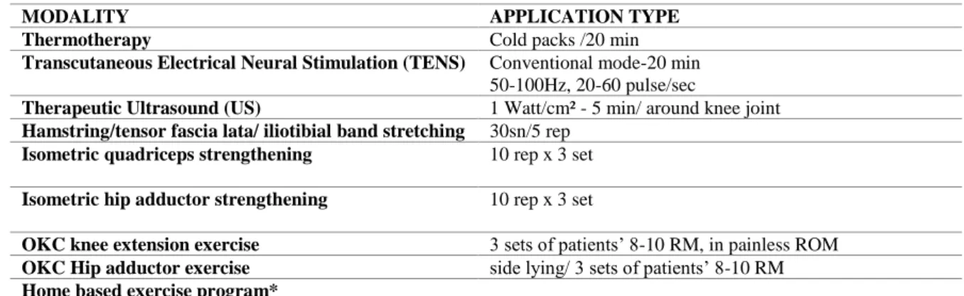
treatment three times a week for 6 weeks. Table 1 shows the details of the multimodal 87
rehabilitation program. 88
Targeted Treatment 89
Patients who did not respond to multimodal treatment were assigned to one of the treatment 90
groups “strong”, “weak and tight”, and “weak and pronated foot”. They then followed a further 91
6 weeks, 45 min targeted intervention program administered three times a week. The targeted 92
treatment program was designed according to the key deficits identified in each patient by the 93
subgrouping clinical assessment tests. The patients in the “strong” subgroup had no muscle 94
strength deficit therefore, the intervention program for this subgroup was targeted at improving 95
neuromuscular control and coordination ability using proprioceptive exercises such as 96
progressive balance exercises, and knee braces46,47 which have been shown to offer 97
improvements in movement control in patients with PFP,41 reductions in patellofemoral 98
reaction forces44 and have been shown to reduce pain at 6 and 12 months during a PFP 99
rehabilitation program.48 In the “weak and tight” subgroup, the exercise program consisted of
5 Closed Kinetic Chain (CKC) muscle strengthening and stretching, and weight management 101
advice, as a larger body mass index was identified as a potentially relevant clinical feature in 102
this subgroup.38 In the “weak and pronated foot” subgroup, muscle weakness and abnormal foot
103
alignment were identified as the key factors. Therefore, the intervention program included CKC 104
strengthening exercises and foot orthoses.5,24 Table 2 shows the details of each of the specific 105
targeted intervention programs. 106
Outcome measures 107
Pain during activity measured using the Visual Analog Scale (VAS) was the primary outcome 108
measure of this study 19. Activity was specified by patients. 109
The Perception of Recovery Scale was measured using a 7-point Likert scale ranging from 110
“completely recovered” to “worse than ever”. Patients were classified as “recovered” if they 111
rated themselves as “completely recovered” or “strongly recovered”. Patients rating themselves 112
in one of the other five categories from “slightly recovered” to “worse than ever" were 113
categorised as “not recovered”.35
114
The EQ-5D-5L was used as a self-reported generic measure of health and quality of life. 115
Patients rated their overall health on the day of the interview on a 0–100 hash-marked, vertical 116
visual analogue scale (EQ-5D-5L-VAS). A higher EQ-5D-5L-VAS score indicating better 117
health status.22 118
Neuropathic Pain was measured using The Self-Administered Leeds Assessment of 119
Neuropathic Symptoms and Signs (S-LANSS) questionnaire. The S-LANSS comprises a 5-120
item questionnaire regarding pain symptoms and two items for clinical signs involving self-121
administered sensory tests for the presence of allodynia and decreased sensation to pinprick. 122
This was used to discriminate the small number of patients who may have neuropathic knee 123
pain from those with nociceptive pain.42 The possible scores range from 0 to 24, with a score
124
of 12 or greater considered to be suggestive of neuropathic pain.28 Finally, a single leg hop test 125
6 was used to determine functional performance.1 Distance was measured from toe to heel and 126
the mean score of three repetitions was recorded.
127
Data analysis 128
A sample size calculation was performed based on the minimal detectable change on the pain 129
VAS. Data from a previous study indicates that the VAS scores in patients with PFP was 4.3 ± 130
1 cm,9 with 30% of the maximum score of the VAS-pain considered to be the detectable change, 131
the sample size for each treatment subgroup was determined to be 8 patients to achieve a 90% 132
power at the 0.05 level of significance. Data were not normally distributed when analysed with 133
the Kolmogorov–Smirnov test Consequently, non-parametric tests were indicated. Therefore 134
the “Wilcoxon signed rank test” was used to compare pre and post treatment outcomes with an 135
alpha value of 0.05. In addition, the mean of rank scores, standard errors and Z scores were 136
reported, along with descriptive statistics to describe the general features of the subjects. All 137
statistical analysis was conducted using SPSS 21.0. 138
139
RESULTS 140
Of the 128 patients who were screened, 95 were included in the present study. Of these 61 141
patients completed the multimodal treatment (Figure 1) (Table 3). Twenty-one patients (36%) 142
demonstrated recovery following multimodal treatment (Phase I) and were discharged. 40 143
Patients (64%) not responding to multimodal treatment were administered a further 6 weeks of 144
targeted intervention designed according to subgroup characteristics (phase 2). Twenty-nine 145
(72.5%) patients demonstrated recovery following targeted intervention (phase II) and 11 146
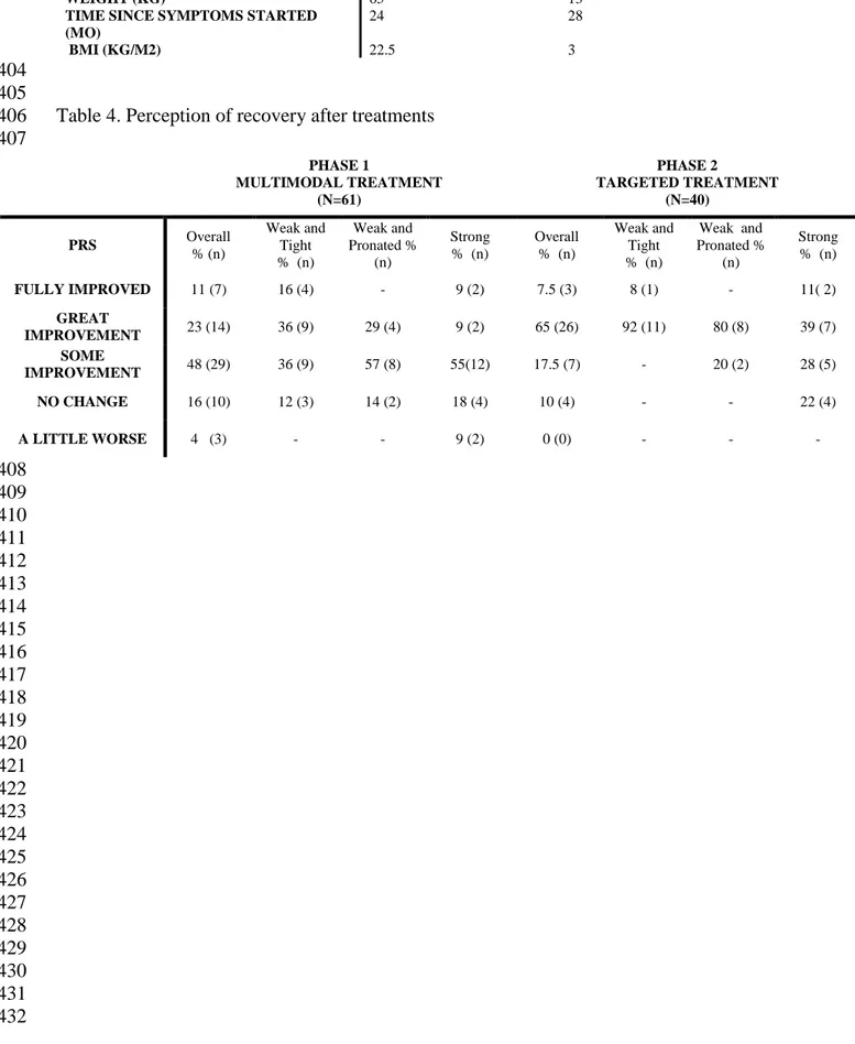
(27.5%) patients did not respond to either of the treatment approaches (Table 4). 147
Pain intensity (VAS) at rest and during activity, and Perceived Recovery Scale (PRS), were 148
significantly improved after targeted intervention (p<0.001) (Table 5). S-LANSS, EQ-5D-5L 149
and EQ5D-5L-VAS scores were significantly improved following targeted intervention 150
7 compared to pre-targeted treatment scores (p = 0.001, p<0.001, p = 0.02), respectively (Table 151
5). 152
Within the three subgroups, the findings showed that PRS score was significantly improved 153
after targeted treatment compared to pre-targeted treatment levels in the “strong”, “weak and 154
tight”, and “weak and pronated foot” subgroups (p= 0.005, p= 0.001, p= 0.004) respectively. 155
VAS pain intensity at rest was also significantly lower after targeted intervention in the “weak 156
and pronated foot” and “weak and tight” subgroups (p=0.011, p= 0.008) respectively, however 157
within the “strong” subgroup, no change was seen between pre-treatment and post treatment (p 158
= 0.245) (Table 6). However, pain intensity during activity was significantly lower after 159
treatment in the “strong” (p=0.006), the “weak and pronated foot” and “weak and tight” 160
subgroups; although these reductions were not statistically significant (p=0.059, p= 0.06) 161
respectively (Table 6). 162
Other measures including quadriceps length test, S-LANSS, EQ5D-5L, and EQ5D-VAS were 163
significantly improved in the “weak and tight” subgroup. S-LANSS, EQ5D-5L, and patellar 164
mobility were significantly improved in the “weak and pronated foot” subgroup. In the “strong” 165
group only gastrocnemius length was significantly different between pre- and post-targeted 166
treatment (p=0.03). Results for outcome measures are shown in Table 7. 167
168
DISCUSSION 169
The results of our study suggest that the TIPPs subgroups and the algorithm used to classify 170
PFP patients as "strong", "weak and tight", "weak and pronated foot" 38 is valid and clinically 171
implementable. The findings from this study were in agreement with previous work13 that 172
reported differential response patterns in outcomes at 12 months in their subgroups. This 173
suggests that targeted interventions based on subgroups, provides an important development in 174
the treatment strategy for patients with PFP.4,52
8 The “strong” subgroup demonstrated a poor response to multimodal treatment but a a 176
significant improvement after targeted treatment was observed. This finding is consistent with 177
Greuel et al.18 and Gallina et al.17 who both reported results confirming that motor control of
178
the quadriceps is problematic in some PFP patients. One explanation for this is improved 179
neuromuscular control in patients classified as “strong”. Since these patients already 180
demonstrated relatively high quadriceps muscle torque, targeted intervention was delivered 181
focusing on progressive development of motor control on unstable surfaces instead of 182
conventional muscle strength exercises. Given that quadriceps strength did not change as a 183
result of the targeted intervention, these progressive balance exercises and patellar bracing has 184
improved motor control and stability.41 In addition, bracing may reduce patellofemoral forces
185
during activities of daily living and sporting tasks44 and improvements within rehabilitation 186
protocols.48 This was reflected in the improvement in the other pain related parameters, 187
However, since the average pre-treatment VAS pain level at rest in this subgroup was already 188
low a decrease from 1.8 to 0.7 has minimal clinical relevance. 189
Clinically the “weak and tight” subgroup appeared to be the most responsive group to treatment 190
overall with a relatively even split of 52% responding to multimodal treatment and all of the 191
remaining patients responding to targeted intervention. This finding was not surprising as 192
multimodal treatment routinely includes strengthening and stretching exercises. However, 193
closer analysis of the outcomes in the "weak and tight" subgroup suggest that although patients’ 194
perception of recovery improved, the VAS activity pain intensity was not significantly 195
decreased after targeted treatment in this subgroup. Considering muscle weakness is the main 196
issue in this subgroup, the probable cause of this unexpected finding is persistent inability to 197
compensate patellofemoral loads especially during relatively high level activities of daily life 198
such as ascending/descending stairs even after the targeted treatment. Targeted intervention 199
consisting of functional strengthening may still be insufficient for high level activities of daily 200
9 living which demand considerable muscular activity, although it caused approximately a 30% 201
increase in muscle torque and a significant improvement in perception of recovery in this 202
subgroup. 203
Findings from the “weak and pronated foot” subgroup suggest that targeted treatment including, 204
foot orthoses and pain free strengthening exercises was also successful in terms of perception 205
of recovery and VAS pain on rest. Although the same improvement was not observed in VAS 206
pain during activity. One explanation for this could be the indirect effect of the foot orthoses 207
on the knee as the patients showed no improvement in strength after targeted treatment. 208
Moreover, optimum correction is very difficult to determine during the intervention of foot 209
orthoses. Special single physiotherapy interventions or combining interventions for patellar 210
taping, mobilisation or manual therapy may have beneficial effects on pain related functional 211
symptoms in PFP.11,30,34 However, the therapeutic effects of these applications remain limited 212
because PFP patients exhibit a wide variety of structural features and biopsychosocial 213
differences. The biomechanical and anthropometric characteristics of patients were not similar. 214
Foot pronation, for example, was noticeably high in some patients, while some had neutral foot 215
alignment. Similarly, quadriceps muscle strength, which is a predisposing factor or a most 216
common symptom in previous studies8,54 has been high in some patients with the remainder 217
having considerable muscle weakness. Therefore, specific applications such as foot orthoses, 218
knee braces, tape, and even exercises may not be required by every patient. 219
The functional hop test is often used in clinics to measure functional capability.51 Considering
220
that there was no increase in quadriceps muscle strength in the “weak and pronated foot”, and 221
“strong” subgroups, an improvement in the hop test scores was not expected. 222
Due to the methodological design of this study, patients received 6 weeks of multimodal 223
treatment before 6 weeks of targeted treatment with no intervening washout period. This is a 224
study limitation since the cumulative effects of the previous treatment (multimodal) were 225
10 ignored. Therefore, the observed difference in some parameters could be the result of regression 226
to the mean. 227
CONCLUSION 228
Both the TIPPs assessment and subgroup classification algorithm are clinically feasible that 229
those with PFP are not a homogeneous group, and have biomechanical and structural 230 differences. 231 232 REFERENCES 233 234
1. Bremander AB, Dahl LL, Roos EM. Validity and reliability of functional performance 235
tests in menisectomised patients with or without knee osteoarthritis. Scand J Med Sci 236
Sports. 2007;17:120-127. 237
2. Brown J. Physiotherapists knowledge of patellofemoral pain syndrome. Br J Ther 238
Rehabil. 2000;7:346–353. 239
3. Callaghan MJ, Selfe J. Has the prevalence of patellofemoral pain in the general 240
population in the United Kingdom been properly evaluated? Phys Ther Sport. 241
2007;8:37-43. 242
4. Callaghan, M., Collins, N., Sheehan F. Patellofemoral pain: proximal, distal, and local 243
factors. 2nd International Research Retreat. JOSPT. 2012;42:A1-A20. 244
5. Collins N, Crossley K, Darnell R, et al. Foot orthoses and physiotherapy in the treatment 245
of patellofemoral pain syndrome: randomised clinical trial. BMJ. 2008;337:1735. 246
6. Collins NJ, Barton CJ, van Middelkoop M, et al. Consensus statement on exercise 247
therapy and physical interventions (orthoses, taping and manual therapy) to treat 248
patellofemoral pain: recommendations from the 5th International Patellofemoral Pain 249
Research Retreat, Gold Coast, Australia, Br J Sports Med. 2018. 250
11 7. Cook C, Hegedus E, Hawkins R, et al. Diagnostic accuracy and association to disability 251
of clinical test findings associated with patellofemoral pain syndrome. Physiother Can. 252
2010;62(1):17-24. 253
8. Cowan SM, Bennell KL, Hodges PW, et al. Delayed onset of electromyographic activity 254
of vastus lateralis compared to vastus medialis obliquus in subjects with patellofemoral 255
pain syndrome. Arch Phys Med Rehabil. 2000;82:83–89. 256
9. Crossley KM, Bennell KL, Cowan SM, et al. Analysis of outcome measures for persons 257
with patellofemoral pain: which are reliable and valid? Arch Phys Med Rehabil. 258
2004;85:815-822. 259
10. Crossley KM, van Middelkoop M, Callaghan MJ, et al. Patellofemoral pain consensus 260
statement from the 4th International Patellofemoral Pain Research Retreat, Manchester. 261
Part 1: Terminology, definitions, clinical examination, natural history, patellofemoral 262
osteoarthritis and patient-reported outcome measures. Br J Sports Med. 2016;50:839-263
843. 264
11. Crossley KM, van Middelkoop M, Callaghan MJ, et al. Patellofemoral pain consensus 265
statement from the 4th International Patellofemoral Pain Research Retreat, Manchester. 266
Part 2: recommended physical interventions (exercise, taping, bracing, foot orthoses and 267
combined interventions). Br J Sports Med. 2016;50:844-852. 268
12. Davis I, Powers C. Patellofemoral pain syndrome: proximal, distal, and local factors. 269
An International Retreat. JOSPT. 2010;40:A1-48. 270
13. Drew BT. Stratification of patellofemoral pain using clinical, biomechanical and 271
imaging features [doctoral dissertation]. University of Leeds; 2018. 272
14. Dvir Z, Halperin N, Shklar A, et aI. Concentric and eccentric torque variations of the 273
quadriceps femoris in patello-femoral pain syndrome. Clin Biomech. 1990;5:68–72. 274
12 15. Dvir Z, Halperin N, Shklar A, et al. Quadriceps function and patellofemoral pain 275
syndrome. Part I: pain provocation during concentric and eccentric isokinetic activity. 276
Isok Exerc Sci. 1991;1:26–30. 277
16. Eng JJ, Pierrynowski MR. The effect of soft foot orthotics on three-dimensional lower-278
limb kinematics during walking and running. Phys Ther. 1994;74:836-44. 279
17. Gallina A, Hunt MA, Hodges PW, et al. Vastus lateralis motor unit firing rate is higher 280
in women with patellofemoral pain. Arch Phys Med Rehabil. 2018;99:907-13. 281
18. Greuel H, Herrington L, Liu A, et al. How does pain influence arthrogenic muscle 282
inhibition and quadriceps torque in individuals with patellofemoral pain. 5th 283
International Patellofemoral Research Retreat. Gold Coast Queensland, Australia 2017. 284
19. Hawker GA, Mian S, Kendzerska T, et al. Measures of adult pain: visual analog scale 285
for pain (vas pain), numeric rating scale for pain (nrs pain), mcgill pain questionnaire 286
(mpq), short‐form mcgill pain questionnaire (sf‐mpq), chronic pain grade scale (cpgs), 287
short form‐36 bodily pain scale (sf‐36 bps), and measure of intermittent and constant 288
osteoarthritis pain (icoap). Arthritis Care Res. 2011;63(11):240-252. 289
20. Heintjes EM, Berger M, Bierma-Zeinstra SMA, et al. Exercise therapy for 290
patellofemoral pain syndrome. Cochrane Database Syst Rev. 2003;4. 291
21. Heintjes EM, Berger M, Bierma-Zeinstra SMA, et al. Pharmacotherapy for 292
patellofemoral pain syndrome. Cochrane Database Syst Rev. 2004;3. 293
22. Herdman M, Gudex C, Lloyd A, et al. Development and preliminary testing of the new 294
five-level version of EQ-5D (EQ-5D-5L). Quality of life research. 2011;20(10):1727-295
1736. 296
23. Hinman, RS, Bowles KA, Payne C, et al. Effect of length on laterally‐wedged insoles 297
in knee osteoarthritis. Arthritis Care Res. 2008;59:144-147. 298
13 24. Hossain M, Alexander P, Burls A, et al. Foot orthoses for patellofemoral pain in adults. 299
Cochrane Database Syst Rev. 2011;1. 300
25. Janssen J. Concepts of patellofemoral pain. In: Selfe J, Janssen J, Callaghan M, eds. 301
Patellofemoral Pain an Evidence Based Clinical Guide. Nova Science; 2017:3-13. 302
26. Jensen R, Hystad T, Baerheim A. Knee function and pain related to psychological 303
variables in patients with long-term patellofemoral pain syndrome. J Orthop Sports Phys 304
Ther. 2005;35:594–600. 305
27. Keays SL, Mason M, Newcombe PA. Individualized physiotherapy in the treatment of 306
patellofemoral pain. Physiother Res Int. 2015;20:22-36. 307
28. Koc R, Erdemoglu AK. Validity and reliability of the Turkish self-administered leeds 308
assessment of neuropathic symptoms and signs (S-LANSS) questionnaire. Pain Med. 309
2010;11:1107-1114. 310
29. Lake DA, Wofford NH. Effect of therapeutic modalities on patients with patellofemoral 311
pain syndrome: a systematic review. Sports health, 2011;3(2):182-189. 312
30. Logan CA, Bhashyam AR, Tisosky AJ, Haber DB, Jorgensen A, Roy A, Provencher 313
MT. Systematic review of the effect of taping techniques on patellofemoral pain 314
syndrome. Sports health. 2017;9(5):456-461. 315
31. Maffiuletti NA. Assessment of hip and knee muscle function in orthopaedic practice 316
and research. J Bone Joint Surg Am. 2010;92(1):220. 317
32. Martimbianco AC, Torloni MR, Andriolo BNG, et al. Neuromuscular electrical 318
stimulation (NMES) for patellofemoral pain syndrome. Cochrane Database Syst Rev. 319
2017;9. 320
33. Nordin M, Frankel V. Basic biomechanics of the musculoskeletal system. London: 321
Lippincott Williams & Wilkins; 1989:176–202. 322
14 34. Page, P. (2011). Effectiveness of elastic resistance in rehabilitation of patients with 323
patellofemoral pain syndrome: what is the evidence?. Sports Health, 3(2), 190-194. 324
35. Rathleff MS, Roos EM, Olesen JL, et al. Lower mechanical pressure pain thresholds in 325
female adolescents with patellofemoral pain syndrome. JOSPT. 2013;3(6):414-421. 326
36. Redmond AC, Crane YZ, Menz HB. Normative values for the foot posture index. J Foot 327
Ankle Res. 2008;1(1):6. 328
37. Selfe J, Callaghan M, Witvrouw E, et al. Targeted interventions for patellofemoral pain 329
syndrome (TIPPS): classification of clinical subgroups. BMJ open. 2013;3(9): e003795. 330
38. Selfe J, Janssen J, Callaghan M, et al. Are there three main subgroups within the 331
patellofemoral pain population? A detailed characterisation study of 127 patients to help 332
develop targeted intervention (TIPPs). Br J Sports Med. 2016; 50(14):873-880. 333
39. Selfe J, Janssen J, Drew B, et al. Anterior knee pain subgroups: the first step towards a 334
personalized treatment. Ann Joint. 2018;3(32). 335
40. Selfe J. Exercises supervised by physiotherapists improve pain and function in patients 336
with patellofemoral pain. J Physiother. 2010;56(1):61. 337
41. Selfe et al. 2011. A clinical study of the biomechanics of step descent using different 338
treatment modalities for patellofemoral pain. Gait & posture. 2011;34(1): 92-96. 339
340
42. Selfe 2017. Chapter 4: Red Flags and Rare pathologies in 1.Selfe J, Janssen J, 341
Callaghan M (2017). Patellofemoral Pain an evidence based Clinical Guide. Nova 342
Science 343
344 345
43. Selhorst M, Rice W, Degenhart T, et al. Evaluation of a treatment algorithm for patients 346
with patellofemoral pain syndrome: a pilot study. Int J Sports Phys Ther. 2015;10:178. 347
44. Sinclair JK., Selfe J, Taylor PJ, Shore HF, Richards JD. Influence of a knee brace 348
intervention on perceived pain and patellofemoral loading in recreational athletes. Clin 349
Biomech 2016; 37: 7-12. 350
15 45. Skalley TC, Terry GC, Teitge RA. The quantitative measurement of normal passive 351
medial and lateral patellar motion limits. Am J Sports Med. 1993;21(5):728-732. 352
46. Smith TO, Drew BT, Meek TH, et al. Knee orthoses for treating patellofemoral pain 353
syndrome. Cochrane Database Syst Rev. 2013(5). 354
47. Syme G, Rowe P, Martin D, et al. Disability in patients with chronic patellofemoral pain 355
syndrome: a randomised controlled trial of VMO selective training versus general 356
quadriceps training. Man Ther. 2009;14:252-263. 357
48. Uboldi FM, Ferrua P, Tradati D, Zedde P, Richards J, Manunta A, Berruto M. Use of 358
an elastomeric knee brace in patellofemoral pain syndrome: short-term 359
results. Joints. 2018;6(02):85-89. 360
49. Van der Heijden RA, Lankhorst NE, van Linschoten R, et al. Exercise for treating 361
patellofemoral pain syndrome. Cochrane Database Syst Rev. 2015;6. 362
50. Weng P, Janssen J, Selfe J, et al. Validity of two clinical knee strength assessments 363
compared to the reference standard. International Journal of Physiotherapy and 364
Research. 2015;11:1264-1270. 365
51. Wilk KE, Romaniello WT, Soscia SM, et al. The relationship between subjective knee 366
scores, isokinetic testing, and functional testing in the ACL-reconstructed knee. JOSPT. 367
1994;20:60-73. 368
52. Witvrouw E, Crossley K, Davis I, et al. 3rd International Patellofemoral Research 369
Retreat, Vancouver, Canada. Br J Sports Med. 2014;48:411-414. 370
53. Witvrouw E, Lysens R, Bellemans J, et al. Intrinsic risk factors fror the development of 371
anterior knee pain in an athletic population: atwo year prospective study. Am J Sports 372
Med. 2000;28(4):480-489. 373
16 54. Witvrouw E, Werner S, Mikkelsen C, et al. Clinical classification of patellofemoral pain 374
syndrome: guidelines for non-operative treatment. Knee Surg Sports Traumatol 375
Arthrosc. 2005;13(2):122-130. 376
55. Yosmaoglu HB, Kaya D, Guney H, et al. Is there a relationship between tracking ability, 377
joint position sense, and functional level in patellofemoral pain syndrome? Knee Surg 378
Sports Traumatol Arthrosc. 2013;21:2564-2571. 379
380 381 382
17 Table 1. Multimodal Treatment Program
383 384 385
MODALITY APPLICATION TYPE Thermotherapy Cold packs /20 min
Transcutaneous Electrical Neural Stimulation (TENS) Conventional mode-20 min 50-100Hz, 20-60 pulse/sec
Therapeutic Ultrasound (US) 1 Watt/cm² - 5 min/ around knee joint
Hamstring/tensor fascia lata/ iliotibial band stretching 30sn/5 rep Isometric quadriceps strengthening 10 rep x 3 set
Isometric hip adductor strengthening 10 rep x 3 set
OKC knee extension exercise 3 sets of patients’ 8-10 RM, in painless ROM OKC Hip adductor exercise side lying/ 3 sets of patients’ 8-10 RM
Home based exercise program*
RM: Repetition Maximum, rep: repetition, ROM: Range of motion, OKC: Open kinetic chain
386
*Home based exercise program included the same applications except TENS, NMES, US 387
388 389
Table 2. Targeted treatment program 390
391
STRONG SUBGROUP
Progressive balance/proprioception exercises Standing on one leg on wobble board 3 sets of 1 min exercise each leg 1-3 sets per session depending on pain
Progression*: Eyes closed, bouncing ball against wall, bouncing ball against wall on an unstable surface
Patellar bracing** Patient was asked to put on knee brace during ADL
Activity modification Activity reduction to fit within envelope of function locally determined and negotiated with individual patient
WEAK AND TIGHT SUBGROUP
CKC strengthening exercises Plie/lunge/single limb squat Pain free ROM
10 reps per set/ 1-3 sets depending on pain
Gastrocnemius and Quadriceps Stretching exercises 30 seconds static stretch x 3 reps x 1 per day
Weight management strategies Locally determined and negotiated with individual patient
WEAK AND PRONATED FOOT SUBGROUP
CKC strengthening exercises Plie/lunge/single limb squat Pain free ROM
10 reps per set/ 1-3 sets depending on pain
Foot orthoses Custom made insole supporting medial longitudinal arch of foot***
Activity modification Improve activity levels locally determined and negotiated with individual patient
ADL: Activity of Daily Life CKC: Closed Kinetic Chain
392
*Progression timing in balance exercise was decided by clinician based on patient pain free achievement
393
** Off the shelf knee support with patellar pad was used (Orthocare© material: 5mm neoprene /SBR /nylon jersey/pk). Brace
394
size was selected by clinician according to patient comfort and patellar coherence (S/M/L/XL sizes were used)
395
*** Custom Made Insoles are tailored individually based on static and dynamic examination of load distribution on foot.
396
using CAT-CAM free step V.1.3.30
397 398 399 400 401
18 Table 3 Demographic data of patients who participated in the study
402 403 PATIENTS (N=61) MEAN SD AGE (YEAR) 27 9 HEIGHT (CM) 170 8 WEIGHT (KG) 65 13
TIME SINCE SYMPTOMS STARTED (MO)
24 28
BMI (KG/M2) 22.5 3
404 405
Table 4. Perception of recovery after treatments 406 407 PHASE 1 MULTIMODAL TREATMENT (N=61) PHASE 2 TARGETED TREATMENT (N=40) PRS Overall % (n) Weak and Tight % (n) Weak and Pronated % (n) Strong % (n) Overall % (n) Weak and Tight % (n) Weak and Pronated % (n) Strong % (n) FULLY IMPROVED 11 (7) 16 (4) - 9 (2) 7.5 (3) 8 (1) - 11( 2) GREAT IMPROVEMENT 23 (14) 36 (9) 29 (4) 9 (2) 65 (26) 92 (11) 80 (8) 39 (7) SOME IMPROVEMENT 48 (29) 36 (9) 57 (8) 55(12) 17.5 (7) - 20 (2) 28 (5) NO CHANGE 16 (10) 12 (3) 14 (2) 18 (4) 10 (4) - - 22 (4) A LITTLE WORSE 4 (3) - - 9 (2) 0 (0) - - - 408 409 410 411 412 413 414 415 416 417 418 419 420 421 422 423 424 425 426 427 428 429 430 431 432
19 Table 5. Outcome measures differences in targeted treatment
433 434
*p<0.05, VAS: Visual Analog Scale, S-LANSS: The Leeds Assessment of Neuropathic Symptoms and Signs, EQ5DL:
435
European Quality 5 Dimension, °: degree
436 437 438 439 440 441 442 443 444 445 446 447 448 449 450 Before Targeted Treatment After Targeted Treatment
Outcome Measures (n=40) Median Min-Max Median Min-Max Z p
Perception of recovery 3 3 - 5 2 1 - 4 -5,034 <0.001* VAS activity (cm) 4.4 0.1 - 8.8 1.8 0 - 7.5 -4.075 <0.001* VAS rest (cm) 1.7 0 - 7.4 0.5 0 - 7.0 -3.599 <0.001* S-LANSS 5 0 - 16 0 0 - 24 -3.449 0.001* EQ5D-5L 7 5 - 10 6 5 - 11 -3.704 <0.001* EQ5D-VAS 80 30 - 95 85 50 - 100 -2.322 0.020* Quadriceps muscle strength
(Nm/kg) 1,1 0,5- 2,1 1,2 0,6 – 2,3 -3.644 <0.001*
Hip abductor muscle strength
(Nm/kg) 1,3 0.7 – 2,6 1,3 0,6 – 1,9 -1.456 0.145
Patellar mobility test (mm) 12 7 - 25 11 2 - 18 -2.062 0.039*
Foot posture index 6 0 - 11 6 0 - 12 -0.372 0.710
Quadriceps length (0) 142.7 115 - 156 145.2 128 - 155 -2.150 0.032
Gastrocnemius length (0) 19.6 8 - 40 20.5 12.3 - 40 -1.358 0.174
20 Table 6. Differences in subgroups before and after targeted treatment (n=40)
451 452
BEFORE TREATMENT AFTER TREATMENT Z P
Median Min-Max Median Min-Max
VAS IN ACTIVITY
Weak and Pronated (n=10)
5.3 0.5 – 8.8 2.7 0.2 – 6.6 -1.886 0.059
Weak and Tight Group (n=12)
3.7 0.4 – 7.7 3 0 – 6.5 -1.883 0.060
Strong Group (n=18)
5.0 0.1- 8.2 2.0 0 – 7.5 -2.741 0.006*
VAS AT REST Weak and
Pronated (n=10)
3.9 0 – 7.1 0.8 0 – 3.4 -2.547 0.011*
Weak and Tight Group (n=12) 1.0 0- 3.5 0.68 0 – 1.6 -2.667 0.008* Strong Group (n=18) 1.8 0 – 7.4 0.7 0 – 7 -1.161 0.245 PRS Weak and Pronated (n=10) 3 3-4 2 2-3 -2.887 0.004*
Weak and Tight Group (n=12) 3 3-4 2 1-2 -3.213 0.001* Strong Group (n=18) 3 3-5 2.5 1-4 -2.830 0.005*
*p<0.05, VAS: Visual Analog Scale, PRS: Perception of Recovery Scale
21 Table 7. Outcome measures in subgroups before and after targeted treatment
*p<0.05, VAS: Visual Analog Scale, LANSS: The Leeds Assessment of Neuropathic Symptoms and Signs, EQ5DL: European Quality 5 Dimension, °: degree
Weak and Tight subgroup (n=12) Weak and Pronated subgroup (n=10) Strong subgroup (n=18)
Before Median (Min-Max) After Median (Min-Max) Z p Before Median (Min-Max) After Median (Min-Max) Z p Before Median (Min-Max) After Median (Min-Max) Z p S-LANSS 5 (0- 11) 0 (0 – 6) -2.716 0.007* 6 (0-11) 0 (0 – 10) -2.410 0.016* 5 (0- 169) 1.5 (0 – 24) -0.947 0.344 EQ5D-5L 7.5 (5-10) 6 (5– 9) -2.556 0.011* 9 ( 6- 9) 6 (5– 11) -2.203 0.028* 6 (5-10) 6 (5– 10) -1.613 0.107 EQ5D-VAS 80 (50- 90) 90 (50-95) -2.034 0.042* 80 (50- 90) 80 (50-100) -1.027 0.305 82.5 (30- 95) 82.5 (55-100) -1.444 0.149 Quadriceps muscle strength (Nm/kg) 0.84 (0.5-.1.3) 1.05 (0.6 – 1.4) -3.061 0.002* 1.06 (0,6-2.1) 1.3 (0.7 – 1.6) -1.887 0.059 1.2 (0.9 – 1.6) 1.2 (0.9 – 2.2) -0,893 0.372
Hip abductor muscle
strength (Nm/kg) 0.9 (0.7 – 1.4) 1.1 (0.6 –1.6) -1,844 0.065 1.1 (0.7– 1.6) 1.2 (0.9– 1.6) -0.593 0.553 1.4 (0.9– 2.6) 1.5 (1 –1.9) -0.259 0.796
Patellar mobility test
(mm) 10 (7- 15) 10 (8- 15) -0.103 0,918 15 (11- 22) 12 (2- 18) -2.325 0.020* 12 (8- 25) 11 (7- 17) -0.803 0,422 Foot posture index 5 (0-9) 5.5 (2-10) -1.725 0.084 7.5 (4-11) 7.5 (2-12) -0.679 0.497 5 (0-11) 6 (0-12) -0.178 0.859
Quadriceps length
(0) 137 (115 – 149) 140 (128 -152) -2.134 0.033* 140 (118 – 152) 146 (130 -155) -1.481 0.139 147 (117 – 155) 148 (128 -155) -0.071 0.943
Gastrocnemius
length (0) 18.2 (10-26) 17.4 (12.6-27) -1.295 0.195 21.3 (10-40) 17.3 (12.6-34) -1.244 0.214 19.6 (8-27) 21.5 (12.3-40) -2.120 0.034*