CASE REPORT – OPEN ACCESS
International Journal of Surgery Case Reports 13 (2015) 76–78Contents lists available atScienceDirect
International Journal of Surgery Case Reports
j o u r n a l h o m e p a g e :w w w . c a s e r e p o r t s . c o mThe bladder carcinoma secondary to schistosoma mansoni infection:
A case report with review of the literature
Murat Can Kiremit
a,∗, Asli Cakir
b, Ferhat Arslan
c, Tugrul Ormeci
d, Bulent Erkurt
a,
Selami Albayrak
aaMedipol University, Department of Urology, Istanbul, Turkey bMedipol University, Department of Pathology, Istanbul, Turkey cMedipol University, Department of Infectious Disease, Istanbul, Turkey dMedipol University, Department of Radiology, Istanbul, Turkey
a r t i c l e i n f o
Article history: Received 29 March 2015
Received in revised form 5 May 2015 Accepted 31 May 2015
Available online 3 June 2015 Keywords: Schistosoma mansoni Bladder cancer Trematode Infestation
a b s t r a c t
INTRODUCTION: Schistosomiasis is a rare condition in Turkey but remains second most prevelant parasitic infestation worldwide.
PRESENTATION OF CASE: A 67-years old male patient admitted to a hospital with macroscopic hematuria. Bladder tumor was diagnosed and referred to our department for the treatment. Transurethral resection of bladder tumor was performed and pathological examination revealed high-grade papillary urothelial carcinoma and Schistosoma mansoni eggs. The patient used praziquantel 40 mg orally for the treatment of Schistosomiasis and intravesical immunotherapy was applied 6 weeks along per week. Neither recurrence of tumor nor S. mansoni eggs in the urine were detected at the 18th month.
DISCUSSION: In spite of well-known etiological relationships between Schistosoma haematobium and blad-der cancer, there is very limited number of cases of bladblad-der carcinoma secondary to S. mansoni infestation in the literature. All of the reported 5 cases were from the rural regions of Brazil. On the other hand, it was noticed that pathological examination had been reported in only one of these cases, and the diagnosis was leiomyoma. Therefore, to the best of our knowledge, there is no data in the literature regarding the clinical course of the transitional cell carcinoma secondary to S. mansoni.
CONCLUSION: Regarding the increasing travels all around the world, clinicians should remember that Schistosoma infection is certainly a part of the differential diagnosis of bladder carcinoma, even if the patients are not from endemic regions.
© 2015 The Authors. Published by Elsevier Ltd. on behalf of Surgical Associates Ltd. This is an open access article under the CC BY-NC-ND license (http://creativecommons.org/licenses/by-nc-nd/4.0/).
1. Introduction
Schistosomiasis is a parasitic disease caused by infestation of Schistosoma, a tropical trematode. While it can be seen all over the world, Schistosoma is endemic in the Mediterranean region, partic-ularly in Egypt. There are three types of this parasite that can cause infection in human, namely: Schistosoma haematobium, Schistosoma mansoni, and Schistosoma japonicum[1]. The first contact of Euro-peans to Schistosoma was occurred in 1779 during the 3-year French invasion of Egypt. Many soldiers are believed to be infected and even Napoleon was supposed to has been infestated[2]. Moreover, increasing travels for business, educational or touristic purposes between countries have resulted in unusual Schistosomiasis cases in non-endemic geographic regions[3]. On the other hand,
Schis-∗ Correspondence to: TEM Avrupa Otoyolu Göztepe C¸ıkıs¸ı No: 1 Ba˘gcılar, 34214 ˙Istanbul, Turkey. Tel.: +90 5326722373; fax: +90 2124607070.
E-mail address:[email protected](M.C. Kiremit).
tosoma infestation especially which is due to S. haematobium is still a serious health problem in endemic regions, because of its well-known relation with bladder cancer[4].
In this report, we present a bladder carcinoma case secondary to S. mansoni infestation in a patient who had never been outside of Turkey.
2. Case report
A 67 years old male patient, a farmer who had never been abroad, was admitted to the urology clinic with the complaint of macroscopic hematuria, dysuria and frequency for 10 months. There was no abnormal finding in abdominal and genital physical examination. The prostate was palpated as benign in a digital rectal examination. Ultrasonography revealed an 8 mm stone in the left kidney and mass lesions located on the right wall and dome of the bladder. Non-contrast and contrast-enhanced scans of computer-ized tomography (CT) revealed nodular thickening and punctuate http://dx.doi.org/10.1016/j.ijscr.2015.05.038
2210-2612/© 2015 The Authors. Published by Elsevier Ltd. on behalf of Surgical Associates Ltd. This is an open access article under the CC BY-NC-ND license (http:// creativecommons.org/licenses/by-nc-nd/4.0/).
CASE REPORT – OPEN ACCESS
M.C. Kiremit et al. / International Journal of Surgery Case Reports 13 (2015) 76–78 77
Fig. 1. (a) Thickening and calcification of the bladder wall on the right side
(non-contrast enhanced). (b) Nodular thickening of the bladder wall with punctate calcifications arising from right anterior wall, and prostatic calcifications (contrast enhanced).
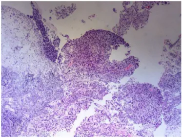
Fig. 2. High-grade papillary urothelial carcinoma, invaded into lamina propria,
con-sisting of papillary structures composed of the cells with prominent nuclei and nuclear atypia.
calcifications located on the right wall of the bladder, as well as prostatic calcifications, preoperatively (Fig. 1a and b).
Multifocal papillary tumoral lesions 4-cm in total diameter were seen on the right wall and dome of the bladder in cystoscopic examination and they were completely resected. In pathological examination of the resected tissue, a high-grade papillary urothe-lial carcinoma (pT1) has been reported with the findings of papillary structures composed of the cells with prominent nuclei and nuclear atypia (Fig. 2). No angio-lymphatic invasion was detected but there
Fig. 3. Schistosoma mansoni egg.
was inflammatory cellular infiltration of lymphoplasmocytes in the lamina propria. On the surface, oval, eosinophilic structures with lytic appearance were observed, indicating Schistosoma eggs (Fig. 3).
A consultation was conducted with the infectious diseases department, and after a detailed medical history of the patient it was discovered that he had been diagnosed and treated for urticaria after a sudden development of itchy skin, a rash with a burning sensation and a mild pain on his leg during garden irrigation. This complaint was accepted as specific dermatitis, a sign of skin pene-tration of cercariae, known as “swimmer’s itch”.
After observing the eggs of S. mansoni in the pathological exam-ination of specimens, IgG levels for S. mansoni and S. haematobium were evaluated and, IgG level for S. mansoni cercariae was 40 (nor-mal <6), and S. mansoni adult IgG was 14 (nor(nor-mal <6). Serology for S. haematobium was negative (IgG < 1/16). There was no find-ing of systemic infection. For the medical treatment of parasite, we administered praziquantel 40 mg/kg twice orally.
Six-week intravesical immunotherapy was administered to treat high-grade papillary urothelial carcinoma invading into lam-ina propria, and no complications were encountered. At the postoperative 3rd month visit, no Schistosoma eggs were detected in the urine analysis, and no tumor recurrence was observed during cystoscopy. In the samples resected from the previous tumor sites, mixed type inflammation with non-keratinized squamous meta-plasia, granulomas caused by immunotherapy, and eosinophils were detected. At the postoperative 18th month visit, there was no sign of tumor recurrence in the cytology or cystoscopy, and no parasitic eggs were found in direct microscopic examination of urine.
Informed consent was obtained from the individual participant included in the study.
3. Discussion
It is known that Schistosoma, a cylindrical trematode, which is 20 mm in length and white or grey in color, causes infesta-tion in 200 million people and 779 million are under risk[5]. The life cycle of Schistosoma begins with the transmission of the eggs from mammalian host to the fresh water supplies. Once the egg comes into contact with water, it transforms into a larva with cilia, called miracidium. Then mature sporocysts begin to develop after miracidia infect snails (intermediate hosts) in the natural or artifi-cial fresh water supplies, irrigation channels or ponds.
CASE REPORT – OPEN ACCESS
78 M.C. Kiremit et al. / International Journal of Surgery Case Reports 13 (2015) 76–78After 4–6 weeks, thousands of mature cercariae leave the snails and migrate into the water. Cercariae whirl around in the water for up to 72 h. When they come into contact with human (definitive host) skin, they penetrate the dermis or buccal mucosa and migrate to the right heart via the lymphatic system through ductus thoraci-cus. Mature cercariae then transmit to the lungs, liver and portal venous system (schistosomula), finally migrating to the mesenteric veins or vesicular venous plexus that drain at the base of the bladder and prostate[6]. While the S. mansoni and S. Japonicum are known as “intestinal Schistosomas” because they settle in mesenteric veins, S. haematobium is known as “genitourinary Schistosoma”[6].
The final diagnosis of genitourinary Schistosomiasis is made by detecting Schistosoma eggs in the urine samples or biopsy materials [6]. The typical appearance of eggs is helpful not only in differential diagnosis between the other helminthes, but also in the subtyping of Schistosomiasis. The serological tests are also diagnostic tools. However, although they have high specificity, they unfortunately can give cross-reactions with other parasitic infestations. In addi-tion, they can test positive for long periods after the treatment of an active infection[7].
Medical treatment of Schistosomiasis has many alternatives such as metrifonate, artesiminin, oxamniquine and praziquantel [8]. Rare and transient side effects (nausea and chills), lack of known long-term toxicity, being oral therapy as well as safety in chil-dren and pregnant women, praziquantel an pyrazinoisoquinoline derivative with activity against adult worms of all schistosome speciesies, has made the most widely used medical treatment of choice nowadays[9].
It is well known today that there is a relation between Schis-tosoma and bladder cancer and this condition was first shown by AR Ferguson in 1911 with a series of 40 cases[10]. Egyptian citi-zens had also been significantly exposed to the other carcinogens (such as smoking and insecticides), so Schistosoma infestation has only been accepted as a risk factor after certain laboratory and epidemiological studies over 100 years[4].
Severe inflammatory reaction that Schistosoma eggs cause in the bladder wall also results in the development of free oxygen radi-cals. These oxygen radicals can cause malign transformation by the way of genetic mutations and formation of certain carcinogenic compounds (N-nitrozamine and policyclic aromatic hydrocarbons, etc.)[11]. Additionally, Shoekir reported that bacterial superinfec-tions in the bladder, which are concomitant to the Schistosomiasis, may cause squamous cell carcinoma[12].
In spite of well-known etiological relationships between S. haematobium and bladder cancer, there is very limited number of cases of bladder carcinoma secondary to S. mansoni infestation in the literature[13,14]. All of the 5 cases reported in the literature were from the rural regions of Brazil. On the other hand, it was noticed that pathological examination had been reported in only one of these cases, and the diagnosis was leiomyoma. Therefore, to
the best of our knowledge, there is no data in the literature regard-ing the clinical course of the transitional cell carcinoma secondary to S. mansoni, or the data delineating the potential differences between these cases and those caused by S. haematobium.
Our patient is of particular importance as he lives in a non-endemic region for S. mansoni. Additionally, it is interesting that the tumor of our case was transitional cell carcinoma, representing a clinical course that is secondary to S. mansoni.
Regarding the increasing travels all around the world, clinicians should remember that Schistosoma infection is certainly a part of the differential diagnosis of bladder carcinoma, even if the patients are not from endemic regions. In addition, when such unusual cases are diagnosed in non-endemic regions, the required measures should be taken by authorities in order to prevent the evolution of the disease to an endemic pathology.
Conflict of interest
The authors declare that they have no conflict of interest. For this type of study formal consent is not required. References
[1] R.C. Neafie, A.M. Marty, Unusual infections in humans, Clin. Microbiol. Rev. 6 (1993) 34–56.
[2] W.D. Ayer, Napoleon Buonaparte and schistomiasis or bilharziasis, N.Y. State J. Med. 66 (1966) 2295–2301.
[3] B. Aytac, I. Sehitoglu, A rare parasitic infection in Turkey: schistosomiasis. Case report, TJPATH 28 (2012) 175–177.
[4] S. Salem, E.R. Mitchell, A.El-A. El-Dorey, A. Joseph, S. Daniel, A. Barocas, Successful control of schistosomiasis and the changing epidemiology of bladder cancer in Egypt, BJU Int. 107 (2011) 206–211.
[5] P. Steinmann, J. Keiser, R. Bos, M. Tanner, J. Utzinger, Schistosomiasis and water resources development: systematic review, meta- analysis, and estimates of people at risk, Lancet Infect. Dise. 6 (2012) 411–425. [6] B. Gryseels, Schistosomiasis, Infect. Dis. Clin. North Am. 26 (2012) 383–397. [7] V.C. Tsang, P.P. Wilkins, Immunodiagnosis of schistosomiasis, Immunol.
Invest. 26 (1997) 175–188.
[8] G.R. Olds, S. Dasarathy, Schistosomiasis, Curr. Treat. Options Infect. Dis. 2 (2000) 88–99.
[9] A.D. Dayan Albendazole, mebendazole and praziquantel: review of non clinical toxicity and pharmacokinetics, Acta Trop. 86 (2003) 141–159. [10] A.R. Ferguson, Associated bilharziasis and primary malignant disease of the
urinary bladder with observations on a series of forty cases, J. Pathol. Bacteriol. 16 (1911) 76–94.
[11] M.P. Rosin, S. Saad el Din Zaki, A.J. Ward, W.A. Anwar, Involvement of inflammatory reactions and elevated cell proliferation in the development of bladder cancer in schistosomiasis patients, Mutat. Res. 305 (1994) 83–92. [12] A.A. Shokeir, Squamous cell carcinoma of the bladder: pathology, diagnosis
and treatment, BJU Int. 93 (2004) 216–220.
[13] E.J. Lopes, T.C. Santos, V. Martins, Esquistossomose mansônica simulando neoplasia da bexiga, Revista Sociedade Brasileira Medicina 39 (2006) 287–288.
[14] M.L. Casella, V.S. Fanni, D.O. Verndl, M.C. Basso, L.F. Mello, S. Glina, Schistosomiasis mansoni of the bladder simulating bladder cancer: a case report, Rev. Soc. Bras. Med. Trop. 42 (September–October (5)) (2009) 581–582.
Open Access
This article is published Open Access atsciencedirect.com. It is distributed under theIJSCR Supplemental terms and conditions, which permits unrestricted non commercial use, distribution, and reproduction in any medium, provided the original authors and source are credited.