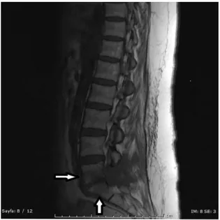
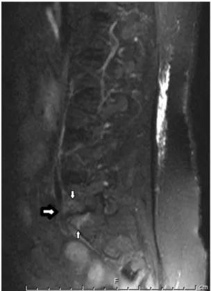
Postoperative Spondylodiscitis and Epidural Abscess Becoming Visible on Magnetic Resonance Imaging before Positive Laboratory Tests
Tam metin
Şekil
Benzer Belgeler
Öğretmen adaylarının eğitimde ölçme ve değerlendirmeye yönelik tutumları üzerinde ölçme ve değerlendirme dersinin etkisini araştırmayı amaçlayan bu
Objective: The aim of this study was to evaluate the ability of diffusion-weighted magnetic resonance imaging (MRI) and its corresponding apparent diffusion coefficient (ADC) values
Masses such as rectal adenocarcinoma, sarcoma, neuroendocrine tumor, leiomyoma, ovarian mass, rectal gastrointestinal stromal tumor (GIST), prostate adenocarcinoma,
Bu çah~mada, akut inme nedeniyle kliniğimize yatınlan ve erken dönemde BBT ile tam ke- sinlefitirilemeyen 39 olguda erken MRG inceleme,si yapılını~tır... SSK TEPECiK
Registration is performed using these expert guided manual anatomical landmarks, and also using the proposed method (both with and without surface refinement step). SSID mea- sures
We show sample image registration results for qualitative evaluation of the proposed method. Figure 4.6 and Fig.4.8 show image slices from two different patients having tumors
T1-weighted sagittal magnetic resonance imaging with contrast of the cervical spine shows the herniated disc and anterior epidural hyperintense signals.... Cervical Disc
Curricula of the Ganja State University and the Azerbaijan State Pedagogical University, which prepare students on the specialty of Informatics Teacher, were compared based on
