This study has been presented in the 24th European Congress of Pathology as an oral presentation and awarded a bursary for attendance the congress.
Address for Correspondence / Yazışma Adresi: Gonca Özgün, MD Başkent University Faculty of Medicine, Department of Pathology Bahçelievler, Çankaya, Ankara 06490 Turkey E-mail:[email protected]
©Telif Hakkı 2017 Gazi Üniversitesi Tıp Fakültesi - Makale metnine http://medicaljournal.gazi.edu.tr/ web adresinden ulaşılabilir. ©Copyright 2017 by Gazi University Medical Faculty - Available on-line at web site http://medicaljournal.gazi.edu.tr/
doi:http://dx.doi.org/10.12996/gmj.2017.71
Histopathological Changes of Neoadjuvant Chemoradiation and Relation with the Pre-Treatment
Tumor Stage in Rectal Carcinoma
Rektum Karsinomlarında Neoadjuvant Kemoradyoterapinin Yarattığı Histopatolojik Değişiklikler ve Tedavi Öncesi
Tümör Evresi ile İlişkisi
Gonca Özgün
1, Fatma Öz Atalay
2, Nesrin Uğraş
2, Ömer Yerci
21 Başkent University Faculty of Medicine, Department of Pathology, Ankara, Turkey 2 Uludağ University Medical Faculty, Department of Pathology, Bursa, Turkey
ABSTRACT
Objective: Neoadjuvant chemoradiotherapy has considered one of the standard treatment modalities for locally advanced rectal cancers. Chemoradiotherapy has a variety of different effects and responses on tumor, tumor bed and peritumoral tissues. The purpose of the present study was to evaluate the stromal responses in tumor bed between the different treatment modalities and different clinical T stages.
Methods: Fifty-seven consecutive patients with median age of 62.4 years were treated for rectal adenocarcinoma between January 2005 and July 2012 in Uludağ University Medical Faculty. Twenty-three of the patients were treated with neoadjuvant chemoradiation therapy and following surgery, 34 patients treated with surgery only.
Results: When we compared the stromal responses in the tumor bed between the two different treatment modalities, the amount of fibrosis and intensity of inflammatory cell infiltration were found considerably marked. The existence of calcification, hemosiderin-laden macrophages and mucin lakes were found also significant marked. There was no difference found in between the patients with different clinical stages which were received neoadjuvant CRT.
Conclusion: The stromal response in the tumor bed increases with the neoadjuvant chemoradiotherapy but the excess of the response doesn’t have any relation with the clinical T stage.
Key Words: Rectum, adenocarcinoma, neoadjuvant chemoradiotherapy, stromal response, tumor bed, T stage
Received: 03.09.2017 Accepted: 09.05.2017
ÖZET
Amaç: Neoadjuvant kemoradyoterapi lokal ileri rektal kanserlerdeki standart tedavi seçeneklerinden biridir. Kemoradyoterapi tümöral dokularda ve çevre dokularda çok çeşitli değişikliklere ve cevaplara neden olmaktadır. Bu çalışmanın amacı farklı tedavi seçeneklerinin tümör yatağında yarattığı değişiklikleri incelemektir.
Gereç ve Yöntemler: Uludağ Üniversitesi'nde Ocak 2005-Temmuz 2012 arasında rektal adenokarsinom nedeniyle tedavi edilmiş, ortalama yaşları 62.4 olan 57 hasta değerlendirildi. Hastaların 23 tanesi pre-operatif kemoradyoterapi sonrası cerrahi rezeksiyon yapılan, 34 tanesi ise sadece cerrahi rezeksiyon yapılan hastalardı.
Bulgular: Tümör yatağında stromal yanıtlar, neoadjuvant kemoradyoterapi sonrası cerrahi rezeksiyon yapılan hastalar ile sadece cerrahi rezeksiyon yapılan hastalar arasında değerlendirildiğinde, fibrozis ve inflamatuvar hücre infiltrasyonu açısından istatistiksel olarak anlamlı farklar bulundu. Kalsifikasyon, hemosiderin yüklü makrofajlar ve müsin gölcüklerinin varlığı açısından da belirgin farklar saptandı. Neoadjuvant kemoradyoterapi alan hasta grubunda, klinik T evreleri arasında değişkenler açısından fark mevcut değildi.
Sonuç: Tümör yatağındaki stromal cevap neoadjuvant kemoradyoterapi ile artış göstermektedir ancak bu artmış yanıtın tümör evresi ile ilişkisi saptanmamıştır.
Anahtar Sözcükler: Rektum, adenokarsinom, neoadjuvant kemoradyoterapi, stromal yanıt, T evresi, tümör yatağı
Geliş Tarihi: 09.03.2017 Kabul Tarihi: 05.09.2017
INTRODUCTION
Rectal cancer is the third most frequent malignancy in males and second in females, accounting for about 1.2 million new cases per year worldwide (1).
Neoadjuvant treatment has become the standard of care for locally advanced gastrointestinal tumors (2). The modern treatment of rectal cancer needs a multidisciplinary approach and a cooperative effort between the medical departments (3). Based on the randomized clinical trials, neoadjuvant chemoradiotherapy (CRT) followed by surgery is established as the standard treatment in locally advanced rectal tumor including clinically T3/T4 and/or
clinically node positive stage cancers (4). It has been demonstrated in several studies, however, that clinical outcome depends not only on the initial stage of the tumor, but also on the CRT-induced tumor response which varies among individual patients (1). The purpose of the neoadjuvant CRT are both downsizing and downstaging the tumor. Downsizing the tumor fascilitates the surgical resection, enables sphincter-preserving procedure and improve local control (5).
A growing body of evidence indicates that pathological response to neoadjuvant treatment can be measured with the histopathological tumor regression grade (TRG) (6). Rectal cancers that respond to neoadjuvant treatment undergo significant changes, which may result in complete disappearance of carcinoma cells and replacement of the tumor by fibrous or fibroinflammatory tissue (7).
Neoadjuvant chemoradiation therapy in rectal cancer is associated with significant tumor response and downstaging. Several systems for tumor response have been advocated (8,9), and a modified Ryan scheme is suggested, which has been shown to provide good inter-observer reproducibility provide prognostic significance (10).
According to the CAP protocol for the examination of specimens from patients with primary carcinoma of the colon and rectum, the tumor regression score (TRS) was established as follows: Score 0: No viable cancer cells (complete response), Score 1: Single cells or rare small groups of cancer cells (near complete response), Score 2: Residual cancer with evident tumor regression, but more than single cells or rare small groups of cancer cells (partial response), Score 3: Extensive residual cancer with no evident tumor regression (poor or no response) (11).
The aim of this study is to find out the stromal responses in the tumor bed between the different treatment modalities and between the different clinical T stages.
MATERIALS and METHODS
Patients
A total of 57 consecutive patients were treated for rectal adenocarcinoma between January 2005 and July 2012 at Uludağ University. The patients were selected retrospectively from the pathology archive. The mean age of 57 patients was 62,4±12,2 (range, 28-81 years). Pre-treatment workup consisted of digital rectal examination, rectosigmoidoscopy, biopsy, abdominopelvic computed tomography and chest X-ray. All the 57 patients were categorized according to the pre-treatment clinical T stage.
Twenty-three of the patients were treated with pre-operative CRT and surgery, while the remaining 34 patients treated with surgery only. In all patients which treated with neoadjuvant CRT, 3D planned conformal radiotherapy was carried out with belly board. Primary tumor as well as lymph nodes at risk was covered and received 45 Gy in 25 fractions over a period of 5 weeks. As a concomitant chemotherapy 500 mg/m2 5-flourourasil continuous infusion was administered.
Surgical resection was performed 6-9 weeks after the completion of the CRT. The main demographic data of all patients is given in Table 1. The remaining 34 patients treated with surgery only had chosen as a control group.
Histopathological evaluation
Pathological response to neoadjuvant CRT in the tumor bed was determined by histopathological evaluation of resected specimens. Evaluation of the TRS, modified Ryan scheme was used.
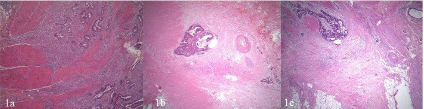
To assess the stromal responses in tumor bed, we evaluated fibrosis and inflammatory cell infiltration around the tumor islands and within the tumor bed. Intensity of fibrosis (Figure 1) and presence of inflammatory cells (Figure 2) were classified as low (Grade 1), intermediate (Grade 2), and high (Grade 3). While evaluating the inflammatory cells, we counted both the lymphocytes and the polymorphonuclear leukocytes. Other variables that we noted while evaluating the changes in the tumor bed were occurrence of calcification, hemosiderin-laden macrophages, and mucin lakes.
Figure 1: Grading the desmoplastic stromal response based on the amount of the collagen around the tumor islands; a) Grade 1 (H&E x40), b) Grade 2 (H&E x40), c) Grade 3 (H&E x25).
Figure 2: Grading the inflammatory response based on the intensity of the inflammatory cells; a) Grade 1 (H&E x40), b) Grade 2 (H&E x100), c) Grade 3 (H&E x40).
GMJ
Statistical analysis
Statistical analysis was performed with SPSS statistical software for Windows, version 13.0 (SPSS Inc, Chicago, IL, USA). Differences between groups were analyzed using Student T-test for continuous variables. ANOVA test was used to test the difference between radiotherapy group. The normality of data was checked before the statistical analysis was performed. A P value of less than 0.05 was considered to be whereas statistically significant.
RESULTS
Low anterior resection or very low anterior resection were performed to all patients. There were 23 patients treated with neoadjuvant CRT and surgery (Group 1), while other 34 patients treated with surgery only (Group 2). Patients clinically staged before treatment. In Group 1, 4 patients were found cT2, 15
patients cT3, 4 patients cT4, respectively. In Group 2, 7 patients were found cT2,
21 patients cT3, 6 patients cT4, respectively. Pathological evaluation of the rectum
resections, response to neoadjuvant CRT revealed complete response (TRS 0) in 4, near complete response (TRS 1) in 7, partial response (TRS 2) in 8 and, poor or no response (TRS 3) in 4 of 23 patients.
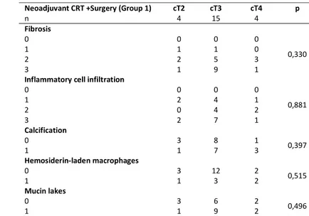
As shown in Table 2, the intensity of fibrosis and inflammatory cell infiltration in the tumor bed were found significantly marked in Group 1 (p<0.01). The occurrence of calcification, hemosiderin-laden macrophages and mucin lakes in the tumor bed were also found significantly distinctive in Group 1 (p<0.01).
We evaluated the same variables between different clinical T stages in Group 1. There were no differences found in stromal responses between different clinical stages (Table 3).
Table 1: The main demographic data of the patients.
DISCUSSION
Neoadjuvant CRT is indicated for all or almost all patients with cT3/cT4 rectal
cancer determined either by trans rectal ultrasound (TRUS) or magnetic resonans imaging (MRI). Relative indications for neoadjuvant CRT include the presence of clinically node-positive rectal cancer in a patient with an MRI or TRUS staged T1/T2 rectal cancer (12). Physical examination, endoscopy, and imaging are used
to define local tumor characteristics and to identify distant disease. Although conventional computed tomography is accurate in identifying invasion of neighboring structures, it is not able to demonstrate the layers of the rectal wall accurately. Investigations currently best suited for the determination of T classification and N status are EUS and MRI (3). In our institution, pretreatment rectal cancer staging was done with computed tomography evaluation. The main deficiency of this study was pretreatment clinical staging that evaluated by using the computed tomography images only.
Table 2: Stromal response variables between the two different treatment modalities.
It is very well known that the CRT has effects on both the tumor itself and the stroma also. Stromal changes include fibrosis, inflammation, mucin lakes (without tumor cells) and the presence of hemosiderin-laden macrophages. Calcification of the necrotic tumor cells can also be seen (12). In our study, all of the stromal changes were seen in both Group 1 and Group 2. Although desmoplastic stromal response, inflammatory cell infiltration, calcification and mucin lakes can be seen against to the tumor development but disappearance of tumor cells, replacement of the tumor by the fibrous tissue and hemosiderin-laden macrophages are changes that related with the CRT (7). In our study, desmoplastic stromal response and the inflammatory cell response were found significantly marked around tumor areas and in the tumor bed in Group 1. Calcification and mucin lakes were also found pronounced in the same patient group. In our study, there was no evidence of hemosiderin-laden macrophages in Group 2. We thought that this finding also supports the evidence of the CRT related findings. Occurrence of hemosiderin-laden macrophages is very helpful to find the tumor bed especially the tumor is totally disappeared because of the neoadjuvant treatment (13).
Table 3: Stromal response variables between the neoadjuvant CRT received cT2,
cT3, and cT4 groups.
Treatment prosedure Age Sex Clinical T Stage Total (n)
mean Male Female cT2 cT3 cT4
nCRT + Surgery (Group 1) 59,7±14,1 12 11 4 15 4 23
Surgery only (Group 2) 64,2±10,6 26 8 7 21 6 34
Neoadjuvant CRT + Surgery (Group 1) Surgery only (Group 2) p n 23 34 Fibrosis 0 0 3 0,000 1 2 14 2 10 15 3 11 2
Inflamatory cell infiltration
0 0 1 0,010 1 7 17 2 6 12 3 10 4 Calcification 0 12 31 0,001 1 11 3 Hemosiderin-laden macrophages 0 17 34 0,001 1 6 0 Mucin lakes 0 11 27 0,013 1 12 7
Neoadjuvant CRT +Surgery (Group 1) cT2 cT3 cT4 p
n 4 15 4 Fibrosis 0 0 0 0 0,330 1 1 1 0 2 2 5 3 3 1 9 1
Inflammatory cell infiltration
0 0 0 0 0,881 1 2 4 1 2 0 4 2 3 2 7 1 Calcification 0 3 8 1 0,397 1 1 7 3 Hemosiderin-laden macrophages 0 3 12 2 0,515 1 1 3 2 Mucin lakes 0 3 6 2 0,496 1 1 9 2
GMJ
2017; 28: 235-238
Özgün et al.
Neoadjuvant chemoradiation
237
Although the stromal changes were significantly different between Group 1 and Group 2, there was not any difference in stromal responses between the neoadjuvant CRT received cT2, cT3 and cT4 stages.
CONCLUSION
Neoadjuvant chemoradiation therapy makes a stromal response in tumor bed by fibrosis, inflammatory cell infiltration, and accumulation of hemosiderin-laden macrophages. These changes occur in every tumor bed constantly and independently from the clinical/pathological T stage according to our study, Conflict of interest
No conflict of interest was declared by the authors.
REFERENCES
1. Farkas R, Pozsgai E, Schally AV, Szigeti A, Szigeti E, Laszlo Z et al. Possible predictors of histopathological response to neoadjuvant chemoradiotherapy for rectal cancer. J Cancer Res Clin Oncol 2012; 138:387-95.
2. Colorectal Cancer Colloborative Group. Adjuvant radiotherapy for rectal cancer: a systemic overview of 8,507 patients from 22 randomised trials. Lancet 2001; 20;358:1291-304.
3. Wu JS. Rectal cancer staging. Clin Colon Rectal Surg 2007; 20:a48-57.. 4. Bosset JF, Collette L, Calais G, Mineur L, Maingon P, Radosevic-Jelic L et al. Chemotherapy with preoperative radiotherapy in rectal cancer. N Engl J Med 2006; 14;355:1114-23.
5. Sauer R. Adjuvant and neoadjuvant radiotherapy and concurrent radiochemotherapy for rectal cancer. Pathol Oncol Res 2002; 8:7-17.
6. Dhadda AS, Dickinson P, Zaitoum AM, Gandhi N, Bessell EM. Prognostic importance of Mandard tumour regression grade following pre-operative chemo/radiotherapy for locally advanced rectal cancer. Eur J Cancer 2011;47:1138-45.
7. O’Neil M, Damjanov I. Histopathology of colorectal cancer after neoadjuvant chemoradiation therapy. The Open Pathology Journal 2009;:91-98.
8. Dworak O, Keilholz L, Hoffmann A. Pathological features of rectal cancer after preoperative radiochemotherapy. Int J Colorect Dis 1997; 12:19-23.
9. Mandard AM, Dalibard F, Mandard JC, Marnay J, Henry-Amar M, Petiot JF et al. Pathologic assessment of tumor regression after preoperative chemoradiotherapy of esophageal carcinoma. Clicopathologic correlations. Cancer 1994; 1;73:2680-6.
10. Ryan R, Gibbons D, Hyland JMP, Treanor D, White A, Mulcahy HE et al. Pathological response following long-course neoadjuvant chemoradiotherapy for locally advanced rectal cancer. Histopathology 2005; 47: 141-6.
11. Tang LH, Berlin J, Branton P, Burgart LJ, Carter DK, Fitzgibbons P et al. Protocol for the examination of specimens from patients with primary carcinoma of the colon and rectum. College of American Pathologist Cancer Protocol Templates
2013 version. Available from: URL:
http://www.cap.org/ShowProperty?nodePath=/UCMCon/Contribution%20Fold ers/WebContent/pdf/cp-colon-16protocol-3400.pdf
12. Chetty R, Gill P, Govender D, Bateman A, Chang HJ, Driman D et al. A multi-centre pathologist survey on pathological processing and regression grading of colorectal cancer resection specimens treated by neoadjuvant chemoradiation. Virchows Arch 2012; 460:151-5.
13. Vecchio FM, Valentini V, Minsky BD, Padula GD, Venkatraman ES, Balducci M et al. The relationship of pathologic tumor regression grade (TRG) and outcomes after preoperative therapy in rectal cancer. Int J Radiat Oncol Biol Phys 2005;62:752-60.