Cukurova Medical Journal
Cukurova Med J 2018;43(2):530-531ÇUKUROVA ÜNİVERSİTESİ TIP FAKÜLTESİ DOI: 10.17826/cumj.334877
Yazışma Adresi/Address for Correspondence: Dr. Alper Parlakgümüş, Baskent University School of Medicine, Adana Teachingand Research Center, Department of General Surgery, Adana, Turkey E-mail: [email protected] Geliş tarihi/Received: 16.08.2017 Kabul tarihi/Accepted: 23.09.2017
EDITÖRE MEKTUP / LETTER TO THE EDITOR
Breast columnar alteration with prominent apical snouts and secretions
Belirgin apikal sunut ve sekresyonları ile giden meme kolumnar değişikliği
Ali Ezer
1, Alper Parlakgümüş
11BaskentUniversity School of Medicine, Adana Teaching and Research Center, Dept. of General Surgery, Adana, Turkey Cukurova Medical Journal 2018;43(2):530-531
Dear Editor,
Columnar cell breast lesions result from changes in the configurations of columnar cells occuring right in and around the terminal duct lobular unit (TDLU) of the breast. Columnar cells are a normal part of functional breast ducts and TDLU’s, but sometimes they develop in unusual ways, or grow more rapidly than one would expect. One such lesion, described by Fraser et al1, is columnar alteration with prominent apical snouts and secretions (CAPSS). This is a term concerning a spectrum of lesions from columnar alteration of lobules to low-grade ductal carcinoma in situ (DCIS) at the high end. Fraser et al notified that DCIS was twice as likely to be associated with CAPSS with atypical features on core neele biopsies (CNB) than as it was with CAPSS without atypical features1. In the literature, there is limited avaible data about algorithm of treatment for these type of lesions. Due to a case of a patient, we would like to discuss approach for these lesions.
A 36 year old gravida 1, para 1 female presented with bilateral mastalgia for a month of time. During physical examination revealed slightly increased nodularity but no palpable lesions on both breasts. On Breast Ultrasonography, hypervascular solid lesions were seen on left outer upper quadrant with a size of 4x10 milimeters (mm) and on right inner upper quadrant with a size of 5x9 mm. In the direction of these results breast magnetic resonance (MRI) examination was requested. MRI revealed diffusion restriction on T1 and T2 weigted images for these lesions (Figure 1a). Tru-cut biopsies revealed intra-ductal papillary lesions. Following,
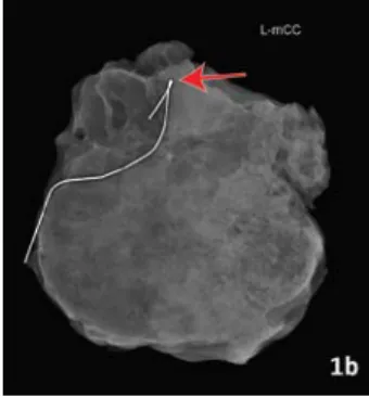
wire localised biopsies were performed, a specimen radiography was taken and demonstrated these two lesions were excised (Figure 1b). Permenant pathology revealed proliferative breast lesion on right breast, while a CAPSS with atypia on the left. According to these results, 6 month follow-up perids were recommended.
Figure 1a. Breast magnetic resonance (MRI) examination revealed diffusion restriction for the masses on the right and left (red arrows).
Figure 1b. a Specimen radiography demonstrated the lesion was excised fort he left defined lesion (red arrow).
Ezer ve Parlakgümüş Cukurova Medical Journal
531
Guerra-Wallace et al. evaluated the incidence of cancer (in situ/invasive) in core biopsies and in subsequent surgical biopsy specimens after an initial core biopsy identifying CAPSS of 195 patients. The rate of breast cancer for CAPSS lesions without atypia was 7.0% , while 12.0% for CAPSS lesions with atypia. This suggests that a detailed examination for atypia in all biopsy specimens containing CAPSS lesions is important in guiding further therapy and direct the surgeon to make right decision.
Therefore, they suggest that a CNB showing a CAPSS lesion with atypical features should undergo
surgical biopsy and after patient should be closely followed-up.
REFERENCES
1. Fraser JL, Raza S, Chorny K, et al. Columnar alteration with prominent apical snouts and secretions: a spectrum of changes frequently present in breast biopsies performed for microcalcifications. Am J Surg Pathol. 1998;22:1521–7.
2. Guerra-Wallace MM, Christensen WN, White RL Jr. A retrospective study of columnar alteration with prominent apical snouts and secretions and the association with cancer. Am J Surg. 2004;188:395-8.