EEvva
allu
ua
attiio
on
n o
off tth
he
e R
Re
ella
attiio
on
nssh
hiip
p B
Be
ettw
we
ee
en
n TTh
hyyrro
oiid
d
FFu
un
nccttiio
on
nss a
an
nd
d D
Drryy EEyye
e S
Syym
mp
ptto
om
mss iin
n P
Pa
attiie
en
nttss w
wiitth
h
H
Ha
assh
hiim
mo
otto
o TTh
hyyrro
oiid
diittiiss
H
Ha
assh
hiim
mo
otto
o TTiirro
oiid
diittii TTa
an
nııllıı H
Ha
asstta
alla
arrd
da
a K
Ku
urru
u G
Gö
özz S
Se
em
mp
ptto
om
mlla
arrıın
nıın
n
TTiirro
oiid
d FFo
on
nkkssiiyyo
on
nlla
arrıı iille
e O
Olla
an
n İİlliişşkkiissiin
niin
n D
De
eğ
ğe
errlle
en
nd
diirriillm
me
essii
Kanuni Sultan Suleyman Research and Training Hospital, Department of Internal Medicine, Division of Endocrinology and Metabolic Diseases, Istanbul, Turkey *Kanuni Sultan Suleyman Research and Training Hospital Department of Internal Medicine, Istanbul, Turkey
**Kanuni Sultan Suleyman Research and Training Hospital, Department of Ophthalmology, Istanbul, Turkey
Purpose: Dry eye is the most common symptom of thyroid-associated ophthalmopathy in Hashimoto thyroiditis. Ocular surface inam-mation, tear hyperosmolarity and overproduction of inammatory cytokines are responsible for the development of this process. In this study, we aimed to investigate the relationship between thyroid functions and dry eye symptoms in patients with Hashimoto thyroiditis. Materials and Methods: The patients, diagnosed with Hashimoto thyroiditis (n:125), were included in this monocenter cross-sectional study. The study population was divided into three groups: euthyroidism (Group–1, n:48), subclinical hypothyroidism (Group–2, n:38) and overt hypothyroidism (Group–3, n:39). The groups were compared based on the presence of dry eye syndrome, Schirmer tests, OSDI scores of patients with dry eye diagnosis and tear break-up time. The results were analyzed with ANOVA and Spearman’s correlation test. Results: No signicant difference was found in the frequency of dry eye syndrome between the groups (p = 0.1). There was no difference in the Schirmer scores and tear break-up time measurements (p = 0.6 and 0.2). The severity of OSDI scores was signicantly milder in the patients with dry eye diagnosis, in the euthyroid group (p = 0.03). Serum fT3 levels positively correlated with tear break up time measure-ments (p = 0.004, r = 0.302) and negatively correlated with presence of dry eye syndrome (p = 0.03, r = –0.981).
Discussion: Although dry eye symptoms tend to be milder in euthyroid patients, the frequency of dry eye syndrome is similar in patients with Hashimoto thyroiditis, independent of thyroid function status.
Keywords: Dry eye, OSDI, Hashimoto thyroiditis
Amaç: Hashimoto tiroiditi tanısı olan hastalarda tiroid fonksiyonlarının durumu ile kuru göz semptomlarının şiddeti arasındaki ilişkinin de-ğerlendirilmesi amaçlanmıştır.
Gereç ve Yöntemler: Prospektif klinik çalışmaya 125 Hashimoto tiroiditi tanılı hasta dahil edilmiştir. Hastalar tiroid fonksiyon durumlarına göre ötiroid (Grup–1, n:48), subklinik hipotiroid (Grup–2, n:38) ve aşikar hipotiroid (Grup–3, n:39) olmak üzere üç gruba ayrılmıştır. Kuru göz tanısı, Schirmer testi, gözyaşı kırılma zamanı ve OSDI anket sonuçlarına göre konulmuştur. Kuru göz tanısı olan hastalarda semptomların şiddeti OSDI anketlerine gore belirlenerek tiroid fonksiyon durumu ile ilişkisi araştırılmıştır. Verilerin analizinde ANOVA ve post-hoc Tukey testi, korelasyon analizinde Spearman Korelasyon analizi kullanılmıştır.
Bulgular: Gruplar arasında kuru göz tanısının sıklığı arasında anlamlı fark izlenmedi (p = 0.1). Ötiroid grupta OSDI skorları belirgin olarak daha düşük izlendi (p = 0.03). Serum sT3 düzeyleri ile gözyaşı kırılma zamanı arasında pozitif korelasyon (p = 0.004, r = 0.302) tespit edildi. Ayrıca serum sT3 düzeylerinin kuru göz sıklığı ile negatif korelasyon gösterdiği izlendi (p = 0.03, r = –0.981).
Tartışma: Ötiroid hashimoto tiroiditi tanılı hastalarda, göz kuruluğu semptomları daha haf seyretmekte olsa da kuru göz sıklığının has-himoto tiroiditi tanılı hastalarda tiroid fonksiyon durumundan bağımsız olarak benzer oranlarda görüldüğü tespit edilmiştir.
Anahtar kelimeler: Kuru göz, OSDI, Hashimoto tiroiditi
Address for Correspondence: Gülşah Yenidünya Yalın, Baskent University Istanbul Research Hospital, Department of Internal Medicine, Division of Endocrinology and Metabolism Disorders, Istanbul, Turkey
Phone: +90 212 214 20 00 E-mail: [email protected] Received: 15.06.2017 Accepted: 08.11.2017
®Copyright 2017 by Turkish Journal of Endocrinology and Metabolism Association Turkish Journal of Endocrinology and Metabolism published by Türkiye Klinikleri
Introduction
Dry eye is one of the most common clinical problems with several adverse effects on the quality of life (1, 2). This disorder may be a manifestation of several systemic or autoimmune disorders in-cluding autoimmune thyroiditis and hypothyroidism (3). Dry eye syndrome may affect visual acuity and daily activities, along with social and physical skills, resulting in significant decrease in the quality of life (4, 5). Any defect in the lacrimal gland, eyelid, and the ocular surface may lead to this syndrome (6).
Dry eye is also the most common finding of thyroid-associated ophthalmopathy (TAO) (7) in thyroid disorders such as Graves’ dis-ease and Hashimoto thyroiditis (8). TAO is defined as an autoim-mune disease of the extraocular muscle tissue and the lacrimal glands, characterized by extraocular myopathy, optic neuropathy, and ocular surface inflammation (9). The exact etiopathogenesis has been not established currently and several factors are consid-ered to be responsible for the development of this disorder. The most common factors are T cell-dependent ocular surface inflam-mation, increased tear film evaporation and hyperosmolarity due to proptosis, especially in Graves’ disease. Additionally, the effect of several inflammatory cytokines such as IL1-α, IL-4, and IGF-1 may play a role in this process (8). Recently, in addition to these factors, thyroid hormone receptors β-1 (Thrb-1), which are located on lacrimal glands, were found to be responsible for the development of dry eye (10). These receptors are known to cause susceptibility toward hypothyroidism, by converting the lacrimal glands as a tar-get tissue for thyroid hormones.
Although there exist a large number of studies involving the pres-ence of dry eye in patients with Graves ophthalmopathy (7,9,11,12,), the number of such studies in patients with Hashimoto thyroiditis is still very limited. In this study, we aimed to investigate the rela-tionship between thyroid functions and dry eye symptoms in pa-tients with diagnosis of Hashimoto thyroiditis.
M
Ma
atte
erriia
allss a
an
nd
d M
Me
etth
ho
od
dss
Study Group: A total of 125 patients (Females:115 and Males:10, with a median age of 38 years, ranging between 18–75 years) who were admitted to endocrinology and metabolism outpatient clinic, between March 1, 2016 and June 1, 2016, with the diagnosis of Hashimoto thyroiditis, were included in the study. The patients with a history of thyroidectomy, eye or eyelid surgery, eye or eyelid ab-normalities (proptosis, exophthalmos), prior diagnosis of dry eye or additional eye pathology, the presence of a rheumatologic dis-ease associated with dry eye syndrome (Sjogren’s disdis-ease, rheumatoid arthritis) were excluded from the study groups. The pa-tients were divided into three groups according to their thyroid function levels and clinical situations such as: euthyroid patients (Group-1; asymptomatic with normal free T4 (fT4) and thyroid stim-ulating hormone (TSH) levels, n:49, F/M:46/2), subclinical hypothy-roid patients ( Group-2; asymptomatic with normal fT4 and elevated TSH levels (5–10 mIU/L), n:38 , F/M:33/5) and patients with overt hypothyroidism (Group-3; symptomatic with normal fT4 and elevated TSH levels (>10 mIU/L) or decreased fT4 and elevated TSH levels (>5 mIU/L), n:39, F/M:36/3). The diagnosis of Hashimoto
thy-roiditis was based on the standard clinical criteria, thyroid function tests of autoantibody levels, and thyroid ultrasonography imaging (13). Serum free T3 (fT3) and free T4 (fT4) were measured with a competitive enzyme immunoassay. Serum TSH, thyroid peroxidase antibody (TPOAb) and thyroglobulin antibody (TgAb) levels were measured using two-site immunoenzymometric assay. The pres-ence of dry eye syndrome was compared between each group. Ocular Surface Disease Index (OSDI ) questionnaire, tear break-up time (TBUT ) and Schirmer I tests were applied for the clinical eval-uation of dry eye syndrome.
Ocular Surface Disease Index (OSDI ) questionnaire: The presence and the severity of the dry eye symptoms related to ocular irritation were evaluated with the scoring system of OSDI questionnaire. OSDI questionnaire consisted of twelve questions and three cate-gories namely, ocular symptoms, visional functions, and environ-mental factors. The patients scored each symptom in all the categories, between 0–4 according to their frequencies and sever-ities (0: never, 4: most often) (14). OSDI score was calculated with the standardized formula: Total score 25/number of valid ques-tions. The scores were defined as normal (0–12.5) or abnormal (>12.5), with mild (12.5–27.8), moderate (27.8–75) to severe (75– 100) dry eye symptom, in a scoring system having a range of 0– 100. The mean OSDI score was compared between each group Measurement of tear break-up time (TBUT ): A strip moistened with fluorescein sodium was applied to the inferior conjunctival region in each eye of the patient. After the strip was removed, the patient was asked to blink for three times in order to gain a homogenous distribution. The time interval between the initial break-up and the formation of the first rupture of the tear film was recorded three times and the average of the measurements was recorded as TBUT score.
Measurement of Schirmer I: A no. 41 Whatman filter strip was placed on the mid and lateral junction of the inferior fornix and the process was carried out in a dim environment without stimulant of light. No topical anesthesia was used prior to the evaluation. After 5 min, the strips were removed and the wet part of the strip was measured in millimeters to define Schirmer scores. The results below 5 mm were accepted as a strong indication for dry eye syn-drome and results below 10 mm were defined as aqueous tear deficiency. Diagnosis of dry eye syndrome was established in pa-tients with abnormal OSDI scores (>12.5) and Schirmer I result below 10 mm or TBUT below 10 s; or patients with normal Schirmer I and TBUT results but who had moderate or severe levels of OSDİ scores; or patients with a Schirmer score of below 5 mm. OSDI questionnaires, TBUT, and Schirmer I tests were evaluated and per-formed at a single center by the same ophthalmologist, for each patient. This study was approved by the ethics committee of our hospital and a written informed consent was obtained from all par-ticipants. All the procedures were performed in accordance with the Declaration of Helsinki.
Statistical Analysis
The distribution of variables was evaluated using the Kolmogorov Smirnov test. The mean, standard deviation [SD], median, mini-mum, maximini-mum, frequency and percentage values were reported
for each data. All the between-group comparisons were analyzed using ANOVA and post-hoc Tukey‘s test. Kruskal-Wallis test, chi-square test, and Spearman’s test were performed for the analysis of OSDI scores, categorical variables, correlation, respectively, with a statistical significance of p <0.05. All the statistical calculations were performed with SPSS 22.0 Package.
Results
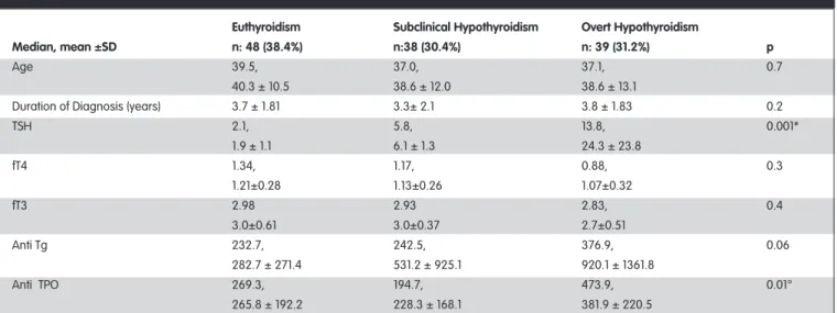
The study included patients who were recently diagnosed with Hashimoto thyroiditis (n:125, F:115, M:10) and had euthyroidism (n:48, 38.4%), subclinical hypothyroidism (n:38, 30.4%) or overt hypothyroidism (n:39, 31.2%) according to their baseline thyroid functions. The mean age of the patients was 39.3 ±11.8 (18–75; 38 years) and mean TSH level was 10.1 ±16.4 (0.1– 100; 5 mIU/L) (mean ±s.d, min-max; median). The distribution of age, duration of diagnosis and levels of TSH, fT4, fT3, and autoantibody
ac-cording to thyroid function status, are summarized in Table 1. The median value of OSDI questionnaire score was 9 (24.4 ±19.5; 0.0–90). OSDI was evaluated as normal in 33.6% of the patients (n:42) while in the remaining, the scores indicated the presence of mild (29.6%, n:37) or moderate (36.8%, n:46) dry eye symp-toms. There were no significant differences between the Schirmer test results (p = 0.6) and TBUT values (p = 0.2) between the three groups (Table 2). Mean values of OSDI scores were similar in euthyroid (23.7), subclinical (24.8) and overt hy-pothyroid patients (25.1). However, when the severity of OSDI scores was compared between the three groups of patients with dry eye diagnosis, euthyroidism tended to be mostly related with milder scores, while the patients with subclinical or overt hy-pothyroidism were more prone to having higher results (p=0.03) in the OSDI questionnaire (Table 2). Dry eye syndrome diagnosis based on OSDI score, Schirmer test and tear break-up time, did not show any significant difference between the three groups
Euthyroidism Subclinical Hypothyroidism Overt Hypothyroidism
Median, mean ±SD n: 48 (38.4%) n:38 (30.4%) n: 39 (31.2%) p
Age 39.5, 37.0, 37.1, 0.7
40.3 ± 10.5 38.6 ± 12.0 38.6 ± 13.1
Duration of Diagnosis (years) 3.7 ± 1.81 3.3± 2.1 3.8 ± 1.83 0.2
TSH 2.1, 5.8, 13.8, 0.001* 1.9 ± 1.1 6.1 ± 1.3 24.3 ± 23.8 fT4 1.34, 1.17, 0.88, 0.3 1.21±0.28 1.13±0.26 1.07±0.32 fT3 2.98 2.93 2.83, 0.4 3.0±0.61 3.0±0.37 2.7±0.51 Anti Tg 232.7, 242.5, 376.9, 0.06 282.7 ± 271.4 531.2 ± 925.1 920.1 ± 1361.8 Anti TPO 269.3, 194.7, 473.9, 0.01° 265.8 ± 192.2 228.3 ± 168.1 381.9 ± 220.5
TSH: Thyroid stimulating hormone, fT4: free T4, fT3: free T3, Anti Tg: antithyroglobulin antibody, Anti TPO: anti-thyroid peroxidase antibody. *post hoc analysis is signicant between overt hypothyroidism-euthyroidism and overt hypothyroidism-subclinical hypothyroidism. ° post hoc analysis is signicant between overt hypothyroidism and subclinical hypothyroidism.
Table 1. The distribution of age, duration of diagnosis, and laboratory results according to thyroid status.
Euthyroidism Subclinical Hypothyroidism Overt Hypothyroidism
Median, mean ±SD n: 48 (38.4%) n:38 (30.4%) n: 39 (31.2%) p
OSDI score in patients with 13.5, 17.7, 32.2, 0.03*
dry eye diagnosis 19.7 ± 20.3 28.6 ± 19.1 38.1 ± 19.5
Schirmer's (mm) 24, 23, 21, 0.6
20.6 ± 8.7 21.6 ±9.9 19.2 ±10.2
Tear Break-Up Time (sec.) 10.5, 10, 10, 0.2
10.4 ±2.0 10.1 ±2.3 9.7 ±2.4
Palpebral Angle (mm) 10.4, 10.1, 9.8 0.2
11.04 ±1.22 10.69 ±1.49 10.38 ±2.03
OSDI: Ocular Surface Disease Index, * post hoc analysis is signicant between overt hypothyroidism and euthyroidism.
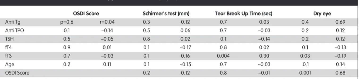
(p=0.1). Spearman’s correlation analysis revealed the presence of negative correlation between fT3 levels and dry eye diagnosis (p = 0.03, r= –0. 98) and plasma fT3 levels were seen to positively correlate with TBUT values (p = 0.004, r = 0.30). The decrease in fT3 levels resulted in a decrease of TBUT scores and an increase in the frequency of dry eye diagnosis (Table 3). Even though there was no significant correlation between OSDI scores and the re-sults of Schirmer or TBUT tests, a significant correlation (p = 0.001, r = 0.685) was seen between OSDI scores and dry eye diagno-sis, as predicted (Table 3).
D
Diissccu
ussssiio
on
n
Dry eye syndrome has been linked to Hashimoto thyroiditis, with decreased Schirmer test results, TBUT measurements and higher OSDI scores (15). The risk factors for the development of dry eye are decreased fT4 levels and the presence of proptosis. In order to decrease the prevalence of dry eye in these patients, levothyroxine treatment was formerly recommended (15). This study revealed a similar frequency of dry eye syndrome diagnosis among patients with hashimoto thyroiditis irrespective of their thyroid function sta-tus. Similarly, the frequency of the presence of dry eye was com-parable between the groups, which was independent of the patient’s thyroid function status. Additionally, there was no signifi-cant correlation between dry eye and fT4 levels, but fT3 levels showed a significant negative correlation with dry eye syndrome and positive correlation with TBUT. The decrease in fT3 levels re-sulted in lower TBUT measurements and increased dry eye diag-nosis. This relation may be explained by the fact that most of the effects of thyroid hormones on peripheral receptors are mediated by fT3 levels (16).
The etiopathogenesis of dry eye syndrome related with TAO have been investigated in several recent studies (7, 9–12). One of the main factors is an increase in tear film evaporation due to propto-sis which subsequently causes tear hyperosmolarity and a de-creased TBUT (17,18). Tear film hyperosmolarity has been shown to induce dry eye syndrome via inflammatory processes by induc-ing several inflammatory cytokines such as Interleukin-1 (IL- 1), TNF-α and MMP-9 (19). It has been reported that tear hyperosmolarity causes dry eye syndrome because of ocular surface damage,
which activates mitogen active protein kinase signal (MAPK) path-ways and induces MMP-9 expression (19, 20). In 1983 Gilbard et al. showed that tear osmolarity was significantly elevated in patients with dry eye syndrome and TAO (12). Proptosis and increase in palpebral angle are few of the many responsible factors in the de-velopment of the dry eye in TAO, particularly in Graves’s disease. However, a previous study demonstrated that the presence of sig-nificant ocular surface inflammation and dry eye symptoms in pa-tients with Graves’s disease was not dependent on either the degree of proptosis or the palpebral angle measurement (21). The presence of inflammation was seen with diffused lymphocyte infil-tration and grade 2–3 metaplasia, in the temporal interpalpebral region. This proved that ocular inflammation is also responsible for the development of dry eye syndrome, which is independent of the degree of proptosis (21).
The majority of the studies pertaining to dry eye syndrome and TAO have been conducted with Graves patients (7,9,11,12) and it has been seen that the studies on Hashimoto thyroiditis are rather limited (12,15). Previous experiments have shown that chronic deficiency of thyroid hormones causes ocular surface ab-normalities and dry eye syndrome (10). In 2007, Dias et al. sug-gested that lacrimal glands express receptors of thyroid hormone β-1 (Thrb), due to which they become a target tissue for thyroid hormones and thereby are susceptible to hypothyroidism. Fur-thermore, the chronic deficiency of thyroid hormones has been shown to be related to a modification in Thrb receptor expres-sion, leading to diminished tear secretion and dry eye syndrome (10).
Thyroid hormone regulates the oxidative metabolism through its nuclear Thrb receptors and therefore chronic hypothyroidism re-sults in increased oxidative stress (22,23). Immunohistochemical analysis indicates the presence of Thrb receptors in the acinar and ductal cells of lacrimal glands and in the nuclei of epithe-lial cells that line the corneal and conjunctival surface and it has been seen that these receptors are upregulated during hypothy-roidism (10). Hence, the presence of hypothyhypothy-roidism may have an adverse effect on lacrimal gland as well as corneal and conjuncti-val epithelial cells which are susceptible to low thyroid hormone levels (via Thrb receptors). The upregulation of these receptors is considered to be a compensatory mechanism, which is irreversible OSDI Score Schirmer's test (mm) Tear Break Up Time (sec) Dry eye
Anti Tg p=0.6 r=0.04 0.3 0.12 0.7 0.03 0.4 0.69 Anti TPO 0.1 –0.14 0.5 0.06 0.7 –0.03 0.2 0.12 TSH 0.5 –0.05 0.8 0.02 0.1 –0.14 0.2 0.12 fT4 0.9 0.01 0.1 –0.17 0.8 0.02 0.1 –0.13 fT3 0.7 –0.03 0.1 0.16 0.004 0.30 0.03 –0.19 Age 0.2 0.11 0.1 –0.15 0.7 –0.03 0.1 0.14 OSDI Score 0.2 0.12 0.8 –0.01 0.001 0.68
Anti Tg: antithyroglobulin antibody, Anti TPO: anti-thyroid peroxidase antibody
fT4: free T4, fT3: free T3, TSH: thyroid stimulating hormone, OSDI: Ocular Surface Disease Index.
in nature, as the number of receptors does not return to baseline levels even after the improvement of hypothyroidism (10). This could be the reason for the similar rates of dry eye syndrome that was observed in our patients who had different thyroid hormone lev-els. The similar frequencies of dry eye symptoms in euthyroid pa-tients may be due to the persistent upregulation of the Thrb receptors which had occurred during an early period of hypothy-roidism.
Moreover, our study also revealed that the patients with euthy-roidism, who were also diagnosed with dry eye syndrome, were more prone to obtain lower scores on OSDI questionnaire, whereas patients with subclinical or overt hypothyroidism tended to get higher scores that indicated severe dry eye symptoms. This implied that even though the frequency of dry eye syndrome was similar between each group, euthyroid patients had milder dry eye symptoms with lower OSDI scores. The amelioration in the symp-toms might have been due to improvement in Thrb receptor up-regulation. Nevertheless, further clinical and experimental studies with wider patient series are needed in order to reach definitive conclusions.
The limitation of our study was that exophthalmometer meas-urements of the patients could not be performed because of technical difficulties. It is possible that the improvement in the proptosis also had an effect on the amelioration of dry eye symptoms. It has been shown that the improvement of thyroid functions, which results in lowering of intraorbital glycosamino-glycan levels, may resolve the proptosis and palpebral angle widening (8). However, no statistically significant difference was found in the palpebral angle measurements between the three groups.
Conclusion
Dry eye is a common clinical problem seen during the manage-ment of hypothyroidism. Thus, ocular symptoms and signs should also be evaluated carefully in the surveillance of patients with Hashimoto thyroiditis. Levothyroxine replacement therapy not only improves several clinical factors but also provides ben-efit in the clinical care of these patients by alleviating the dry eye symptoms and improving daily activities. Further studies with wider population groups are needed in order to understand and improve the quality of life in patients with Hashimoto thy-roiditis.
A
Au
utth
ho
orr C
Co
on
nttrriib
bu
uttiio
on
nss
Idea/Concept: Gülşah Yalın, Hüseyin Çelik; Design: Fuat Cenik, Gülşah Yalın; Control/Supervision: Abdülbaki Kumbasar; Data Col-lection and/or Processing: Nihat Sayın; Analysis and/or Interpre-tation: Fuat Cenik, Gülşah Yalın; Literature Review: Gülşah Yalın, Hüseyin Çelik; Writing the Article: Gülşah Yalın; Critical Review: Ab-dülbaki Kumbasar; References and Fundings: Fuat Cenik; Materi-als: Nihat Sayın.
Conflict of Interest: The authors declare that they have no conflict of interest. Financial Disclosure: There is no organization that funded our research.
References
1. Lemp MA. Report of the National Eye Institute/Industry workshop on clinical trials in dry eyes. CLAO J. 1995;21:221-232.
2. Moss SE, Klein R, Klein BE. Prevalence of and risk factors for dry eye syndrome. Arch Ophthalmol. 2000;118:1264-1268.
3. Shashikala P. Prevalence of dry eye in hypothyroidism. Int J Clin Case Invest. 2013;46:51.
4. Pflugfelder SC. Prevalence, burden, and pharmacoeconomics of dry eye disease. Am J Manag Care. 2008 14:102-106.
5. Schiffman RM, Walt JG, Jacobsen G, Doyle JJ, Lebovics G, Sumner W. Utility assessment among patients with dry eye disease. Ophthal-mology. 2003 110:1412-1419.
6. Stern ME, Beuerman RW, Fox RI, Gao J, Mircheff AK, Pflugfelder SC. The pathology of dry eye: the interaction between the ocular surface and lacrimal glands. Cornea. 1998 17:584-589.
7. Nowak M, Marek B, Kos-Kud a B, Kajdaniuk D, Siemi ska L. [Tear film profile in patients with active thyroid orbitopathy]. Klin Oczna. 2005;107:479-482.
8. Yamada M, Li AW, Wall JR. Thyroid-associated ophthalmopathy: clin-ical features, pathogenesis, and management. Crit Rev Clin Lab Sci. 2000;37:523-549.
9. Burch HB, Wartofsky L. Graves’ ophthalmopathy: current concepts re-garding pathogenesis and management. Endocr Rev. 1993;14:747-793.
10. Dias AC, Módulo CM, Jorge AG, Braz AM, Jordão AA Jr, Filho RB, de Paula JS, Rocha EM. Influence of thyroid hormone on thyroid hormone receptor beta-1 expression and lacrimal gland and ocular surface morphology. Invest Ophthalmol Vis Sci. 2007;48:3038-3042. 11. Eckstein AK, Finkenrath A, Heiligenhaus A, Renzing-Köhler K, Esser J,
Krüger C, Quadbeck B, Steuhl KP, Gieseler RK. Dry eye syndrome in thyroid-associated ophthalmopathy: lacrimal expression of TSH re-ceptor suggests the involvement of TSHR-specific autoantibodies. Acta Ophthalmol Scand. 2004;82:291-297.
12. Gilbard JP, Farris RL. Ocular surface drying and tear film osmolarity in thyroid eye disease. Acta Ophthalmol (Copench). 1983;61:108-116. 13. Klee GG, Hay ID. Biochemical testing of thyroid function. Endocrinol
Metab Clin North Am. 1997;26:763-775.
14. Sullivan BD, Whitmer D, Nichols KK, Tomlinson A, Foulks GN, Geerling G, Pepose JS, Kosheleff V, Porreco A, Lemp MA. An objective ap-proach to dry eye disease severity. Invest Ophthalmol Vis Sci. 2010 51:6125-6130.
15. Kan E, Kılıçkan E, Ecemiş G, Beyazyildiz E, Çolak R. Presence of dry eye in patients with Hashimoto’s thyroiditis. J Ophthalmol. 2014;2014:754923.
16. Larsen PR, Silva JE, Kaplan MM. Relationships between circulating and intracellular thyroid hormones: physiological and clinical impli-cations. Endocr Rev. 1981;2:87-102.
17. Bartley GB, Fatourechi V, Kadrmas EF, Jacobsen SJ, Ilstrup DM, Garrity JA, Gorman CA. Clinical features of Graves’ ophtalmopathy in an in-cidence cohort. Am J Ophtalmol. 1996;121:284-290.
18. Perros P, Kendall-Taylor P. Pathogenic mechanisms in tyroid-associ-ated ophtalmopathy. J Intern Med. 1992;231:205-211.
19. Pflugfelder SC, Farley W, Luo L, Chen LZ, de Paiva CS, Olmos LC, Li DQ, Fini ME. Matrix metalloproteinase-9 knockout confers resistance to corneal epithelial barrier disruption in experimental dry eye. Am J Pathol. 2005;166:61-71.
20. Luo L, Li DQ, Doshi A, Farley W, Corrales RM, Pflugfelder SC. Experi-mental dry eye stimulates production of inflammatory cytokines and MMP-9 and activates MAPK signaling pathways on the ocular sur-face. Invest Ophthalmol Vis Sci. 2004;45:4293-4301.
21. Gürdal C, Saraç O, Genç I, Kırımlıoğlu H, Takmaz T, Can I. Ocular sur-face and dry eye in Graves’ disease. Curr Eye Res. 2011;36:8-13. 22. Lazar MA. Thyroid hormone receptors: multiple forms, multiple
pos-sibilities. Endocr Rev. 1993;14:184-193.
23. Brent GA. The molecular basis of thyroid hormone action. N Engl J Med. 1994;331:847-853.