DOI: 10.5455/annalsmedres.2019.12.860 2020;27(2):688-94
The relationship between Thiol/Disulphide Homeostasis
and Fibromyalgia
Bilge Ekinci1, Pervin Baran2, Cemile Koca Bicer3, Sema Haliloglu4, Hulya Uzkeser5, Ayse Carlioglu6 1Erzincan Binali Yildirim University, Faculty of Medicine, Department of Physical Medicine and Rehabilitation, Erzincan, Turkey 2Ataturk Training and Research Hospital, Clinic of Biochemistry, Ankara, Turkey
3Yildirim Beyazit University, Faculty of Medicine, Department of Biochemistry, Ankara, Turkey
4Kartal Training and Research Hospital, Department of Physical Medicine and Rehabilitation, Istanbul, Turkey 5Ataturk University, Faculty of Medicine, Department of Physical Medicine and Rehabilitation, Erzurum, Turkey 6Lokman Hekim University, Faculty of Medicine, Department of Endocrinology, Ankara,Turkey
Copyright © 2020 by authors and Annals of Medical Research Publishing Inc. Abstract
Aim: The main purpose of this study was to examine how a change in thiol/disulfide balance, which plays a significant role in cellular
activities like detoxification, antioxidant protection, apoptosis, and cell growth, affects fibromyalgia (FM) patients.
Material and Methods: This study was executed in two groups, included 46 female FM patients and 46 healthy female subjects. The
American College of Rheumatology (ACR) standard was used to diagnose patients with fibromyalgia. The automated measurement method was used to examine the native thiol-disulfide exchanges in both groups.
Results: After determining the parameters of thiol/disulphide balance for groups, it was discovered that the mean total thiol (p =
0.009), native thiol levels (p = 0.001), native thiol/total thiol (p < 0.001) percent ratios were higher in control group than FM group. Conversely, the disulphide levels (p = 0.026), disulphide/native thiol percent ratios (p < 0.001), disulphide/total thiol percent ratios (p < 0.001) were lower in control group than FM group.
Conclusion:The study demonstrates that the thiol/disulfide equilibrium could be a new oxidative stress marker that can be used for FM. However, it is considered that there needs to be more confirmatory studies to use thiol/disulfide balance in fibromyalgia as a stress marker.
Keywords: Fibromyalgia; thiol/disulfide ratio; oxidative stress; thiol oxidation
Received: 20.12.2019 Accepted: 25.02.2020 Available online: 10.03.2020
Corresponding Author: Bilge Ekinci, Erzincan Binali Yildirim University, Faculty of Medicine, Department of Physical Medicine and Rehabilitation, Erzincan, Turkey E-mail: [email protected]
INTRODUCTION
Fibromyalgia (FM) is a disorder characterized by pervasive chronic musculoskeletal pain, tenderness, and stiffness of specific anatomical sites, which are known as tender points. Headaches fatigue, irritable bowel syndrome, depression, and sleep disorders usually go along with this disease (1). The American College of Rheumatology (ACR) reported that FM prevalence ranges from two to eight percent of Canada and the United States population (2). Additionally, females, the elderly, and those with low-income levels are more likely to develop FM (3). Although the pathophysiology and etiology of FM are not yet completely understood, oxidative stress (OS) plays a significant role in FM recently (4).
Reactive oxygen species (ROS) and free oxygen radicals are continuously produced in the body and removed by
antioxidant mechanisms. ROS are the main molecules that cause oxidative damage to various cellular and tissue structures. These are produced because of the redox reactions, which include lipid peroxidation and protein degradation, and controlled by antioxidant defense systems (5). Antioxidants can be enzymatic, as in catalase (CAT), glutathione peroxidase (GSHPx) and superoxide dismutase (SOD). It can also be non-enzymatic, as in alpha-tocopherol, ascorbic acid, and glutathione (GSH) (6). Under normal circumstances, there is homeostasis between oxidative damage and antioxidant defense. However, when there is excessive production of ROS or insufficiency in the antioxidant defense systems, this balance is disturbed, and OS occurs. Thiols (SH) are organic compounds which include a sulfhydryl group (-SH), they are formed of hydrogen and a sulfur atom added with a carbon atom (7). They have many functions,
such as, is playing a significant role in antioxidant defense mechanisms (7). The plasma SH level is primarily created of protein thiols (PSH), albumin and, a lesser amount of low-molecular-weight SH like cysteine (cys), cysteinyl glycine, homocysteine, Gama-glutamylcysteine and GSH (8). The free radicals, which are a result of oxidation reaction, attack the cell, causing dysfunctions and chain reactions in the deoxyribonucleic acid and certain molecules that can even lead to cell death. The hydrophilic amino acids have a special structure due to their disulfide (SS) bonds. The SS bonds are important for increasing the stability of proteins. SHs can undergo oxidation reactions through oxidants, and as a result of that, disulfide bonds will be formed (9). OS occurs when cys residues exposed to oxidation; this causes the reversible structure of mixed SS between PSH groups and low-molecules-mass SHs. Since SS bonds can be diminished back to SH groups, SH-SS balance is preserved (10).
In this study, the change in the balance of SH/SS was evaluated in FM patients. This study aimed to investigate the antioxidant-oxidative stress balance, which underlies the etiology of many rheumatic diseases in fibromyalgia patients. In this study, the main hypothesis was to determine that the thiol/disulfide measures were different in the study groups. Therefore, an automated and new analysis method that is developed by Erel and Neselioglu was used (11). Thus, thiol-disulfide balance, which is the best indicator of oxidative stress, was investigated.
MATERIAL and METHODS
This study was carried out between January to April 2015 at Physical Therapy and Rehabilitation Clinic of Erzurum Regional Training and Research Hospital. Forty-six female patients who diagnosed with fibromyalgia according to the ACR 2010 diagnostic criteria were included in the study. The healthy control group included 46 volunteers who were similar in terms of age, weight, and height to the study group. Persons with additional comorbidities such as the history of diabetes mellitus, hypertension, thyroid function disorders, liver, and renal dysfunction, infections, anemia, osteoporosis, malignancy, cerebrovascular disease, heart disease, pregnant, and inflammatory autoimmune disease were excluded from the study. Additionally, participants who used corticosteroids, lipid-lowering drugs, antiepileptics, anti-inflammatory drugs, antioxidants, and antidepressants, or vitamin supplements were also excluded from the study. The demographic features of the participants, which included their body mass index (BMI), the duration of their disease, and smoking habits, were documented. Additionally, their accompanying symptoms such as fatigue, sleep disorders, paresthesia, headaches, and irritable bowel syndrome were also recorded. The patients were examined by a physician to verify the presence of widespread chronic pain and to determine tender point numbers. The FM Impact Questionnaire (FIQ) was used for the evaluation of FM patients (12). FIQ is an assessment tool that inquires with ten items, such as work difficulty, morning tiredness, fatigue, pain, stiffness, anxiety, and depression.
The Ethical Committee of the hospital accepted the present study, and informed consent was taken from all individuals included in the study. Blood samples from the control group and FM patients were obtained in the morning after eight hours of fasting. The plasma was separated from the serum, and the samples were rapidly centrifuged for 10 minutes at 1500 rpm. The serums were preserved at -80oC till they were analyzed. The complete blood count, erythrocyte sedimentation, C-reactive proteins, thyroid, liver, and kidney function, total protein, albumin, and total cholesterol (including triglycerides, high-density, and low-density lipoprotein cholesterols) as well as glucose levels were determined for all participants.
The SH/SS balance was determined with a new method developed by Erel and Neselioglu (11). Measurements were obtained by using a chemical analyzer (Cobas c501, Roche Diagnostic, Mannheim, Germany). The values of Serum SH/disulfide balance were given in mmol / L. Briefly, and disulfide bonds were reduced with sodium borohydride (NaBH4) to form free functional SH groups. Unused reducing NaBH4 was used to prevent the reduction of 5,5’ditiobis- (2-nitrobenzoic acid) (DTNB) and removed with formaldehyde and all SH groups, including reduced and natural SH groups, were determined after reaction with 5. 5’-dithiobis- (2-nitrobenzoic acid). The chemicals used in the study were obtained from Merc Chemicals (Darmstadt, Germany) and Sigma Aldrich Chemie (Milwaukee, Wisconsin, USA). Besides, when determining total SH and natural SH levels, half of the difference between total SH and SH groups was taken, and the dynamic SS was determined. After that, SS/total SH percent ratios, SH/total SH percent ratios, and SS/SH percent ratios were calculated by multiplying with 100 (11).
Statistical Analysis: IBM SPSS 22 (Armonk, NY: IBM Corp.) was used for analysis. Results were presented as mean ± standard deviation, median (minimum-maximum) for continuous variables. The normality assumption was checked by using the Kolmogorov-Smirnov normality test. For the comparison between groups Students t-test or nonparametric alternative Mann Whitney U test was used. Pearson’s or Spearman’s correlation was used to determining the correlation between measurements according to normality. A p-value was smaller than 0.05accepted significant.
Ethical approval: This study was conducted in conformity with the ethical standards of the research committee and the Helsinki Declaration (Ethics committee decisions; Date: 20.01.2015, Decision no:2015/02-16).
RESULTS
Ninety-two women, 46 of them were FM patients, and 46 of them were healthy, were participated in the study. Average of the age was 32.25±7.04 years in the FM group and 31.48±6.76 in the control group (p=0.873). The demographic features and biochemical findings were shown in Table 1.
Table 1.Demographic characteristics and laboratory findings of the study population Patients (n=46) Controls (n=46) P Age (y) 32.25 ± 7.04 31.48 ± 6.76 0.873* 33.5(20-51) 33(18-45) BMI (kg/m2) 26.94 ± 5.39 26.65 ± 5.12 0.787 26.6(17.6-37.7) 27(15.2-37.2) Waist circumference (cm) 85.96 ± 11.52 84.50 ± 11.99 0.534 87(67-108) 83.5(64-108) FIQ 60.35 ±10.46 - -60.58(39.31-94.19) WBC (x103/uL) 6.93 ± 1.62 7.69 ± 2.18 0.061 6.99(4.11-11.23) 8.195(4.5-12.3) Hemoglobin (g/dL) 14.20 ± 1.36 13.61 ± 1.51 0.438* 14.69(10.58-16.8) 14.44(11.8-15.95) Hemotocrit (%) 43.51 ± 3.46 41.57 ± 3.89 0.132* 44(35.41-50) 42.88(36.4-47.95) Platelet (x103/uL) 290.69 ± 56.34 282.30 ± 76.81 0.573 293(163-430) 262(196-486) PDW (%) 19.77 ± 0.85 19.28 ± 1.85 0.142 19.8(17.57-21.73) 19.65(18.43-23.5) RDW (%) 12.67 ± 1.91 12.83 ± 1.31 0.098* 11.9(10.9-19.6) 12(11.1-13.9) MPV (fL) 7.76 ± 1.24 7.83 ± 1.51 0.813 7.6(4.59-10.7) 7.42(5.35-9.6) TSH (mlU/L) 1.84 ± 1.43 1.48 ± 0.87 0.306* 1.505(0.13-88) 0.97(0.8-1.8) Creatinine (mg/dL) 0.70 ± 0.17 0.64 ± 0.11 0.064* 0.68(0.28-1.5) 0.6(0.4-0.8) Total cholesterol (mg/L) 183.47 ± 48.34 177.41 ± 35.96 0.904* 172(126-339) 172(66-255) HDL (mgl/L) 48.31 ± 11.71 51.76 ± 8.66 0.385 51(31.5-69) 55(33.32-66) LDL (mg/L) 119.64 ± 43.56 103.58 ± 33.01 0.292* 109(75-263) 95(34.8-278) Trigliseride (mg/L) 123.47 ± 61.87 98.50 ± 47.73 0.111* 107(55-284) 71.5(44-209) Albumin (g/dL) 4.32 ± 0.24 4.28 ± 0.24 0.629* 4.285(3.93-4.9) 4.275(3.5-15) Total protein (g/dL) 7.69 ± 0.42 7.67 ± 0.44 0.820* 7.7(6.4-8.32) 7.5(4.5-13)
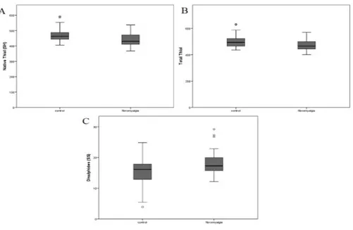
After reviewing the SH/SS parameters for the groups, it was determined that the mean SH (p=0.001), total SH (p=0.009) levels, and SH/total SH (p<0.001) percent ratios were higher in the control group than the FM group. Conversely the SS levels (p=0.026), SS/SH percent ratios (p<0.001) and SS/total SH percent ratios (p < 0.001) were higher in the FM group than the control group (Table 2). The SH, total SH and SS levels can be found in Figure 1.
The SS/total SH, SS/SH, and SH/total SH percent ratios can be found in Figure 2.
The relationship between SH/SS homeostasis concentrations and other variables were evaluated. It was determined that age is slightly negatively correlated with SH (r=-0.37, p<0.001), Low Density Lipid (LDL) (r=-0.34, p=0.017), FIQ (r=-0.34, p=0.002) and slightly positively correlated with White Blood Cell (WBC) (r=0.27,
Table 2. Thiol and Disulphides levels in study groups
Patients (n=46) Controls (n=46) P
Native thiol (SH)(µmol/L) 438.62 ± 39.77 471.89 ± 45.84 0.001*
430.5(367.3-537.0) 462.5(405.4-592.1)
Total thiol (µmol/L) 474.57 ± 42.43 502.84 ± 50.07 0.009*
465.7(400.9-570.0) 493.9(435.6-631.7) Disulphides (SS)(µmol/L) 17.96 ± 3.72 15.47 ± 4.86 0.007 17.3(12.1-29.2) 16.1(3.9-24.8) %SS/SH 4.10 ± 0.80 3.27 ± 0.97 0.001* 3.97(2.61-6.63) 3.36(0.84-5.04) %SS/Total SH 3.78 ± 0.68 3.05 ± 0.87 0.001* 3.67(2.48-5.85) 3.14(0.83-4.58) %SH/Total SH 92.42 ± 1.36 93.88 ± 1.74 0.001* 92.64(88.29-95.04) 93.71(90.84-98.34)
*Mann Whitney U test was used, otherwise Students t-test was used.
Results were presented as mean±standart deviation, median (minimum-maximum)
AST (U/L) 18.21 ± 5.03 16.79 ± 3.43 0.150* 18(11-32) 17(13-31) ALT (U/L) 16.71 ± 7.05 14.21 ±5.12 0.180* 13.5(6-31) 14(6-26) GGT (U/L) 15.19 ± 8.25 15.27 ± 8.26 0.711* 14(4-50) 12(6-58) Glucose (mg/dL) 94.73 ± 14.83 89.16 ± 15.34 0.056* 90(79-158) 86(61-138) CRP (mg/dL) 0.44 ± 0.37 1.11 ±1.2 0.190* 0.358(0.09-1.99) 0.35(0.1-3.79)
*Mann Whitney U test was used, otherwise Students t-test was used.
Results were presented as mean±standart deviation, median (minimum-maximum).
BMI, body mass index; FIQ, Fibromyalgia Impact Questionnaire; WBC, white blood cell; PDW, platelet distribution width; RDW, red blood cell distribution width, MPV, mean platelet volume; TSH thyroid-stimulating hormone; HDL, high-density lipoprotein; LDL, low-density
p=0.039) and Free T3 (FT3) (r=0.31, p=0.035). Total SH was found to have a slightly negative correlation with age (r=-0.35, p=0.001), LDL (r=-0.34, p<0.018), FIQ (r=-0.28, p=0.010) and a slightly positive correlation with WBC (r=0.31, p=0.016). Finally, it was discovered that SS has a moderately positive correlation with Alkaline Phosphatase (ALP) (r=0.40, p=0.006). It has also a slightly positive correlation with Neutrophil (NEU) (r=0.27, p=0.042), Lymphocyte (LYM) (r=0.28, p=0.035) and WBC (r=0.31, p=0.018) and has a slightly negatively correlated with Free T4 (FT4) (r=-0.28, p<0.049). Other parameters had slightly positive or negative correlations.
DISCUSSION
FM, which is, characterized by widespread tenderness and pain, is accompanied by various symptoms. Although several causes have been proposed, the etiopathogenesis of FM is complex and remains unclear (13).
Recently, the oxidant/antioxidant homeostasis and its effect on the organism have begun to attract attention. Recent studies have shown that OS may play a remarkable role in the pathogenesis of FM. When there is an imbalance between antioxidant defense mechanisms and ROS, OS occurs. High levels of OS can cause local tissue
(A) Native Thiol, (B) Total Thiol, (C) Disulphide
Figure 1. Oxidation variables according to groups
(A) Disulphide/Native Thiol (%), (B) Disulphide/Total Thiol (%), (C) Native Thiol/Total Thiol (%)
injuries, organ dysfunction, and many other disorders like inflammation, carcinogenesis, diabetes mellitus, rheumatoid arthritis, lung and/or pancreatic diseases, peptic ulcers, and atherosclerosis (14,15). When there is an emotional and/or a physically traumatic event, OS, which may lead to the development of FM, occurs. Fidan et al. reported that serum SS levels were reduced, and SH levels were increased in FM patients (16). According to this result, FM may be attributable to an imbalance between antioxidants and oxidants.
The main purpose of our study was to investigate the SH/SS balance in FM patients. Furthermore, the current study is the first one to use Erel and Neselioglu’s method to investigate how the SH/SS homeostatic status and oxidative balance may be attributable to the development of FM.
The data indicated that the SS levels (p=0.026), SS/ SH (p<0.001), and SS/total SH percent ratios (p<0.001) were higher in the FM group than in the control group. Additionally, the SH levels (p=0.001), total SH levels (p=0.009), and SH/total SH percent ratios (p<0.001) were lower in the FM group than in the control group. Due to the increased SS and decreased SH levels, the results indicated that the SH/SS balance shifted toward SS, which suggests that imbalanced SH and SS levels may contribute toward the etiopathogenesis of FM (11). However, there is a limited number of studies that have investigated the SH/SS homeostatic status in FM patients. In one of the studies of Eisenger et al., it was reported that there are significantly decreased plasma GSH in FM patients when compared with controls (17). Glutathione is an antioxidant molecule that captures free radicals and neutralizes them. Sendur et al. found decreased glutathione and catalase levels (18), and Alasehirli et al. reported that there is no relationship between serum nitric oxide levels and FM in the Turkish population (19). In contrast to these three studies, Akbas et al. found that SOD activity was higher in the FM group when compared with control, while GSHPx activity was not statistically significant between groups (20). Recently, Cordero et al. have studied how mitochondrial dysfunction might affect the etiopathogenesis of FM (21). As mitochondria are the strong producers of ROS, there may be a relationship between the pathogenic mechanisms of numerous diseases, including FM.
Additionally, many in vitro studies have indicated that when SS and SH levels are unbalanced, abnormal proliferation or apoptosis occurred at the cellular level (22,23). In the past, SH/SS homeostasis could only be measured unilaterally (24). However, Erel and Neselioglu developed a method that the levels of both substances may be measured separately, additively, and may be evaluated both individually and wholly (11). Previously, low-molecular-weight SH compounds, which are cys, reduced GSH, and glutathione disulfide (GSSG), could
usually be measured (25,26). Nevertheless, only a small fraction of the total SH in an organism consists of low molecular weight SHs while the majority of the total SHs are primarily albumin and other proteins (27). Therefore, it is likely that the SS and SH levels that were determined in the earlier studies do not reflect the SS and total SH amounts in the body; therefore, they do not accurately reflect the SH/SS homeostatic status.
Bozkurt et al. observed that there was not a significant difference between study groups’ serum total antioxidant status (28). The results of our study demonstrated that the homeostasis between antioxidant status and oxidant has deteriorated in female FM patients when compared with the control group. Additionally, our study identified that OS levels were increased in FM patients. Similarly, Wang et al. advocate that OS is associated with FM symptoms (29). Thus, when treating FM, the focus should be placed on reducing the patients’ oxidative loads, which would ultimately reduce their OS levels.
Our study is one of the few studies which use this new method to investigate thiol-disulfide balance in FM. Our results suggest that oxidative stress, which is thought to have a role in order rheumatic diseases, may also be involved in the etiopathogenesis of FM. However, our study did not perform on a sufficient number of patients. In order to fully understand the etiopathogenesis of FM, there needs to be studies that executed with the same method, but involving more patients should be done in different centers.
CONCLUSION
The data gathered during this study indicated that increased SS and decreased SH levels may be associated with the presence of OS in FM patients. We believe that this study is one of the limited numbers of studies to investigate the shift of dynamic SH/SS homeostasis toward SS form in FM patients. Additionally, this study demonstrates that the mechanisms that involve OS contribute to the development of FM. Moreover, by using Erel and Neselioglu’s newly developed method, this study verified that the SH/SS homeostatic status had been weakened in FM patients. This method will help determine individual SH and SS levels; it will also provide an easy, safe, fast, and inexpensive means to monitor dosage and time, as well as test the effectiveness of nutrients and medicines rich in SH. Further researches are needed to understand the pathophysiologic mechanisms of FM clearly. The loss of the SH group of proteins is the major molecular mechanism leading to structural and functional changes. Our study showed that in FM patients, these molecules shifted to the SS side, and the OS level was higher than the control group. With the addition of SH-containing molecules, reduction of the negative effect of OS in FM patients may be considered.
Competing interests: The authors declare that they have no competing interest.
Financial Disclosure: There are no financial supports.
Ethical approval:This study was conducted in conformity with the ethical standards of the research committee and the Helsinki Declaration (Ethics committee decisions; Date: 20.01.2015, Decision no:2015/02-16). Bilge Ekinci ORCID: 0000-0002-7520-0205
Pervin Baran ORCID: 0000-0001-8552-832X Cemile Koca Bicer ORCID: 0000-0001-7937-4475 Sema Haliloglu ORCID: 0000-0002-5356-6687 Hulya Uzkeser ORCID: 0000-0002-1364-2657 Ayse Carlioglu ORCID: 0000-0002-5622-9563
REFERENCES
1. Goldenberg DL, Burckhardt C, Crofford L. Management of fibromyalgia syndrome. Jama 2004;292:2388-95. 2. Wolfe F, Smythe HA, Yunus MB et al. The American
College of Rheumatology 1990 criteria for the classification of fibromyalgia. Arthritis Rheum 1990;33:160-72.
3. Cobankara V, Unal UO, Kaya A, et al. The prevalence of fibromyalgia among textile workers in the city of Denizli in Turkey. Int J Rheum Dis 2011;14:390-4. 4. Mease P, Fibromyalgia syndrome: review of clinical
presentation, pathogenesis, outcome measures, and treatment. J Rheumatol Suppl 2005;75:6-21.
5. Ates I, Ozkayar N, Topcuoglu C, et al. Relationship between oxidative stress parameters and asymptomatic organ damage in hypertensive patients without diabetes mellitus. Scand Cardiovasc J 2015;49:249-56.
6. Fridovich I. The biology of oxygen radicals. Science 1978;201:875-80.
7. Sen CK, Packer L. Thiol homeostasis and supplements in physical exercise. Am J Clin Nutr 72(2 Suppl) 2000;653-69.
8. Turell L, Radi R, Alverez B. The thiol pool in human plasma: the central contribution of albumin to redox processes. Free Radic Biol Med 2013;65:244-53. 9. Cremers CM, Jakob U. Oxidant sensing by reversible
disulfide bond formation. J Biol Chem 2013;288:26489-96.
10. Jones DP, Liang Y. Measuring the poise of thiol/ disulfide couples in vivo. Free Radic Biol Med 2009;47:1329-38.
11. Erel O, Neselioglu S, A novel and automated assay for thiol/disulphide homeostasis. Clin Biochem 2014;47:326-32.
12. Burckhardt CS, Clark SR, Bennett RM. The fibromyalgia impact questionnaire: development and validation. J Rheumatol 1991;18:728-33.
13. Furness PJ, Vogt K, Ashe S, et al. What causes fibromyalgia? An online survey of patient perspectives. Health Psychol Open 2018;5:1-11.
14. Southern PA, Powis G. Free radicals in medicine. Involvement in human disease. Mayo ClinicProc 1988;63:390-408.
15. Ma TY, Hollender T, Freeman D. Oxygen free radical injury of IEC 18 small intestinal cell monolayers. Gastroenterology 1991;100:1533-43.
16. Fidan F, Alkan BM, Ugurlu FG, et al. Dynamic Thiol/ Disulphide Homeostasis in Patients With Fibromyalgia. Arch Rheumatol 2017;32:112-7.
17. Eisinger J, Gandolfa C, Zakarian H, et al. Reactive oxygen species, antioxidant status and fibromyalgia. J Musculoskeletal Pain 1997;5:5-15.
18. Sendur OF, Turan Y, Tastaban E, et al. Serum antioxidants and nitricoxide levels in fibromyalgia: a controlled study. Rheumatol Int 2009;29:629-33. 19. Alaşehirli B, Demiryürek S, Arica E, et al. No
evidence for an association between the Glu298Asp polymorphism of endothelial nitric oxide synthase gene and fibromyalgia syndrome. Rheumatol Int 2007;27:275-80.
20. Akbas A, Inanir A, Benli I, et al. Evaluation of some antioxidant enzyme activities SOD and GPX) and their polymorphisms (MnSOD2 Ala9Val, GPX1 Pro198Leu) in fibromyalgia. Eur Rev Med Pharmacol Sci 2014:18:1199-203.
21. Cordero MD, de Miguel M, Moreno-Fernandez AM. Mitochondrial dysfunction in fibromyalgia and implication in the pathogenesis of disease. MedClin (Barc) Mar 2011;136:252-6.
22. Kirlin WG, Cai J, Thompson SA, et al. Glutathione redox potential in response to differentiation and enzyme inducers. Free Radic Biol Med 1999;27:1208-18. 23. Nkabyo YS, Ziegler TR, Gu LH, et al. Glutathione
and thio redox in redox during differentiation in human colon epithelial Caco-2) cells. Am J Physiol Gastrointest Liver Physiol 2002;283:1352-9.
24. Ellman G, Lysko H. A precise method for the determination of whole blood and plasma sulfhydrylgroups. Anal Biochem 1979;93:98-102. 25. Winther JR, Thorpe C. Quantification of thiol and
disulfides. Biochem Biophys Acta 2014;1840:838-46. 26. Glowacki R, Bald E. Fully automated method for
simultaneous determination of total cysteine ,cysteinylglycine, glutathione and homocysteine in plasma by HPLC with UV absorbance detection. J Chromatogr B Analyt Technol Biomed Life Sci 2009;877:3400-4.
27. Chen W, Zhao Y, Seefeldt T, et al. Determination of thiols and disulfides via HPLC quantification of 5-thio-2- nitrobenzoicacid. J Pharma Biomed Anal 2008;48:1375-80.
28. Bozkurt M, Caglayan M, Oktayoglu P, et al. Serum prolidase enzyme activity and oxidative status in patient with fibromyalgia. Redox Rep 2014;19:148-53. 29. Wang ZQ, Porreca F, Cuzzocrea S, et al. A newly
idetified role for superoxide in inflammatory pain. J Pharmacol Exp Ther 2004;309:869-78.