634 See related article, p 605.
P
aroxysmal atrial fibrillation (AF) is considered to carry a similar risk of ischemic stroke compared with persistent AF,1,2 and the management algorithm in terms of choosing the appropriate antithrombotic regimen is not different for both these types of AF.3 The widespread use of ambulatory cardiac moni-toring,4–6 together with advances in implantable devices7–9 and arrhythmia recognition algorithms, has not only increased the detection rate of high-risk atrial tachyarrhythmias like persistent and paroxysmal AF but also made it possible to identify other aberrations such as short-lasting (<30 seconds) irregular runs of nonsustained supraventricular tachycardia in patients with isch-emic stroke.10 Despite their resemblance to AF, these rhythms cannot be formally classified as paroxysmal AF because of their nonsustained nature.11 More importantly, although shown to be predictive of future conversion to chronic AF,12,13 it is currentlyunknown whether nonsustained AF episodes play a similar role in stroke pathophysiology like their persistent and paroxysmal counterparts. Previous studies have revealed a close relation-ship between total AF burden and embolic complications; data obtained from recordings in patients with implanted pacemak-ers show an increase in the incidence of embolic events when the duration of AF is >5 minutes, and this risk further escalates when the episodes last >24 hours.13–16 This information, how-ever, does not answer the question of whether more brief epi-sodes are enough to trigger the formation of intracardiac thrombi and thereby result in further embolic complications.
The ideal approach to understand the pathophysiologic role of nonsustained AF in stroke would be to perform pro-spective population-based studies in which the risk of isch-emic stroke is compared between cohorts with and without such an arrhythmia. Until this information becomes available Background and Purpose—The widespread use of ambulatory cardiac monitoring has not only increased the detection of high-risk arrhythmias like persistent and paroxysmal atrial fibrillation (AF), but also made it possible to identify other aberrations such as short-lasting (<30 seconds) irregular runs of supraventricular tachycardia. Ischemic stroke phenotype might be helpful in understanding whether these nonsustained episodes play a similar role in stroke pathophysiology like their persistent and paroxysmal counterparts.
Methods—In a consecutive series of patients with ischemic stroke, we retrospectively determined clinical and imaging features associated with nonsustained AF (n=126), defined as <30-second-lasting supraventricular tachyarrhythmias with irregular RR interval on 24-hour Holter monitoring, and compared them to patients with persistent/paroxysmal AF (n=239) and no AF (n=246).
Results—Patients with persistent/paroxysmal AF significantly differed from patients with nonsustained AF by a higher prevalence of female sex (odds ratio [95% confidence interval], 1.8 [1.1–2.9]), coronary artery disease (1.9 [1.1–3.0]), and embolic imaging features (2.7 [1.1–6.5]), and lower frequency of smoking (0.4 [0.2–0.8]) and hyperlipidemia (0.5 [0.3–0.8]). In contrast, patients with no AF were younger (0.5 [0.4–0.6] per decade) and more likely to be male (1.7 [1.0– 2.8]) in comparison with nonsustained AF population. The prevalence of nonsustained AF was similar among cryptogenic and noncryptogenic stroke patients (32% versus 29%). Voxel-wise comparison of lesion probability maps revealed no significant difference between cryptogenic stroke patients with and without nonsustained AF.
Conclusions—Clinical features of patients with nonsustained AF exhibited an intermediary phenotype in between patients with persistent/paroxysmal AF and no AF. Furthermore, imaging features did not entirely resemble patterns observed in patients with longer durations of AF. (Stroke. 2015;46:634-640. DOI: 10.1161/STROKEAHA.114.006396.)
Key Words: atrial fibrillation ◼ electrocardiography, ambulatory ◼ magnetic resonance imaging
Ischemic Stroke Phenotype in Patients With
Nonsustained Atrial Fibrillation
Ethem M. Arsava, MD; Demet F. Bas, MD; Enver Atalar, MD; Arzu C. Has, BSc;
Kader K. Oguz, MD; Mehmet A. Topcuoglu, MD
Received June 7, 2014; final revision received September 22, 2014; accepted October 14, 2014.
From the Department of Neurology, Faculty of Medicine (E.M.A., D.F.B., M.A.T.), Department of Cardiology, Faculty of Medicine (E.A.), and Department of Radiology, Faculty of Medicine (K.K.O.), Hacettepe University, Ankara, Turkey; and National Magnetic Resonance Research Center, Bilkent University, Ankara, Turkey (A.C.H.).
The online-only Data Supplement is available with this article at http://stroke.ahajournals.org/lookup/suppl/doi:10.1161/STROKEAHA.114. 006396/-/DC1.
Correspondence to Ethem Murat Arsava, MD, Department of Neurology, Faculty of Medicine, Hacettepe University, 06100 Sihhiye, Ankara, Turkey. E-mail arsavaem@hotmail.com
© 2015 American Heart Association, Inc.
Stroke is available at http://stroke.ahajournals.org DOI: 10.1161/STROKEAHA.114.006396
by guest on September 29, 2017 http://stroke.ahajournals.org/ Downloaded from by guest on September 29, 2017 http://stroke.ahajournals.org/ Downloaded from by guest on September 29, 2017 http://stroke.ahajournals.org/ Downloaded from by guest on September 29, 2017 http://stroke.ahajournals.org/ Downloaded from by guest on September 29, 2017 http://stroke.ahajournals.org/ Downloaded from by guest on September 29, 2017 http://stroke.ahajournals.org/ Downloaded from by guest on September 29, 2017 http://stroke.ahajournals.org/ Downloaded from by guest on September 29, 2017 http://stroke.ahajournals.org/ Downloaded from by guest on September 29, 2017 http://stroke.ahajournals.org/ Downloaded from by guest on September 29, 2017 http://stroke.ahajournals.org/ Downloaded from by guest on September 29, 2017 http://stroke.ahajournals.org/ Downloaded from by guest on September 29, 2017 http://stroke.ahajournals.org/ Downloaded from by guest on September 29, 2017 http://stroke.ahajournals.org/ Downloaded from by guest on September 29, 2017 http://stroke.ahajournals.org/ Downloaded from by guest on September 29, 2017 http://stroke.ahajournals.org/ Downloaded from by guest on September 29, 2017 http://stroke.ahajournals.org/ Downloaded from by guest on September 29, 2017 http://stroke.ahajournals.org/ Downloaded from by guest on September 29, 2017 http://stroke.ahajournals.org/ Downloaded from
Arsava et al Nonsustained AF and Ischemic Stroke 635 in the literature, some clues obtained by looking into the
stroke phenotype might be helpful in providing preliminary answers to the question. If indeed nonsustained AF exhibits a similar behavior like persistent or paroxysmal AF in terms of stroke risk, then it would be reasonable to hypothesize that phenotypic features like stroke risk factors, stroke pathogen-esis, and lesion patterns would not differ significantly among patients with nonsustained and longer durations of AF. In this study, we therefore determined clinical and imaging features of patients with ischemic stroke harboring nonsustained AF on 24-hour Holter monitoring and compared them to those patients with persistent/paroxysmal AF and no AF to obtain some insight into the pathophysiologic role of nonsustained AF in ischemic stroke.
Methods
This was a retrospective analysis of patients with ischemic stroke consecutively admitted to a tertiary care center over a period of 3 years. The analyses were restricted to patients who had undergone 24-hour Holter ECG monitoring for determination of stroke patho-genesis. In addition, patients with stroke with either a history of per-sistent or paroxysmal AF or newly documented AF on ECG strips or during inpatient heart rhythm monitoring were also included into the study. To be included into the imaging analyses, patients had to have undergone an MRI study within 72 hours after symptom onset. The flowchart of patients included to and excluded from the study is shown in Figure I in the online-only Data Supplement. The study was approved by the local institutional review board.
A 24-hour Holter monitoring was performed with a 3-elec-trode recorder with standard and identical settings in all patients (Lifecard CF, Spacelabs Healthcare, Washington, USA). The pres-ence of supraventricular runs with >3 beats and lasting <30 seconds, where RR interval was irregular and no evident p-waves detectable, was considered as nonsustained AF. Longer, self-terminating runs of fibrillation were considered as paroxysmal AF. The evaluation of Holter recordings primarily relied on the original clinical reports and the readjudication of ECG strips present on the enclosed re-port summaries. MRI, performed by a 1.5-T scanner (Magnetom TIM, Siemens, Erlangen, Germany), included axial T2-weighted (W) turbo spin echo (TR/TE; 3900/100 ms), FLAIR (TR/TE/ TI; 8900/100/2000 ms) imaging, and diffusion-weighted imaging (DWI) (single-shot echo planar, TR/TE; 5100/137 ms; with a
maxi-mum of 1,000 s/mm2) together with isotropic diffusion images and
apparent diffusion coefficient maps calculated online immediately after completion of the scan.
Comparison of Clinical Stroke Features
The purpose of this analysis was to compare clinical stroke features among 3 groups of patients: (1) patients with chronic or persistent/ paroxysmal AF determined either by history, conventional ECG, inpatient cardiac monitoring, or 24-hour Holter ECG; (2) patients without evidence of AF lasting ≥30 seconds but with nonsustained AF on 24-hour Holter ECG; and (3) patients without any duration of AF by ECG, cardiac monitoring, and 24-hour Holter ECG. For this purpose, age, sex, stroke risk factors (hypertension, diabetes melli-tus, hyperlipidemia, coronary artery disease, prior history of transient ischemic attack and stroke, current smoking), admission National Institute of Health Stroke Scale score, and stroke pathogenesis were determined in all patients. All patients underwent a thorough evalu-ation regarding the intracranial and extracranial vasculature (either by magnetic resonance angiography, computed tomography angiog-raphy, or carotid/vertebral/transcranial Doppler studies) as part of the standard of care in our institution. The Causative Classification
of Stroke system was used for etiologic subtyping17; as per the
purposes of the study, which basically aims to determine whether nonsustained AF is equivalent to paroxysmal or persistent AF, the
presence or absence of AF episodes <30 seconds was not included into the classification algorithm. In addition, where available, we collected information regarding left ventricular ejection fraction, left atrial diameter, and admission brain natriuretic peptide levels from patient charts. If the null hypothesis tested in the study (nonsustained AF~persistent/paroxysmal AF) is correct, one would expect no sig-nificant differences in clinical and laboratory features between non-sustained AF and persistent/paroxysmal AF groups, while both these groups would differ greatly from the group of patients with no AF. Concordantly, the prevalence of nonsustained AF would be higher among the otherwise cryptogenic patients, in comparison with pa-tients with apparent causes of stroke.
Comparison of Imaging Stroke Features
These analyses were restricted to patients with MRI obtained within 72 hours of symptom onset. The purpose of this analysis was to com-pare imaging stroke features across 3 groups of patients: (1) patients with persistent/paroxysmal AF determined either by history, conven-tional ECG, inpatient cardiac monitoring, or 24-hour Holter ECG; (2) patients with cryptogenic stroke and nonsustained AF on 24-hour Holter ECG; and (3) patients with cryptogenic stroke and no evidence of any duration of AF on 24-hour Holter ECG. Patients with persis-tent/paroxysmal AF and a concomitant stroke pathogenesis (like large artery atherosclerosis, small artery occlusion) or cardiac pathology (like prosthetic valve disease, rheumatic valve disease) were left out of these analyses as these additional pathologies might potentially interfere with lesion patterns on MRI. The initial set of comparisons among these 3 groups focused on the prevalence of imaging and an-giographic features suggestive of cerebral embolism. These features included the number of acute ischemic lesions, presence of isolated acute cortical lesions, and simultaneous acute ischemic lesions in multiple arterial territories on admission DWI, angiographic evi-dence of cutoff or recanalization on magnetic resonance angiography or computed tomographic angiography studies, and chronic ter-ritorial infarcts (excluding deep infarcts suggestive of small vessel disease) on T2W or FLAIR images, and were determined by consen-sus agreement between an experienced neuroradiologist and stroke neurologist. In the second stage, admission DWI of all patients were coregistered to MNI152 T1 template using the FSL-FLIRT (Oxford Centre for Functional Magnetic Resonance Imaging of the Brain Software [FSL, www.fmrib.ox.ac.uk/fsl] Linear Image Registration Tool).18,19 After coregistration, acute ischemic lesions on DWI were
outlined using a semiautomated segmentation algorithm (MRIcro software; University of Nottingham, UK, www.mricro.com) to cre-ate region of interest masks. In addition to calculation of admission DWI lesion volumes, these region of interests in each group were used to calculate group-wise lesion distribution probability maps by the add and divide commands in FSL. The randomize command was then used to perform voxel-wise comparisons of lesion
distribu-tions among these 3 groups of patients.19 All image analyses were
performed while blinded to clinical information of patients. Similar to the analyses mentioned above focusing on clinical stroke features, the null hypothesis of the study would be rejected if imaging features differed significantly between patients with nonsustained AF and per-sistent/paroxysmal AF.
Numeric variables are expressed as median (interquartile range) and categorical variables as n (%). Kruskal–Wallis and Mann– Whitney U tests were used to assess the difference between numeric variables, and χ2 test to assess differences with respect to categorical
variables among study groups. A multinomial regression model was performed to assess clinical characteristics independently associated with the 3 study groups (no AF, nonsustained AF, and persistent/par-oxysmal AF) which constituted the dependent variable in this mul-tivariate model; baseline demographic and clinical characteristics (age, sex, history of hypertension, diabetes mellitus, hyperlipidemia, coronary artery disease, prior stroke and transient ischemic attack, current smoking) were included in the model as independent vari-ables. Nonsustained AF group comprised the reference category in the model. A P<0.05 was considered statistically significant. SPSS version 16.0 was used for statistical analyses.
by guest on September 29, 2017
http://stroke.ahajournals.org/
636 Stroke March 2015
Results
The study population consisted of 611 patients; 239 (39%) of these patients had evidence of persistent or paroxysmal AF (≥30 seconds) detected either by ECG, inpatient routine car-diac monitoring, or 24-hour Holter ECG. On the other hand, 126 (21%) patients had no arrhythmia on ECG or cardiac monitoring, while episodes of AF lasting <30 seconds were present on 24-hour Holter monitoring. The remaining 246 (40%) patients had no documented episode of AF, regardless of duration, on ECG, cardiac monitoring, and 24-hour Holter monitoring. Holter monitoring was performed after a median (interquartile range) delay of 12 (7–18) days after the onset of stroke symptoms.
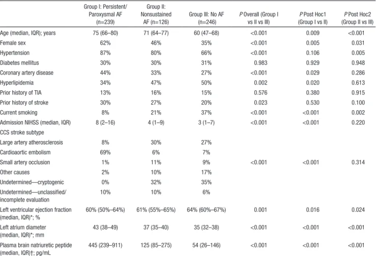
Table 1 summarizes the clinical and laboratory features of the study cohort. Overall, patients with nonsustained AF exhibited an intermediary phenotype between patients with persistent/paroxysmal AF and without AF. The mean patient age, prevalence of female sex, hypertension, coronary artery disease, and prior history of stroke demonstrated a sequen-tial stepwise increase from no-AF group to nonsustained AF group and finally to persistent/paroxysmal AF group (P=0.023 for history of stroke and P<0.001 for the remaining). A similar but inverse relationship was present with respect to hyperlipidemia (P=0.002) and current smoking (P<0.001).
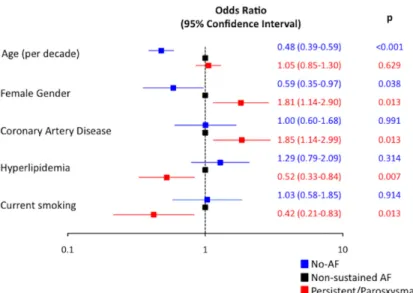
In multivariate analysis, younger (odds ratio [OR] 0.5 per decade, 95% confidence interval [CI], 0.4–0.6; P<0.001) and male (OR, 1.7; 95% CI, 1.0–2.8; P=0.038) patients were more likely to exhibit no-AF phenotype in comparison to nonsus-tained AF. On the other hand, female patients (OR, 1.8; 95% CI, 1.1–2.9; P=0.013) and those with coronary artery disease (OR, 1.9; 95% CI, 1.1–3.0; P=0.013) were more likely to have persistent/paroxysmal AF. In addition, current smoking (OR, 0.4; 95% CI, 0.2–0.8; P=0.013) and hyperlipidemia (OR, 0.5; 95% CI, 0.3–0.8; P=0.007) were factors significantly and negatively associated with persistent/paroxysmal AF in com-parison to nonsustained AF (Figure 1). Patients with persis-tent/paroxysmal AF had more severe strokes when compared with patients with nonsustained AF or no AF (P<0.001). The median (interquartile range) left ventricular ejection frac-tion and left atrial diameter were 60% (50–64) and 43 mm (38–49), 61% (55–65) and 37 mm (35–40), and 64% (60–67) and 35 mm (32–38) in patients with persistent/paroxysmal AF, nonsustained AF, and no AF, respectively (P<0.001). Among patients with an admission plasma brain natriuretic peptide level available, there was again a sequential distribution, with highest levels observed in persistent/paroxysmal AF patients and lowest levels in patients without any AF (P<0.001). The distribution of stroke subtypes differed significantly between Table 1. Comparison of Clinical Stroke Features and Laboratory Findings Among Patients With Persistent/Paroxysmal AF,
Nonsustained AF and No AF Group I: Persistent/ Paroxysmal AF (n=239) Group II: Nonsustained AF (n=126) Group III: No AF (n=246) P Overall (Group I vs II vs III) P Post Hoc1 (Group I vs II) P Post Hoc2 (Group II vs III)
Age (median, IQR); years 75 (66–80) 71 (64–77) 60 (47–68) <0.001 0.009 <0.001
Female sex 62% 46% 35% <0.001 0.005 0.031
Hypertension 87% 80% 66% <0.001 0.106 0.005
Diabetes mellitus 30% 30% 31% 0.983 0.929 0.948
Coronary artery disease 44% 33% 27% <0.001 0.029 0.286
Hyperlipidemia 34% 47% 50% 0.002 0.020 0.613
Prior history of TIA 13% 16% 15% 0.576 0.380 0.915
Prior history of stroke 30% 27% 20% 0.023 0.530 0.100
Current smoking 8% 21% 37% <0.001 <0.001 0.002
Admission NIHSS (median, IQR) 8 (2–16) 4 (1–9) 3 (1–7) <0.001 <0.001 0.220
CCS stroke subtype
Large artery atherosclerosis 8% 30% 27%
Cardioaortic embolism 69% 6% 7%
Small artery occlusion 1% 11% 9% <0.001 <0.001 0.314
Other causes 2% 10% 17%
Undetermined—cryptogenic 0% 32% 35%
Undetermined—unclassified/ incomplete evaluation
10% 10% 6%
Left ventricular ejection fraction (median, IQR)*; %
60% (50%–64%) 61% (55%–65%) 64% (60%–67%) 0.001 0.016 0.024
Left atrium diameter (median, IQR)*; mm
43 (38–49) 37 (35–40) 35 (32–38) <0.001 <0.001 <0.001
Plasma brain natriuretic peptide (median, IQR)†; pg/mL
445 (239–911) 125 (85–275) 54 (26–146) <0.001 <0.001 <0.001
Analyses limited to 532* and 241† patients. AF indicates atrial fibrillation; CCS, Causative Classification of Stroke; IQR, interquartile range; NIHSS, National Institute of Health Stroke Scale; and TIA, transient ischemic attack.
by guest on September 29, 2017
http://stroke.ahajournals.org/
Arsava et al Nonsustained AF and Ischemic Stroke 637
persistent/paroxysmal AF patients and patients in the other 2 categories (Table 1). When the prevalence of nonsustained AF was evaluated among noncardioembolic stroke subtypes, no statistically significant difference was observed (P=0.445; Table 2). Specifically, nonsustained AF was not more common among cryptogenic stroke patients, compared with patients with other identified causes of stroke.
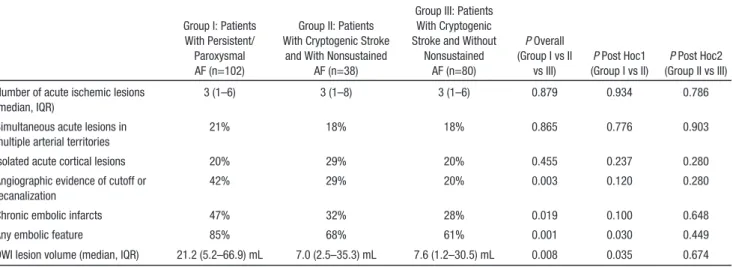
Table 3 summarizes the imaging features of patients with persistent/paroxysmal AF and patients with cryptogenic stroke with and without nonsustained AF. The 3 groups were not sig-nificantly different with respect to the number of acute ischemic lesions, presence of simultaneous acute lesions in multiple arterial territories, and isolated cortical lesions. On the other hand, patients with persistent/paroxysmal AF had larger acute ischemic lesions on DWI, had more chronic embolic infarcts, and were more likely to have angiographic features suggestive of embolism. In addition, when the presence or absence of any embolic imaging features was evaluated as a composite imaging signature, it was observed that these features were significantly more common among patients with persistent/ paroxysmal AF (Table 3). The multivariate model, which took into account clinical features significantly related to type of AF (age, sex, coronary artery disease, hyperlipidemia, current smoking; per prior analyses), showed a significantly higher prevalence of any embolic feature among persistent/parox-ysmal AF patients in comparison to nonsustained AF group (OR [95% CI], 2.7 [1.1–6.5]; P=0.035). The lesion distribu-tion probability maps of acute ischemic lesions on DWI are shown in Figure 2. No significant difference was present in lesion distributions among cryptogenic stroke patients with
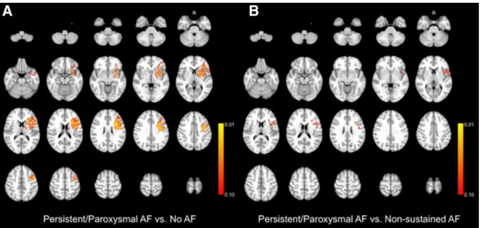
and without nonsustained AF. On the other hand, the distribu-tion pattern was significantly different between patients with persistent/paroxysmal AF and cryptogenic stroke patients without nonsustained AF (Figure 3A), with a propensity for left striatal and insular lesions in the former group. There was also a higher likelihood of left insular lesions when persis-tent/paroxysmal AF patients were compared with cryptogenic stroke patients with nonsustained AF; however, the signifi-cance level was between 0.05 and 0.10 in all of the relevant voxels (Figure 3B).
Discussion
Our findings show that patients with nonsustained AF show an intermediary phenotype with respect to clinical, laboratory, and echocardiographic features in between patients with persistent/ paroxysmal AF and no AF. We were not able to demonstrate a selective variability in the prevalence of <30 seconds-long AF episodes among various stroke subtypes. Furthermore, lesion patterns in cryptogenic stroke patients with nonsustained AF resembled to those patients without any AF, while patients with persistent/paroxysmal AF segregated significantly from both of these groups in terms of lesion volume, lesion distribution, and imaging features suggestive of embolism.
Studies performed by various long-term ECG monitoring tools like inpatient cardiac telemetry, Holter ECG, and external or implantable loop recorders have shown that a new diagnosis of AF can be established in up to 28% of patients presenting with ischemic stroke.4–9,20,21 Nonetheless, most of these studies, which show significant variation in terms of type, timing, and duration of monitoring; ECG analysis algorithm; and patient cohort characteristics, generally focus on the detection of AF episodes lasting ≥30 seconds, which is well known to alter the therapeutic management plan once if identified. On the other hand, knowledge is limited regarding the role of brief episodes of AF in the ischemic stroke setting. Outpatient cardiac moni-toring studies performed in cryptogenic and noncryptogenic stroke cohorts have revealed that these shorter runs of AF are encountered much more commonly than the conventional, ≥30-second-lasting AF episodes.10,22–24 The yield of 24-hour Holter ECG was 2% in terms of detecting ≥30-second-lasting Table 2. Prevalence of Nonsustained AF Among
Noncardioembolic Stroke Subtypes
Large artery atherosclerosis (n=123) 31%
Small artery occlusion (n=37) 38%
Other causes (n=59) 22%
Cryptogenic causes (n=125) 32%
Unclassified causes (n=37) 24%
P=0.445. AF indicates atrial fibrillation.
Figure 1. Results of the multivariate model with
nonsustained atrial fibrillation (AF) group as the reference category, and persistent/paroxysmal AF and no-AF groups constituting the other dependent variables. Bars show the odds ratio and 95% confi-dence intervals.
by guest on September 29, 2017
http://stroke.ahajournals.org/
638 Stroke March 2015
paroxysmal AF episodes in our cohort, while 31% of patients undergoing Holter monitoring had <30-second-lasting episodes (data not shown). In terms of clinical characteristics, prior stud-ies that involved patients with nonsustained AF generally have not evaluated them as separate cohorts, but rather combined them with group of patients that had longer durations of AF; their findings highlight that patients with any duration of AF are more likely to be older10,24 and have a history of diabetes mel-litus10 compared with patients devoid of AF. Our study, which not only includes the largest cohort of patients with brief dura-tions of AF reported in the literature, but also analyzes them separately from patients with longer durations of AF, suggests that combining patients with short and long durations of AF might not be entirely a correct approach. Demographic, clini-cal, and laboratory features that are well known to be related to the interplay between ischemic stroke and AF are more com-monly observed in patients with nonsustained AF with respect to patients with normal findings on Holter monitoring, but are still not as common as those observed in longer durations of AF.25–27 All these findings fit well into the recent observations that short supraventricular runs designate initial stages of left atrial remodeling and therefore are a predictor of future AF.12,13
Leaving aside the prognostic value in predicting conver-sion into persistent AF, the more critical question is whether nonsustained AF plays a similar role in stroke pathophysiol-ogy like its persistent or paroxysmal equivalents. One way to answer this question might be to assess the presence of non-sustained AF in various stroke subtypes and look for a higher prevalence of this arrhythmia in cryptogenic strokes. In con-cordance with this hypothesis, the yield of long-term rhythm monitoring for conventionally defined AF episodes lasting ≥30 seconds is higher in patients with cryptogenic stroke, suggesting that ≥30-second AF episodes are causally linked to the ischemic event and underlie the otherwise cryptogenic pathophysiology in a proportion of these patients.21 However, this is not the case for nonsustained AF; neither our findings, nor previous reports in the literature,24 were able to identify a higher rate of nonsustained AF episodes in cryptogenic stroke patients. An alternative clue regarding the pathogenic role of nonsustained AF might come from analyses involving imaging features of patients with stroke; the identification of embolic stroke features and characteristic lesion patterns in these patients might provide the missing link between non-sustained AF and ischemic stroke pathophysiology. Some of Table 3. Comparison of Imaging Features Among Patients With Persistent/Paroxysmal AF and Cryptogenic Stroke
Group I: Patients With Persistent/
Paroxysmal AF (n=102)
Group II: Patients With Cryptogenic Stroke
and With Nonsustained AF (n=38)
Group III: Patients With Cryptogenic Stroke and Without
Nonsustained AF (n=80) P Overall (Group I vs II vs III) P Post Hoc1 (Group I vs II) P Post Hoc2 (Group II vs III) Number of acute ischemic lesions
(median, IQR)
3 (1–6) 3 (1–8) 3 (1–6) 0.879 0.934 0.786
Simultaneous acute lesions in multiple arterial territories
21% 18% 18% 0.865 0.776 0.903
Isolated acute cortical lesions 20% 29% 20% 0.455 0.237 0.280
Angiographic evidence of cutoff or recanalization
42% 29% 20% 0.003 0.120 0.280
Chronic embolic infarcts 47% 32% 28% 0.019 0.100 0.648
Any embolic feature 85% 68% 61% 0.001 0.030 0.449
DWI lesion volume (median, IQR) 21.2 (5.2–66.9) mL 7.0 (2.5–35.3) mL 7.6 (1.2–30.5) mL 0.008 0.035 0.674
AF indicates atrial fibrillation; DWI, diffusion-weighted imaging; and IQR, interquartile range.
Figure 2. The lesion distribution probability maps of acute ischemic lesions on diffusion-weighted imaging in patients with persistent/
paroxysmal atrial fibrillation (AF; A), cryptogenic stroke with nonsustained AF (B), and cryptogenic stroke without nonsustained AF (C).
Highlighted regions signify voxels with acute ischemic lesions present in >10% of patients.
by guest on September 29, 2017
http://stroke.ahajournals.org/
Arsava et al Nonsustained AF and Ischemic Stroke 639
the previous studies, not independently analyzing patients with <30- and ≥30-second-long AF, have suggested that a new diagnosis on AF on prolonged monitoring was related to anterior circulation infarcts,5 and acute cortical and chronic infarcts on computerized tomography or MRI,23 while oth-ers were not able to identify any difference in terms of lesion topogprahy.10,24 Our analyses which separately evaluated patients with nonsustained and persistent/paroxysmal AF have shown that lesion patterns in nonsustained AF did not resemble those patterns in patients with longer durations of AF. Presence of imaging and angiographic features suggestive of embolism and distribution of acute ischemic lesions were not significantly different between patients with and without nonsustained AF. Therefore, neither the analyses focusing on the distribution of nonsustained AF among various stroke sub-types nor the stroke-related imaging features were supportive of an exact similarity between nonsustained AF and persis-tent/paroxysmal AF in ischemic stroke. These findings can be considered as concordant with previous reports suggesting that left atrial stunning, the inciting event of atrial appendicu-lar thrombus formation, is relatively uncommon before 15 to 20 minutes after the onset of AF episode.28
Several limitations of our study merit consideration. An inherent selection bias is unavoidable because of the ret-rospective nature of the study; although it is a standard of care to perform Holter monitoring to all stroke patients with no apparent AF on ECG or inpatient rhythm monitoring (regardless of the presence or absence of alternative stroke pathogeneses), there were still patients that were not able to undergo Holter monitoring because of various reasons like early mortality, physician discretion, and early discharge with loss to follow-up. There were however no significant differences in terms of age and baseline cardiovascular risk factors among patients with and without Holter monitoring. Excluded patients primarily resembled those patients with no evidence of AF on Holter monitoring, except for a higher number of patients with cryptogenic stroke in the latter group. This variability might hinder the applicability of our analyses regarding the relationship between stroke pathogen-esis and nonsustained AF to the general stroke population. Another source of selection bias arose from the restriction of imaging analyses to patients who had undergone MRI within 72 hours of symptom onset. Nonetheless, none of the demographic and clinical variables, including admission
stroke severity—which is closely related to lesion volume and location—differed substantially between patients with and without MRI. The presence or absence of nonsustained AF was defined per 24-hour Holter monitoring; it is highly probable that runs of AF lasting either <30 or ≥30 seconds would be detected in a certain amount of these patients if they were monitored for longer durations or by other tools. Still, 24-hour Holter monitoring is the most widely avail-able ambulatory monitoring tool, and we therefore think that our approach reflects the everyday clinical practice. We only evaluated left atrial diameter and left ventricular ejection fraction as echocardiographic parameters in our study; however, many other measures of left atrium func-tion determined either by transthoracic or transesophageal echocardiography are gaining importance in predicting AF and the associated stroke risk,29,30 and therefore should also be studied in this context. Finally, our analyses comparing lesion distribution probability maps have shown a borderline difference between persistent/paroxysmal AF and nonsus-tained AF patients, and no difference between nonsusnonsus-tained AF and no-AF patients; although the size of patient groups was considerably sufficient for voxel-wise analyses, future studies performed with larger number of patients might iden-tify additional disparities in lesion patterns that could have been missed in our study.
In conclusion, our findings suggest that clinical and imag-ing characteristics observed in patients with nonsustained AF do not entirely resemble patterns observed in patients with longer durations of AF. Because of the retrospective nature of the study and absence of a control group, these findings should be considered as hypothesis generating at best, and not be used to refute the causative role of nonsustained AF in stroke. For now, these findings, together with the already published literature, can be interpreted such that patients with ischemic stroke and nonsustained AF should be followed up closely for conversion to persistent AF, but may not necessar-ily need to be treated as patients with persistent/paroxysmal AF in terms of stroke prophylaxis. Considering the possible rise in recognition of these arrhythmias in the near future by advances in heart rhythm monitoring technologies and their ease of accessibility, we definitely need further studies to clarify the causative role of nonsustained AF during ischemic stroke and how they should be handled regarding secondary stroke prevention.
Figure 3. Voxel-wise comparison of
lesion distribution probability maps among patients with persistent/paroxys-mal atrial fibrillation (AF) vs cryptogenic
stroke without nonsustained AF (A), and
patients with persistent/paroxysmal AF vs cryptogenic stroke with nonsustained AF (B). Highlighted regions signify voxels that
are more commonly involved in patients with persistent/paroxysmal AF with a
P<0.10.
by guest on September 29, 2017
http://stroke.ahajournals.org/
640 Stroke March 2015
Sources of Funding
Dr Arsava received financial support from Turkish Academy of Sciences as part of Young Scientists Award Program (GEBIP).
Disclosures
None.
References
1. Marini C, De Santis F, Sacco S, Russo T, Olivieri L, Totaro R, et al. Contribution of atrial fibrillation to incidence and outcome of ischemic stroke: results from a population-based study. Stroke. 2005;36:1115– 1119. doi: 10.1161/01.STR.0000166053.83476.4a.
2. Hart RG, Pearce LA, Rothbart RM, McAnulty JH, Asinger RW, Halperin JL. Stroke with intermittent atrial fibrillation: incidence and predictors during aspirin therapy. Stroke Prevention in Atrial Fibrillation Investigators. J Am Coll Cardiol. 2000;35:183–187.
3. Hohnloser SH, Pajitnev D, Pogue J, Healey JS, Pfeffer MA, Yusuf S, et al; ACTIVE W Investigators. Incidence of stroke in paroxysmal versus sustained atrial fibrillation in patients taking oral anticoagulation or com-bined antiplatelet therapy: an ACTIVE W Substudy. J Am Coll Cardiol. 2007;50:2156–2161. doi: 10.1016/j.jacc.2007.07.076.
4. Gladstone DJ, Spring M, Dorian P, Panzov V, Thorpe KE, Hall J, et al; EMBRACE Investigators and Coordinators. Atrial fibrillation in patients with cryptogenic stroke. N Engl J Med. 2014;370:2467–2477. doi: 10.1056/NEJMoa1311376.
5. Jabaudon D, Sztajzel J, Sievert K, Landis T, Sztajzel R. Usefulness of ambulatory 7-day ECG monitoring for the detection of atrial fibrilla-tion and flutter after acute stroke and transient ischemic attack. Stroke. 2004;35:1647–1651. doi: 10.1161/01.STR.0000131269.69502.d9. 6. Liao J, Khalid Z, Scallan C, Morillo C, O’Donnell M. Noninvasive
car-diac monitoring for detecting paroxysmal atrial fibrillation or flutter after acute ischemic stroke: a systematic review. Stroke. 2007;38:2935–2940. doi: 10.1161/STROKEAHA.106.478685.
7. Sanna T, Diener HC, Passman RS, Di Lazzaro V, Bernstein RA, Morillo CA, et al; CRYSTAL AF Investigators. Cryptogenic stroke and underly-ing atrial fibrillation. N Engl J Med. 2014;370:2478–2486. doi: 10.1056/ NEJMoa1313600.
8. Healey JS, Connolly SJ, Gold MR, Israel CW, Van Gelder IC, Capucci A, et al; ASSERT Investigators. Subclinical atrial fibrillation and the risk of stroke. N Engl J Med. 2012;366:120–129. doi: 10.1056/ NEJMoa1105575.
9. Cotter PE, Martin PJ, Ring L, Warburton EA, Belham M, Pugh PJ. Incidence of atrial fibrillation detected by implantable loop recorders in unexplained stroke. Neurology. 2013;80:1546–1550. doi: 10.1212/ WNL.0b013e31828f1828.
10. Tayal AH, Tian M, Kelly KM, Jones SC, Wright DG, Singh D, et al. Atrial fibrillation detected by mobile cardiac outpatient telemetry in cryptogenic TIA or stroke. Neurology. 2008;71:1696–1701. doi: 10.1212/01.wnl.0000325059.86313.31.
11. Calkins H, Brugada J, Packer DL, Cappato R, Chen SA, Crijns HJ, et al; Heart Rhythm Society; European Heart Rhythm Association; European Cardiac Arrhythmia Society; American College of Cardiology; American Heart Association; Society of Thoracic Surgeons. HRS/ EHRA/ECAS expert consensus statement on catheter and surgical abla-tion of atrial fibrillaabla-tion: recommendaabla-tions for personnel, policy, pro-cedures and follow-up. A report of the Heart Rhythm Society (HRS) Task Force on Catheter and Surgical Ablation of Atrial Fibrillation developed in partnership with the European Heart Rhythm Association (EHRA) and the European Cardiac Arrhythmia Society (ECAS); in col-laboration with the American College of Cardiology (ACC), American Heart Association (AHA), and the Society of Thoracic Surgeons (STS). Endorsed and approved by the governing bodies of the American College of Cardiology, the American Heart Association, the European Cardiac Arrhythmia Society, the European Heart Rhythm Association, the Society of Thoracic Surgeons, and the Heart Rhythm Society. Europace. 2007;9:335–379. doi: 10.1093/europace/eum120.
12. Kochhäuser S, Dechering DG, Dittrich R, Reinke F, Ritter MA, Ramtin S, et al. Supraventricular premature beats and short atrial runs predict atrial fibrillation in continuously monitored patients with cryptogenic stroke. Stroke. 2014;45:884–886. doi: 10.1161/STROKEAHA.113.003788. 13. Glotzer TV, Hellkamp AS, Zimmerman J, Sweeney MO, Yee R,
Marinchak R, et al; MOST Investigators. Atrial high rate episodes
detected by pacemaker diagnostics predict death and stroke: report of the Atrial Diagnostics Ancillary Study of the MOde Selection Trial (MOST). Circulation. 2003;107:1614–1619. doi: 10.1161/01. CIR.0000057981.70380.45.
14. Botto GL, Padeletti L, Santini M, Capucci A, Gulizia M, Zolezzi F, et al. Presence and duration of atrial fibrillation detected by con-tinuous monitoring: crucial implications for the risk of thromboem-bolic events. J Cardiovasc Electrophysiol. 2009;20:241–248. doi: 10.1111/j.1540-8167.2008.01320.x.
15. Capucci A, Santini M, Padeletti L, Gulizia M, Botto G, Boriani G, et al; Italian AT500 Registry Investigators. Monitored atrial fibrillation dura-tion predicts arterial embolic events in patients suffering from bradycar-dia and atrial fibrillation implanted with antitachycarbradycar-dia pacemakers. J Am Coll Cardiol. 2005;46:1913–1920. doi: 10.1016/j.jacc.2005.07.044. 16. Boriani G, Botto GL, Padeletti L, Santini M, Capucci A, Gulizia M, et al;
Italian AT-500 Registry Investigators. Improving stroke risk stratification using the CHADS2 and CHA2DS2-VASc risk scores in patients with par-oxysmal atrial fibrillation by continuous arrhythmia burden monitoring. Stroke. 2011;42:1768–1770. doi: 10.1161/STROKEAHA.110.609297. 17. Ay H, Benner T, Arsava EM, Furie KL, Singhal AB, Jensen MB, et al.
A computerized algorithm for etiologic classification of ischemic stroke: the Causative Classification of Stroke System. Stroke. 2007;38:2979– 2984. doi: 10.1161/STROKEAHA.107.490896.
18. Jenkinson M, Bannister P, Brady M, Smith S. Improved optimization for the robust and accurate linear registration and motion correction of brain images. Neuroimage. 2002;17:825–841.
19. Jenkinson M, Beckmann CF, Behrens TE, Woolrich MW, Smith SM. FSL. Neuroimage. 2012;62:782–790. doi: 10.1016/j.neuroimage.2011.09.015. 20. Grond M, Jauss M, Hamann G, Stark E, Veltkamp R, Nabavi D, et al.
Improved detection of silent atrial fibrillation using 72-hour Holter ECG in patients with ischemic stroke: a prospective multicenter cohort study. Stroke. 2013;44:3357–3364. doi: 10.1161/STROKEAHA.113.001884. 21. Seet RC, Friedman PA, Rabinstein AA. Prolonged rhythm monitoring
for the detection of occult paroxysmal atrial fibrillation in ischemic stroke of unknown cause. Circulation. 2011;124:477–486. doi: 10.1161/ CIRCULATIONAHA.111.029801.
22. Flint AC, Banki NM, Ren X, Rao VA, Go AS. Detection of par-oxysmal atrial fibrillation by 30-day event monitoring in crypto-genic ischemic stroke: the Stroke and Monitoring for PAF in Real Time (SMART) Registry. Stroke. 2012;43:2788–2790. doi: 10.1161/ STROKEAHA.112.665844.
23. Alhadramy O, Jeerakathil TJ, Majumdar SR, Najjar E, Choy J, Saqqur M. Prevalence and predictors of paroxysmal atrial fibrillation on Holter monitor in patients with stroke or transient ischemic attack. Stroke. 2010;41:2596–2600. doi: 10.1161/STROKEAHA.109.570382. 24. Rabinstein AA, Fugate JE, Mandrekar J, Burns JD, Seet RC, Dupont SA,
et al. Paroxysmal atrial fibrillation in cryptogenic stroke: a case-control study. J Stroke Cerebrovasc Dis. 2013;22:1405–1411. doi: 10.1016/j. jstrokecerebrovasdis.2013.05.013.
25. Camm AJ, Kirchhof P, Lip GY, Schotten U, et al; European Heart Rhythm Association; European Association for Cardio-Thoracic Surgery. Guidelines for the management of atrial fibrillation: the Task Force for the Management of Atrial Fibrillation of the European Society of Cardiology (ESC). Eur Heart J. 2010;31:2369–2429.
26. Letsas KP, Filippatos GS, Pappas LK, Mihas CC, Markou V, Alexanian IP, et al. Determinants of plasma NT-pro-BNP levels in patients with atrial fibrillation and preserved left ventricular ejection fraction. Clin Res Cardiol. 2009;98:101–106. doi: 10.1007/s00392-008-0728-8.
27. Gage BF, Waterman AD, Shannon W, Boechler M, Rich MW, Radford MJ. Validation of clinical classification schemes for predicting stroke: results from the National Registry of Atrial Fibrillation. JAMA. 2001;285:2864–2870.
28. Sparks PB, Jayaprakash S, Mond HG, Vohra JK, Grigg LE, Kalman JM. Left atrial mechanical function after brief duration atrial fibrillation. J Am Coll Cardiol. 1999;33:342–349.
29. Warraich HJ, Gandhavadi M, Manning WJ. Mechanical discordance of the left atrium and appendage: a novel mechanism of stroke in par-oxysmal atrial fibrillation. Stroke. 2014;45:1481–1484. doi: 10.1161/ STROKEAHA.114.004800.
30. Tanaka K, Koga M, Sato K, Suzuki R, Minematsu K, Toyoda K. Three-dimensional analysis of the left atrial appendage for detecting paroxysmal atrial fibrillation in acute ischemic stroke. Int J Stroke. 2014;9:1045–1051. doi: 10.1111/ijs.12268. http://onlinelibrary.wiley. com/doi/10.1111/ijs.12268/pdf. Accessed September 22, 2014.
by guest on September 29, 2017
http://stroke.ahajournals.org/
Topcuoglu
Ethem M. Arsava, Demet F. Bas, Enver Atalar, Arzu C. Has, Kader K. Oguz and Mehmet A.
Ischemic Stroke Phenotype in Patients With Nonsustained Atrial Fibrillation
Print ISSN: 0039-2499. Online ISSN: 1524-4628
Copyright © 2015 American Heart Association, Inc. All rights reserved.
is published by the American Heart Association, 7272 Greenville Avenue, Dallas, TX 75231 Stroke
doi: 10.1161/STROKEAHA.114.006396
2015;46:634-640; originally published online January 29, 2015;
Stroke.
http://stroke.ahajournals.org/content/46/3/634
World Wide Web at:
The online version of this article, along with updated information and services, is located on the
http://stroke.ahajournals.org/content/suppl/2016/04/10/STROKEAHA.114.006396.DC4 http://stroke.ahajournals.org/content/suppl/2016/04/06/STROKEAHA.114.006396.DC3 http://stroke.ahajournals.org/content/suppl/2016/04/06/STROKEAHA.114.006396.DC2 http://stroke.ahajournals.org/content/suppl/2015/01/29/STROKEAHA.114.006396.DC1
Data Supplement (unedited) at:
http://stroke.ahajournals.org//subscriptions/
is online at: Stroke
Information about subscribing to
Subscriptions:
http://www.lww.com/reprints
Information about reprints can be found online at:
Reprints:
document.
Permissions and Rights Question and Answer
process is available in the
Request Permissions in the middle column of the Web page under Services. Further information about this Once the online version of the published article for which permission is being requested is located, click
can be obtained via RightsLink, a service of the Copyright Clearance Center, not the Editorial Office. Stroke
in
Requests for permissions to reproduce figures, tables, or portions of articles originally published
Permissions:
by guest on September 29, 2017
http://stroke.ahajournals.org/
Supplemental Figure Legends:
16 Stroke日本語版Vol. 10, No. 2
非持続性心房細動患者における虚血性脳卒中の表現型
Ischemic Stroke Phenotype in Patients With Nonsustained Atrial Fibrillation
Ethem M. Arsava, MD 1; Demet F. Bas, MD 1
; Enver Atalar, MD, et al.
1 Department of Neurology; and 2 Department of Cardiology, Faculty of Medicine, Hacettepe University, Ankara, Turkey.
背景および目的: 携帯型心電計が広く使用されるようにな り,持続性および発作性心房細動( AF )などの高リスクの 不整脈の検出率が高くなっただけでなく,短時間持続する (< 30 秒)不規則な上室性頻拍などの異常を同定すること も可能になった。虚血性脳卒中の表現型を知ることは,こ のような非持続性のエピソードが持続性および発作性のエ ピソードと同様に脳卒中の病態生理に関与するか否かを解 明するのに有益と考える。 方法: 虚血性脳卒中患者の連続症例で非持続性 AF 患者(n = 126)の臨床的特性と画像的特性を後ろ向きに検討した。 非持続性 AF は上室性頻拍性不整脈が < 30 秒間持続し, 24 時間ホルター心電図で RR 間隔が不規則なものと定義 し,持続性・発作性 AF 患者(n = 239)および非 AF 患者(n = 246)と比較した。 結果: 持続性・発作性 AF 患者と非持続性 AF 患者は,女 性の患者数が多い[オッズ比( 95% 信頼区間):1.8( 1.1 ∼ 2.9 )],冠動脈疾患[ 1.9( 1.1 ∼ 3.0 )]と塞栓像の有病 率が高い[ 2.7( 1.1 ∼ 6.5 )]こと,および喫煙[ 0.4( 0.2 ∼ 0.8 )]と高脂血症[ 0.5( 0.3 ∼ 0.8 )]の頻度が低いこと で有意に異なっていた。一方,非 AF 患者は非持続性 AF 患者と比べて若年で[年代毎に 0.5( 0.4 ∼ 0.6 )],男性の 方が多かった[ 1.7( 1.0 ∼ 2.8 )]。非持続性 AF の有病率 は原因不明か否かにかかわらず脳卒中患者では同様であっ た( 32% 対 29% )。ボクセル解析による病巣分布確率マッ プ( lesion probability maps )の比較では,非持続性 AF を 有する原因不明の脳卒中患者と非持続性 AF がない原因不 明の脳卒中患者に有意差は認められなかった。 結論: 非持続性 AF 患者の臨床特性は,持続性・発作性 AF 患者と非 AF 患者の中間の表現型であった。また,画 像的特性は長期間 AF を有する患者で認められたパターン とは全く異なっていた。
Stroke 2015; 46: 634-640. DOI: 10.1161/STROKEAHA.114.006396. Abstract 図 2 持続性・発作性心房細動( AF )患者( A ),非持続性 AF のある原因不明の脳卒中患者( B ),および非持続性 AF がない原因不明の脳 卒中患者( C )の拡散強調画像上で認められた急性虚血性病変の病巣分布確率マップ。強調表示した範囲は>10% の患者に認められ た急性虚血性病変のボクセルを示す。 持続性・発作性AF患者 非持続性AF患者 20% 10% 20% 10% 20% 10% 非AF患者 A B C STR-J_10-2_ab2_main.indd 16 STR-J 10-2 ab2 main.indd 16 2015-8-25 14:53:262015-8-25 14:53:26
37
Abstract 14
비지속성 심방세동 환자의 허혈뇌졸중의 임상양상
Ischemic Stroke Phenotype in Patients With Nonsustained Atrial Fibrillation
Ethem M. Arsava, MD; Demet F. Bas, MD; Enver Atalar, MD; Arzu C. Has, BSc; Kader K. Oguz, MD; Mehmet A. Topcuoglu, MD
(Stroke. 2015;46:634-640.)
Key Words: atrial fibrillation ■ electrocardiography, ambulatory ■ magnetic resonance imaging
배경과 목적
활동 심장 모니터링의 광범위한 사용은 지속성 또는 발작성 심방 세동(atrial fibrillation, AF)과 같은 고위험 부정맥의 발견을 증가 시킬 뿐 아니라, 30초 미만으로 짧게 지나가는 불규칙 심실상빈맥 과 같은 이상의 발견도 가능하게 한다. 허혈뇌졸중의 표현형이, 아 마도 이들 비지속성(nonsustained) 사건이 지속성 또는 발작성 심 방세동과 같이 뇌졸중의 병태생리학에 비슷한 역할을 하는지 여부 를 이해하는데 도움이 될 것이다. 방법 허혈뇌졸중 환자를 연속적으로 포함시킨 시리즈에서, 후향적으로 비지속성 AF (n=126)와 관련된 임상, 영상 특성을 분석하였다. 비 지속성 AF는 24시간 홀터 모니터링에서 RR 간격이 불규칙한 심 실상성 부정빈맥이 30초 미만으로 지속되는 경우로 정의하였다. 이들 환자를 지속성/발작성 AF 환자(n=239)와 AF가 없는 환자 (n=246)와 비교하였다. 결과 지속성/발작성 AF 환자는 비지속성 환자와 비교해서 여성이 많았 고(OR [95% CI], 1.8 [1.1-2.9]), 관상동맥질환이 많았으며(1.9 [1.1-3.0]), 영상의 색전성 병변이 많았다(2.7 [1.1-6.5]). 또한 흡 연비율은 낮았고(0.4 [0.2-0.8]), 고지질혈증이 적었다(0.5[0.3-0.8]). 반대로 AF가 없는 환자가 비지속성 AF 환자보다 더 젊었고 (0.5 [0.4-0.6] per decade), 남성이 더 많았다(1.7 [1.0-2.8]). 비 지속성 AF의 유병률은 원인불명(cryptogenic)이나 비특발성 뇌졸 중에서 비슷했다(32% versus 29%). 원인불명 뇌졸중 환자에 비지 속성 AF의 유무를 Voxel-wise comparison of lesion probability maps를 이용해서 비교했을 때 유의한 차이가 없었다. 결론 비지속성 AF 환자의 임상 특성은 지속성/발작성 AF 환자와 AF가 없는 환자의 중간 표현형으로 보였다. 더구나, 영상적 특성은 좀더 긴 시간 동안 지속되는 AF 환자에서 관찰되는 양상과 전혀 비슷하 지 않았다. 638 Stroke March 2015
paroxysmal AF episodes in our cohort, while 31% of patients undergoing Holter monitoring had <30-second-lasting episodes (data not shown). In terms of clinical characteristics, prior stud-ies that involved patients with nonsustained AF generally have not evaluated them as separate cohorts, but rather combined them with group of patients that had longer durations of AF; their findings highlight that patients with any duration of AF are more likely to be older10,24 and have a history of diabetes
mel-litus10 compared with patients devoid of AF. Our study, which
not only includes the largest cohort of patients with brief dura-tions of AF reported in the literature, but also analyzes them separately from patients with longer durations of AF, suggests that combining patients with short and long durations of AF might not be entirely a correct approach. Demographic, clini-cal, and laboratory features that are well known to be related to the interplay between ischemic stroke and AF are more com-monly observed in patients with nonsustained AF with respect to patients with normal findings on Holter monitoring, but are still not as common as those observed in longer durations of AF.25–27 All these findings fit well into the recent observations
that short supraventricular runs designate initial stages of left atrial remodeling and therefore are a predictor of future AF.12,13
Leaving aside the prognostic value in predicting conver-sion into persistent AF, the more critical question is whether nonsustained AF plays a similar role in stroke pathophysiol-ogy like its persistent or paroxysmal equivalents. One way to answer this question might be to assess the presence of non-sustained AF in various stroke subtypes and look for a higher prevalence of this arrhythmia in cryptogenic strokes. In con-cordance with this hypothesis, the yield of long-term rhythm monitoring for conventionally defined AF episodes lasting ≥30 seconds is higher in patients with cryptogenic stroke, suggesting that ≥30-second AF episodes are causally linked to the ischemic event and underlie the otherwise cryptogenic pathophysiology in a proportion of these patients.21 However,
this is not the case for nonsustained AF; neither our findings, nor previous reports in the literature,24 were able to identify
a higher rate of nonsustained AF episodes in cryptogenic stroke patients. An alternative clue regarding the pathogenic role of nonsustained AF might come from analyses involving imaging features of patients with stroke; the identification of embolic stroke features and characteristic lesion patterns in these patients might provide the missing link between non-sustained AF and ischemic stroke pathophysiology. Some of
Table 3. Comparison of Imaging Features Among Patients With Persistent/Paroxysmal AF and Cryptogenic Stroke Group I: Patients
With Persistent/ Paroxysmal AF (n=102)
Group II: Patients With Cryptogenic Stroke and With Nonsustained
AF (n=38)
Group III: Patients With Cryptogenic Stroke and Without
Nonsustained AF (n=80) P Overall (Group I vs II vs III) P Post Hoc1 (Group I vs II) P Post Hoc2 (Group II vs III) Number of acute ischemic lesions
(median, IQR) 3 (1–6) 3 (1–8) 3 (1–6) 0.879 0.934 0.786
Simultaneous acute lesions in
multiple arterial territories 21% 18% 18% 0.865 0.776 0.903
Isolated acute cortical lesions 20% 29% 20% 0.455 0.237 0.280
Angiographic evidence of cutoff or
recanalization 42% 29% 20% 0.003 0.120 0.280
Chronic embolic infarcts 47% 32% 28% 0.019 0.100 0.648
Any embolic feature 85% 68% 61% 0.001 0.030 0.449
DWI lesion volume (median, IQR) 21.2 (5.2–66.9) mL 7.0 (2.5–35.3) mL 7.6 (1.2–30.5) mL 0.008 0.035 0.674 AF indicates atrial fibrillation; DWI, diffusion-weighted imaging; and IQR, interquartile range.
Figure 2. The lesion distribution probability maps of acute ischemic lesions on diffusion-weighted imaging in patients with persistent/ paroxysmal atrial fibrillation (AF; A), cryptogenic stroke with nonsustained AF (B), and cryptogenic stroke without nonsustained AF (C). Highlighted regions signify voxels with acute ischemic lesions present in >10% of patients.
Arsava et al Nonsustained AF and Ischemic Stroke 637
persistent/paroxysmal AF patients and patients in the other 2 categories (Table 1). When the prevalence of nonsustained AF was evaluated among noncardioembolic stroke subtypes, no statistically significant difference was observed (P=0.445; Table 2). Specifically, nonsustained AF was not more common among cryptogenic stroke patients, compared with patients with other identified causes of stroke.
Table 3 summarizes the imaging features of patients with persistent/paroxysmal AF and patients with cryptogenic stroke with and without nonsustained AF. The 3 groups were not sig-nificantly different with respect to the number of acute ischemic lesions, presence of simultaneous acute lesions in multiple arterial territories, and isolated cortical lesions. On the other hand, patients with persistent/paroxysmal AF had larger acute ischemic lesions on DWI, had more chronic embolic infarcts, and were more likely to have angiographic features suggestive of embolism. In addition, when the presence or absence of any embolic imaging features was evaluated as a composite imaging signature, it was observed that these features were significantly more common among patients with persistent/ paroxysmal AF (Table 3). The multivariate model, which took into account clinical features significantly related to type of AF (age, sex, coronary artery disease, hyperlipidemia, current smoking; per prior analyses), showed a significantly higher prevalence of any embolic feature among persistent/parox-ysmal AF patients in comparison to nonsustained AF group (OR [95% CI], 2.7 [1.1–6.5]; P=0.035). The lesion distribu-tion probability maps of acute ischemic lesions on DWI are shown in Figure 2. No significant difference was present in lesion distributions among cryptogenic stroke patients with
and without nonsustained AF. On the other hand, the distribu-tion pattern was significantly different between patients with persistent/paroxysmal AF and cryptogenic stroke patients without nonsustained AF (Figure 3A), with a propensity for left striatal and insular lesions in the former group. There was also a higher likelihood of left insular lesions when persis-tent/paroxysmal AF patients were compared with cryptogenic stroke patients with nonsustained AF; however, the signifi-cance level was between 0.05 and 0.10 in all of the relevant voxels (Figure 3B).
Discussion
Our findings show that patients with nonsustained AF show an intermediary phenotype with respect to clinical, laboratory, and echocardiographic features in between patients with persistent/ paroxysmal AF and no AF. We were not able to demonstrate a selective variability in the prevalence of <30 seconds-long AF episodes among various stroke subtypes. Furthermore, lesion patterns in cryptogenic stroke patients with nonsustained AF resembled to those patients without any AF, while patients with persistent/paroxysmal AF segregated significantly from both of these groups in terms of lesion volume, lesion distribution, and imaging features suggestive of embolism.
Studies performed by various long-term ECG monitoring tools like inpatient cardiac telemetry, Holter ECG, and external or implantable loop recorders have shown that a new diagnosis of AF can be established in up to 28% of patients presenting with ischemic stroke.4–9,20,21 Nonetheless, most of these studies, which show significant variation in terms of type, timing, and duration of monitoring; ECG analysis algorithm; and patient cohort characteristics, generally focus on the detection of AF episodes lasting ≥30 seconds, which is well known to alter the therapeutic management plan once if identified. On the other hand, knowledge is limited regarding the role of brief episodes of AF in the ischemic stroke setting. Outpatient cardiac moni-toring studies performed in cryptogenic and noncryptogenic stroke cohorts have revealed that these shorter runs of AF are encountered much more commonly than the conventional, ≥30-second-lasting AF episodes.10,22–24 The yield of 24-hour Holter ECG was 2% in terms of detecting ≥30-second-lasting Table 2. Prevalence of Nonsustained AF Among
Noncardioembolic Stroke Subtypes
Large artery atherosclerosis (n=123) 31%
Small artery occlusion (n=37) 38%
Other causes (n=59) 22%
Cryptogenic causes (n=125) 32%
Unclassified causes (n=37) 24%
P=0.445. AF indicates atrial fibrillation.
Figure 1. Results of the multivariate model with
nonsustained atrial fibrillation (AF) group as the reference category, and persistent/paroxysmal AF and no-AF groups constituting the other dependent variables. Bars show the odds ratio and 95% confi-dence intervals.
14
2(38)’2015 ЭПИДЕМИОЛОГИЯ И КЛИНИКА
Фенотип ишемического инсульта у пациентов с неустойчивой фибрилляцией
предсердий
Источник: E.M. Arsava, D.F. Bas, E. Atalar, A.C. Has, K.K. Oguz, M.A. Topcuoglu. Ischemic stroke phenotype in patients with nonsustained atrial fibrillation. Stroke 2015;46:3:634–640.
Department of Neurology, Faculty of Medicine, Department of Cardiology, Faculty of Medicine, and Department of Radiology, Faculty of Medicine, Hacettepe University, Ankara, Turkey and National Magnetic Resonance Research Center, Bilkent University, Ankara, Turkey. Дополнительные данные доступны on-line по адресу: http://stroke.ahajournals.org/lookup/suppl/doi:10.1161/ STROKEAHA.114.006396/-/DC1 Предпосылки и цель исследования. Широкое использование кардиомониторинга в амбулаторных условиях привело не только к повы-шению уровня диагностики аритмий с высокой степенью риска, таких как постоянная и пароксизмальная формы фибрилляции предсер-дий (ФП), но также способствовало обнаружению других аберраций, таких как кратковременные (<30 секунд) нерегулярные пробежки суправентрикулярной тахикардии. Определение фенотипа ишемического инсульта может быть полезным для понимании роли эпизодов неустойчивой ФП в патофизиологии инсульта. Методы. У произвольно отобранных пациентов с ишемическим инсультом провели ретро-спективное изучение клинических и визуализационных особенностей, связанных с наличием неустойчивой ФП (n=126). Критерием ФП явилось выявление по результатам суточного холтеровского мониторирования эпизодов суправентрикулярной тахиаритмии с нерегу-лярными интервалами RR продолжительностью <30 секунд. Затем эти характеристики сравнили с характеристиками пациентов с постоянной/пароксизмальной ФП (n=239) и без ФП (n=246). Результаты. В отличие от пациентов с неустойчивой ФП среди пациентов с постоянной/пароксизмальной ФП чаще встречались женщины (отношение шансов 1,8 , 95% доверительный интервал 1,1–2,9), более распространенным сопутствующим заболеванием была ишемическая болезнь сердца (1,9 [1,1–3,0]). У этих пациентов чаще развива-лись тромбоэмболические осложнения (2,7 [1,1–6,5]), но среди них было меньше курильщиков (0,4 [0,2–0,8]) и лиц с гиперлипидемией (0,5 [0,3–0,8]). Пациенты без ФП были моложе (0,5 [0,4–0,6] на десять лет) и чаще мужского пола (1,7 [1,0–2.8]) по сравнению с пациен-тами с неустойчивой ФП. Распространенность неустойчивой ФП практически не отличалась у пациентов с криптогенным и некриптогенным инсультами (32% vs 29%). При повоксельном сравнении вероятностных карт поражения выявили отсутствие значимого различия между пациентами с криптогенным инсультом с и без неустойчивой ФП. Выводы. Клинические особенности пациентов с неустойчивой ФП пред-ставляют собой промежуточный фенотип между пациентами с постоянной/пароксизмальной ФП и без ФП. Кроме того, визуализационные особенности не полностью отражают характер поражения, наблюдаемый у пациентов с более длительными эпизодами ФП.
Ключевые слова: фибрилляция предсердий (atrial fibrillation), электрокардиография, амбулатория (electrocardiography, ambulatory), маг-нитно-резонансная томография (magnetic resonance imaging)
Считается, что при пароксизмальной форме фибрил-ляции предсердий (ФП) риск развития ишемического инсульта (ИИ) аналогичен таковому при постоян-ной форме [1, 2]. Нет различий в алгоритме ведения с точки зрения выбора целесообразного режима при-менения антитромботических препаратов при этих типах ФП [3]. Широкое использование кардиомо-ниторинга в амбулаторных условиях [4–6], наряду с усовершенствованием имплантируемых устройств [7–9] и алгоритмов распознавания аритмии, привело не только к повышению уровня диагностики предсерд-ных тахиаритмий с высокой степенью риска, таких как постоянная и пароксизмальная формы ФП, но также способствовало обнаружению других аберраций, таких как кратковременные (<30 секунд) нерегулярные про-бежки суправентрикулярной тахикардии у пациентов с ИИ [10]. Несмотря на сходство с ФП, эти ритмы нельзя формально классифицировать как пароксизмальную ФП из-за их неустойчивого характера [11]. Еще более важно то, что несмотря на известную предсказатель-ную ценность в отношении будущей трансформа-ции в хроническую ФП [12, 13], в настоящее время неизвестно, играют ли эпизоды неустойчивой ФП такую же роль в патофизиологии инсульта, как посто-янная и пароксизмальная ФП. В ранее проведенных исследованиях выявили тесную связь между наличием ФП и развитием эмболических осложнений. Данные, полученные при расшифровке записей у пациентов с имплантированными кардиостимуляторами, демонс-трируют повышение риска развития эмболии при про-должительности эпизода ФП >5 минут, с дальнейшим повышением риска при продолжительности эпизода >24 часов [13–16]. Эта информация, однако, не дает ответа на вопрос, провоцируют ли краткосрочные эпи-зоды аритмии образование внутрисердечных тромбов, тем самым приводя к развитию эмболических ослож-нений в дальнейшем. Идеальным подходом к пониманию патофизиологи-ческой роли неустойчивой ФП при инсульте являет-ся проведение проспективных популяционных иссле-дований, в которых риск развития ИИ сравнивают между группами пациентов с и без аритмии. До тех пор, пока такая информация не появится в лите-ратуре, некоторые данные, полученные посредством изучения фенотипа инсульта, могут быть полезны в получении предварительных ответов на указанный выше вопрос. Если, действительно, неустойчивая ФП играет роль аналогичную постоянной или пароксиз-мальной ФП в риске развития инсульта, то разум-но предположить, что феразум-нотипические особенразум-ности, © American Heart Association, Inc., 2015
Адрес для корреспонденции: Ethem Murat Arsava, MD, Department of Neurology, Faculty of Medicine, Hacettepe University, 06100 Sihhiye, Ankara, Turkey. E-mail: arsavaem@hotmail.com


