Address for correspondence: Dr. Gokhan Canaz, Department of Neurosurgery, Haseki Research and Training Hospital, 34087 Fatih, İstanbul, Turkey.
E‑mail: gokhancanaz@gmail. com
Access this article online Website: www.asianjns.org DOI: 10.4103/1793-5482.228567
Quick Response Code: Abstract
Objective: Our aim of this study was to determine effective doses of progesterone which has a vasodilatory effect during the early stage of vasospasm. Cerebral vasospasm (CV) is a predominant cause of morbidity and mortality which develops following subarachnoidal hemorrhage (SAH). Etiopathogenesis of CV is multifactorial. Despite many previously performed studies on this issue, the mechanism by which blood and blood products in the subarachnoidal space induce CV has not been clarified yet. Materials and Methods: In our study, we used “Rat Femoral Artery Vasospasm Model” introduced by Okada et al. Thanks to easy procurement and maintenance of rats. Rats were divided into four groups as: Group 1 (n = 8; control group), Group 2 (n = 8; vasospasm group), Group 3 (n = 8; vasospasm + 3 mg/kg progesterone group), and Group 4 (n = 8; vasospasm +15 mg/kg progesterone group). Progesterone which is an endogenously synthesized natural steroid was preferred in our study. Progesterone increases the production of vasodilatory epoxyeicosatrienoic acid by acting on its binding sites termed as pregnane X receptor. It decreases the intracellular influx of Ca2+ by blocking the functioning of L-type channels in smooth muscle cells. It manifests another
vasodilatory effect by decreasing expression of TxA2 receptor. In our study, at the end of the 7th day, where the most intense vasospasm is seen, 1 cm pieces were excised from the femoral
arteries and histopathologically examined under light microscope. Results: Vascular walls of
three vasospasm-induced groups were relatively thicker when compared with the control group. Drug-treated groups were not different from each other. Vascular walls of the groups treated with lower and higher doses of the drug were thinner when compared with the vasospasm group without any statistically significant difference between groups (P > 0.05). Luminal cross-sectional areas of the drug-treated groups did not differ from each other. Mean luminal cross-sectional areas of the control and the drug-treated groups were larger than that of the vasospasm group without any statistically significant intergroup difference (P > 0.05). Conclusion: Based on the results of our study, progesterone did not exert protective effects on vascular wall thickness, while histopathological examination of luminal cross-sectional areas revealed its vasodilatory effects without any statistically significant difference between groups. Starting from the study results obtained, we think that its potential use as a preventive agent against the development of post-SAH CV requires conduction of multicentered, placebo-controlled, randomized, and double-blind studies.
Keywords: Progesterone, subarachnoidal hemorrhage, vasodilation, vasospasm
Morphometric Analysis of Dose-dependent Effect of Progesterone on
Experimental Vasospasm-induced Rat Femoral Arteries
Original Article
Metin Kasap, Huseyin Canaz1, Gokhan Canaz2, Mehmet Tokmak3, Alper Bingul4, Ibrahim Alatas1 Department of Neurosurgery, Bakirkoy Dr. Sadi Konuk Training and Research Hospital,1Department of Neurosurgery,
Florence Nightingale Hospital, Istanbul Bilim University,
2Department of Neurosurgery,
Haseki Training and Research Hospital, 3Department of
Neurosurgery, Faculty of Medicine, Medipol University, Istanbul, 4Department of
Neurosurgery, Kilis State Hospital, Kilis, Turkey
How to cite this article: Kasap M, Canaz H,
Canaz G, Tokmak M, Bingul A, Alatas I. Morphometric analysis of dose-dependent effect of progesterone on experimental vasospasm-induced rat femoral arteries. Asian J Neurosurg 2018;13:271-6.
This is an open access journal, and articles are distributed under the terms of the Creative Commons Attribution-NonCommercial-ShareAlike 4.0 License, which allows others to remix, tweak, and build upon the work non-commercially, as long as appropriate credit is given and the new creations are licensed under the identical terms.
For reprints contact: [email protected]
Introduction
Subarachnoidal hemorrhage (SAH) is the detection of blood between pia mater and arachnoid. The presence of SAH in patients exposed to head trauma increases mortality rate two-fold. When all these factors are taken into consideration, head trauma is the most frequently seen cause of SAH. The most frequent cause of spontaneously developed SAH is an aneurysmal rupture. Besides, many factors which develop without any known etiology, and causative factors related to infectious, and toxic agents, vascular pathologies, bleeding
disorders, and brain tumors have been described.[1]
Some patients die secondary to severe clinical manifestations before the establishment of diagnosis, and sometimes misdiagnosis makes it impossible to estimate actual incidence of SAH. However, SAH is observed in nearly 10% of all cardiovascular diseases, and it is responsible approximately 25% of all deaths due to cerebrovascular mortality.[2]
Twelve percent of the patients who lost their lives after SAH because they did not receive any treatment. Twenty-five percent of the patients lost their lives within the
first 21 h, and another 40–60% of the cases existed within the first 30 days.[3]
Cerebrovascular spasm (cerebral vasospasm [CV]) is a narrowing of the large arteries of the basis cranii induced by accumulation of blood, blood degradation products, and other chemical substances in the cisternas leading to decreased perfusion in the distal segment of the artery.[4]
CV is the most important pathological condition which adversely affects mortality and morbidity.[2,5] Despite
greater number of studies performed on this issue, its etiopathogenesis has not been revealed completely. Currently, medical treatments have not yielded satisfactory results. The main objective of the studies performed so far has been to reveal pathogenesis of SAH and develop effective treatment strategies which may limit the extent of vasospasm.
In this study, dose-dependent effects of an endogenous hormone (medroxyprogesterone acetate) without any serious side effects which conceivably might be an effective vasodilatory agent in the treatment of CV on rat femoral arteries were morphologically examined.
Materials and Methods
In this study, surgical interventions were performed in Istanbul University Laboratory of Experimental Medicine Research Institute, Department of Experimental Animals Biology, and Biomedical Application Techniques. Light microscopic examinations were performed in Division of Pathology, Haseki Training and Research Hospital.
In this study, 32 Sprague-Dawley rats weighing 180–280 g were operated using microsurgical instruments and surgical microscope. As a vasospasm model “Rat Femoral Artery Vasospasm Model” suggested by Okada et al. was used.[6]
The study was approved by the faculty Animal Experiments Local Ethics Committee of Istanbul University Faculty of Medicine, Reference No. 2011/73. This research received no specific grant from any funding agency in the public, commercial, or not-for-profit sectors. We declare no conflict of interest in preparing this article.
Thirty-two rats were equally divided into four groups as follows:
• Group 1 (n = 8 rats) control group; a silastic sheath was wrapped around femoral artery without application of vasospasm or drug therapy
• Group 2 (n = 8 rats) vasospasm group; vasospasm of the femoral artery was performed without the application of progesterone
• Group 3 (n = 8 rats) low-dose drug group; vasospasm of the femoral artery was induced, and progesterone at daily doses of 3 mg/kg was applied
• Group 4 (n = 8 rats) high-dose drug group; vasospasm of the femoral artery was induced, and progesterone at daily doses of 15 mg/kg was applied.
For surgical intervention, the rats were successively anesthetized with percutaneous intraperitoneal 60 mg/kg ketamine HCl (Ketalar flacon 50 mg/ml, Eczacıbaşı, Türkey), and 10 mg/kg xylazine (Rompun 2% Bayer, Turkey), and laid in the supine position on the wooden blocks.
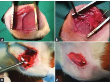
Inguinal regions of the rats were shaved and disinfected with povidone-iodine solution (Batticon solution 10%, Adeka, Turkey). A 3 cm long skin incision was made, and under microscopic examination, femoral neurovascular bundle was exposed [Figure 1a]. Avoiding traumatization of the femoral artery, femoral artery was dissected away from the femoral vein and nerve [Figure 1b]. A silastic sheath was wrapped around 1.5 cm segment of the femoral artery and sutured circumferentially [Figure 1c].
In Groups 2, 3, and 4, 1 cm proximal of the tip of the rat tails was excised and using insulin injector 0.1 cc blood sample was drawn and injected into silastic sheath wrapped around femoral artery to construct an SAH model. The tail was ligated proximal to the excised segment. Following hemostasis layers were closed in proper order.
Rats in all groups recovered normally from anesthesia under room temperature. The rats were fed with standard rat pellets in their cages heated to appropriate ambient temperature for 7 days. The day of surgery was considered as day 1, and once daily doses of 1 ml isotonic saline solution were administered through intramuscular route for 7 days.
The day of surgery was considered as day 1, and once daily doses of 1 ml isotonic saline solution containing 3 mg/kg and 15 mg/kg progesterone were administered through intramuscular route for 7 days in Groups 3 and 4, respectively.
At the end of the 7 days, the rats were reanesthetized with 60 mg/kg percutaneous intraperitoneal ketamine
Figure 1: (a) Neurovascular bundle. (b) Dissection and exposure of femoral artery. (c) Silastic sheath was wrapped around the artery so as to separate them from surrounding tissues. (d) Silastic sheath discerned under granulation tissue on postoperative 7 day
a b
HCl (Ketalar flacon 50 mg/ml, Eczacıbaşı, Turkey), and 10 mg/kg xylazine (Rompun 2% Bayer, Turkey), and laid in the supine position on wooden blocks. The former incisions were opened, and the silastic sheath around femoral artery was approached [Figure 1d]. Silastic sheath was opened, and femoral arteries were exposed.
Meanwhile, the skin over the sternum was shaved, and following cleansing and disinfecting the surgical field with a povidone-iodine solution (Batticon solution 10%, Adeka, Turkey), sternum was dissected away from its attachments with ribs using dissection scissors, and thorax was explored [Figure 2]. The pericardium was opened, and the left ventricle was entered through a created puncture hole. Serum set was connected to the tip of the puncture catheter. With the aid of a large volume injector, 300 ml isotonic solution was delivered into the left ventricle under physiologic artery pressure. After circulation of the delivered solution within the vascular system, it was drained from the opened right atrium. Evacuation of all the delivered blood volume was ensured, and then perfusion procedure was initiated. For perfusion, a solution containing a mixture of 100 ml 0.003 M phosphate buffer (pH: 7.4) + 200 ml 4% formaldehyde and 1% gluteraldehyde was perfused as a bolus from a height of 1 m. Circulation of all delivered perfusion fluid through all vascular system and evacuation through excised right atrium were ensured. At the end of this procedure, crystal clear perfusion fluid was drained wholly throughout the right ventricle. Nearly, 1.5 cm length femoral artery segment was excised, examined under a light microscope, and immersed in buffered 10% formaldehyde solution for analysis.
In the pathology laboratory from each vascular specimen, at least two 3 mm thick vascular segments were cut and immersed in buffered 10% formaldehyde solution. Then, cut sections were stored in cassettes. On the same day in the automated tissue processing device, the samples were subjected through processes of fixation with formaldehyde,
dehydration with grade alcohol, made transparent with xylene, and embedded in paraffin blocks. Tissue samples were left in paraffin for 24 h, and then taken out of tissue processing device, and embedded in paraffin blocks. Five-micron thick cut samples were placed on slides. Following deparaffinization procedure in the incubator at 60°C for 1 h, the slides were rinsed thrice with xylene solution. The specimens were treated with grade alcohol, rehydrated, and rinsed with water. The slides were stained with hematoxylin-eosin and examined under Nikon surgical microscope (Nikon Instruments Europe BV, Amsterdam / Netherlands) at ×100, ×200, and ×400 magnification. Vascular wall thickness and luminal cross-sectional areas were measured using micrometers.
The light microscopic examination was performed in Division of Pathology, Haseki Training and Research Hospital. DP2-BSW program was used during measurements of vascular wall thickness and luminal cross-sectional areas.
Results
Histological changes
• Group 1: In the control group, light microscopic examination of the femoral artery revealed vessels with thin and smooth vascular endothelium, thin and uncurled internal elastic lamina, and concentrically arrayed smooth muscle cells [Figure 3a]
• Group 2: In the vasospasm group, light microscopic examination of the femoral artery disclosed markedly stenotic lumen and thickened vascular wall, disruption of the endothelial integrity, curling of internal elastic lamina, and vacuolization in the muscular layer [Figure 3b]
• Group 3: In the vasospasm plus low-dose progesterone group, in the light microscopic examination of the
Figure 2: Exploration of thorax and heart
Figure 3: (a) Control group: Microscopic appearance of the vascular lumen under a light microscope (×200). (b) Vasospasm group, light microscope. (c) Low-dose progesterone group under a light microscope. (d) High-dose progesterone group under light microscope
a b
femoral artery, when compared with the vasospasm group, thinner vascular endothelium, patchy areas of curled internal elastic lamina, and concentrically arrayed smooth muscle cells were detected [Figure 3c] • Group 4: In the vasospasm plus high-dose progesterone
group, in the light microscopic examination of the femoral artery, similar to the vascular structure detected in the control group, thin and smooth endothelium, thin and occasionally curled internal elastic lamina, and concentrically arrayed smooth muscle cells were observed [Figure 3d].
Morphometric analysis
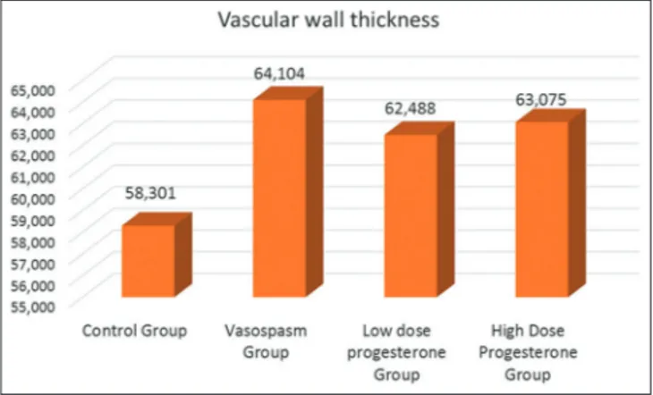
Femoral arteries of four groups were harvested. Mean (±standard deviation [SD]) arterial wall thickness and luminal cross-sectional areas of femoral arteries were calculated. The values obtained were statistically analyzed. When groups were compared as for mean arterial wall thickness, the smallest and the highest mean values were seen in the control and vasospasm groups, respectively. Mean values of low- and high-dose progesterone groups were closer to those of the vasospasm group.
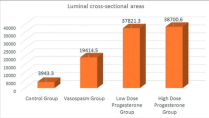
When groups were compared as for mean values for luminal cross-sectional areas, the lowest and the highest mean values were detected in the vasospasm and control groups, respectively. Mean values of low- and high-dose progesterone groups were closer to those of the control group.
Method of statistical analysis used
For the statistical evaluation of study data, Statistical Package for Social Sciences for Windows 18.0 (IBM, New York, USA) program was used.
In the evaluation of study data, in addition to descriptive statistical methods as means and SD, in the intergroup comparison of quantitative data among more than two groups, parameters with normal distribution were compared with “One-way ANOVA test” and in the detection of the group which caused intergroup difference “Tukey HSD test” was used.
In the comparison of quantitative data between two groups, parameters without normal distribution were compared using “Mann–Whitney U-test.” In the comparison of mean
values of more than two groups, “Kruskal–Wallis test” was used. The results were evaluated within 95% confidence interval and at a significance level of P < 0.05.
Statistical values
In the study, four groups each consisting of eight cases were analyzed.
Maximum and minimum mean wall thicknesses were detected in the control and the vasospasm groups, respectively [Figure 4]. One-way variance analysis was used to determine if any difference between mean vascular wall thickness of the groups exists. Since one-way variance analysis could not yield homogenous results in assumptions as an alternative Kruskal–Wallis test was applied [Table 1]. As a result of statistical analysis, any statistically significantly difference between mean vascular wall thickness of the groups was not found (P = 0.948 > 0.05). Mean values for luminal cross-sectional areas of the groups were also analyzed [Table 2]. The highest and lowest mean values were noticed in the vasospasm and control groups, respectively [Figure 5]. Accordingly, the mean value for luminal cross-sectional area of the control group was observed higher than those of the other groups, while the relevant mean values in progesterone groups were nearly two-fold higher than those of the vasospasm group without any statistically significant intergroup difference because of increased SDs estimated in one-way variance tests (P = 0.618 > 0.05).
Table 1: Descriptive statistics related to distribution of vascular wall thickness among groups Mean±SD
Control group Vasospasm group Low‑dose progesterone group High‑dose progesterone group
Vascular wall thickness 58.301±20.376 64.104±10.640 62.488±7.617 63.075±10.527
SD – Standard deviation
Table 2: Distribution of luminal cross‑sectional areas among groups Mean±SD
Control group Vasospasm group Low‑dose progesterone group High‑dose progesterone group
Luminal cross-sectional areas 39,436.30±44,329.70 19,414.51±22,655.36 37,821.30±36,697.38 38,700.64±33,128.05
Excluding the control group, we concluded that mean values for luminal cross-sectional areas estimated in vasospasm and progesterone groups did not differ statistically significantly (P = 0.398 > 0.05).
Discussion
CV occurring following subarachnoidal bleeding is one of the most important pathological events adversely affecting morbidity and mortality. CV can be defined as narrowing of cerebral vasculature caused by blood, metabolites, and chemical substances of blood which accumulate in the subarachnoidal space.[7] Still, its pathophysiology has not
been elucidated so far.
Its etiopathogenesis is multifactorial including main oxyhemoglobin and spasmogenic agents.
In the experimental studies, blood injected into subarachnoidal space has induced CV. Still in clinical studies, computed tomography sections obtained immediately after aneurysmal bleeding revealed that amount of blood is the most important indicator of the development of late-term ischemic deficit.[8,9]
In a study performed by Okada et al., the authors injected whole blood and washed erythrocytes and demonstrated that the most severe reaction had developed at the end of the 7th
day. The outcomes seen in vasospasm and periadventitial blood were compared, and the consequences of femoral vasospasm observed in rats were similar to those found in their experiment as for the degree of severity, specificity, progression in time, and histological characteristics. Since silastic material applied around adventitia had no effect on vasospasm, lower cost, and easy maintenance of rats, “Rat Femoral Artery Vasospasm Model” used by Okada et al. was found to be applicable in our study.[6]
Cerebral arteries (CAs) were analyzed under light microscope immediately after SAH, and thickening of the vascular wall, internal elastic lamina, narrowing of the luminal diameter of CAs, disruption of endothelial cellular structure and integrity, vacuoli in smooth muscle, migration of proliferated myointimal cells into intima, and development of periadventitial inflammation associated with
perivascular axon loss were demonstrated.[10] Moreover, in
our study, in compliance with literature findings, alterations in the vascular structure of CAs were observed.
Undoubtedly, mainly oxyhemoglobin and free oxygen radicals are responsible for the development of vasospasm occurring after SAH.[11-13] Oxidative stress stimulates
hypertrophy and proliferation of smooth muscle cells, and accelerates apoptotic processes. These changes are associated with vascular contractile responses.[14]
Correlations between the development of CV and increased levels of superoxide anions in the cerebrospinal fluid have been already reported. Oxyhemoglobin increases the amount of phospholipase C (PLC). PLC indirectly promotes the intracellular influx of Ca2+ leading to vasoconstriction.
PLC also increases the levels of diacylglycerol resulting in increased amounts of protein kinase C. However, PKC decreases the production of NO and consequently prevents vasodilation.
Still, the most potent vasoconstrictor endothelin-1 activates RhoA/Rho kinase and protein kinase-C which reinforces contraction of cerebrovascular smooth muscles are under the influence of oxyhemoglobin.[15]
Previous studies demonstrated that progesterone decreases the expression of TxA2 receptor which is a potent vasoconstrictor, blocks L-type Ca2+ channels leading to
decrease in intracellular Ca2+ content, and increases the
production of vasodilatory agent epoxyeicosatrienoic acid. In our study, endogenous substance progesterone was used which was thought to have lesser side effects.
A histological and statistically significant difference was not detected between vasospasm and low-dose progesterone groups (P = 0.948 > 0.05). Vascular walls of all these three groups were found to be thicker when compared with the control group.
Mean luminal cross-sectional areas of the vessels in the control and progesterone groups were found to be markedly higher relative to the vasospasm group. However, in statistical analysis because of higher SDs, the intergroup difference was deemed to be devoid of any significance (P = 0.618 > 0.05).
These outcomes of our study, though statistically insignificant, have suggested a vasodilatory effect of progesterone on the cross-sectional area of the vascular lumen. Thanks to potent vasodilatory effects of progesterone, especially over Ca2+ metabolism, we think
that its clinical use in the treatment of CV deserves merit, and consequently experimental and clinical studies should be maintained.
Conclusion
Two different doses of progesterone which is an endogenous hormone with potent vasodilatory efficacy synthesized in the body without any known serious side
effect were analyzed in the experimental SAH model which was induced by injecting autologous blood on the periphery of rat femoral artery.
In our study, we used “Rat Femoral Artery Vasospasm Model” suggested by Okada et al. to obtain parameters of vascular wall thickness and cross-sectional areas of vascular lumens which were then subjected to statistical analysis.
Analysis of vascular wall thickness and luminal cross-sectional areas could not demonstrate any significant difference between Group 4 (15 mg/kg/day progesterone group) and Group 3 (3 mg/kg/day progesterone group). Mean values for luminal cross-sectional areas of all three groups were higher than those of the vasospasm group without any statistically significant intergroup difference. We think that progesterones potential use as a preventive agent against the development of post-SAH CV requires conduction of multicentered, placebo-controlled, randomized, and double-blind studies.
Financial support and sponsorship
Nil.
Conflicts of interest
There are no conflicts of interest.
References
1. Weir B. Aneurysms Affecting the Nervous System. Baltimore: Williams and Wilkins; 1987.
2. Akdemir H. Subaraknoid kanama. In: Aksoy K, editor. Temel Nöroşirürji. Ankara: Turkish Neurosurgical Society; 2005. p. 441-7.
3. Ingall T, Asplund K, Mähönen M, Bonita R. A multinational comparison of subarachnoid hemorrhage epidemiology in the WHO MONICA stroke study. Stroke 2000;31:1054-61.
4. Chehrazi BB, Giri S, Joy RM. Prostaglandins and vasoactive amines in cerebral vasospasm after aneurysmal subarachnoid hemorrhage. Stroke 1989;20:217-24.
5. Kılıç T, Konya D. Temel Nöroşirürji. Ankara: Turkish Neurosurgical Society; 2005. p. 191-2.
6. Okada T, Harada T, Bark DH, Mayberg MR. A rat femoral artery model for vasospasm. Neurosurgery 1990;27:349-56.
7. Kassell NF, Torner JC, Haley EC Jr., Jane JA, Adams HP, Kongable GL. The ınternational cooperative study on the timing of aneurysm surgery. Part 1: Overall management results. J Neurosurg 1990;73:18-36.
8. Fisher CM, Kistler JP, Davis JM. Relation of cerebral vasospasm to subarachnoid hemorrhage visualized by computerized tomographic scanning. Neurosurgery 1980;6:1-9.
9. Kırış T, Sencer A. Subaraknoid kanamanın komplikasyonları. In: Aksoy K, editor. Temel Nöroşirürji. Ankara: Turkish Neurosurgical Society; 2005.
10. Gules I, Satoh M, Clower BR, Nanda A, Zhang JH. Comparison of three rat models of cerebral vasospasm. Am J Physiol Heart Circ Physiol 2002;283:H2551-9.
11. Gaetani P, Lombardi D. Brain damage following subarachnoid hemorrhage: The imbalance between anti-oxidant systems and lipid peroxidative processes. J Neurosurg Sci 1992;36:1-10. 12. Kim DE, Suh YS, Lee MS, Kim KY, Lee JH, Lee HS, et al.
Vascular NAD(P) H oxidase triggers delayed cerebral vasospasm after subarachnoid hemorrhage in rats. Stroke 2002;33:2687-91. 13. Macdonald RL, Weir BK. Cerebral vasospasm and free radicals.
Free Radic Biol Med 1994;16:633-43.
14. Griendling KK, Ushio-Fukai M. Redox control of vascular smooth muscle proliferation. J Lab Clin Med 1998;132:9-15. 15. Lan C, Das D, Wloskowicz A, Vollrath B. Endothelin-1
modulates hemoglobin-mediated signaling in cerebrovascular smooth muscle via RhoA/Rho kinase and protein kinase C. Am J Physiol Heart Circ Physiol 2004;286:H165-73.