Original Article
Morphometric and ultrastructural analysis of
the effect of bromocriptine and cyclosporine
on the vasospastic femoral artery of rats
Mehmet Tokmak1, Kahan Başocak2, Hüseyin Canaz3, Gökhan Canaz4, Celal İplikçioğlu51Department of Neurosurgery, Medipol University, Istanbul; 2Department of Neurosurgery, Bakırköy Dr. Sadi
Konuk Reseach and Training Hospital, Istanbul; 3Şişli Florence Nightingale Hospital, Istanbul Bilim University,
Spina Bifida Research Center, Istanbul; 4Department of Neurosurgery, Haseki Research and Training Hospital,
Istanbul; 5Department of Neurosurgery, Harran University Medicine Faculty, Istanbul
Received March 15, 2015; Accepted September 28, 29; Epub October 15, 2015; Published October 30, 2015 Abstract: Vasospasm is the main causes of mortality and morbidity in patiens with subarachnoid hemorrhage (SAH). The arterial narrowing mechanism that develops after SAH is not yet fully understood but many studies showed that hypotension, neurogenic reflexes, clots in the subarachnoidal space, spasmogenic agents, humor-al and celluler immunity play a role in the etiology. In this study we investigate the effects of Bromocriptine and Cyclosporine A in vasospasm secondary to SAH on rat femoral artery from ultrastructural and morphometric per-spectives. 120 male Sprague-Dawley rats divided into 12 groups: Vasospasm (V), control (K), surgical control (CK) groups, vasospasm+Bromocriptine and/or Cyclosporine-A groups (VCyA, VBr, VBr+CyA), Bromocriptine and/ or Cyclosporine-A control groups (CK, BK, Br+CyAK), Bromocriptine and/or Cyclosporine-A surgical control groups (BCK, CyCK, Br+CyACK). In order to create SAH model, 0, 1 cm3 blood injected into silastic sheath wrapped rat
femoral artery. Bromocriptine (2 mg/kg/d) and Cyclosporine A (10 mg/kg/d) combinations applied to control, sur-gical control and vasospastic models. Light microscopy, transmission electron microscopy and scanning electron microscopy used during this study. Statistical evaluation of the morphometric measurement data concerning vascu-lar wall thickness and luminal cross-sectional areas of all groups were performed using Mann-Whitney U, Wilcoxon-signed rank, and Student-t tests. Cyclosporine A, whose effects in the prevention of vasospasm have been dem-onstrated in previous studies. In this study we discovered that Bromocriptine demdem-onstrated strong effects similar to Cyclosporine-A. Bromocriptine and Cyclosporine A markedly prevent the development of chronic morphologic vasospasm following SAH. The combined use of both drugs does not change this preventive effect.
Keywords: Bromocriptine, cyclosporine-a, subarachnoid hemorrhage, vasospasm
Introduction
Vasospasm is among the most predominant causes of mortality and morbidity in patients who present subarachnoid hemorrhage (SAH) secondary to aneurysmal bleeding. Vasospasm can present itself along with head traumas, tumors, and arteriovenous malformation (AVM). Although pathogenesis of cerebral vasospasm is not fully understood, it constitutes a serious problem for neurosurgery [1].
Cerebral vasospasm emerges after SAH. SAH can be defined as the pathological narrowing of basal cerebral vessels caused by a collection of blood, blood metabolites, and some
chemi-cal substances within cisterna. Although the arterial narrowing mechanism that develops after subarachnoidal bleeding is not yet fully understood, degradation of erythrocytes, hypo-tension, neurogenic reflexes, clots in the sub-arachnoidal space, and spasmogenic agents released from degradation products (i.e., PGE2, PGF2, arachidonic acid,serotonin, tromboxan A2, epinephrine, norepinephrine, histamine, angiotensin, thrombin, plasmine, fibrin degra-dation products, and potassium) are known to play a role in the etiology of arterial narrowing [2-4]. In addition to other mechanisms, respon-siveness to cellular immunity, followed by humoral immunity and eicocanosid reactions,
have been demonstrated to have a role in the development of cerebral vasospasm [5-13]. According to this autoimmune theory, it is accepted that erythrocytes appearing after minor bleedings behave like an autoantigen and lead to the formation of an immune com-plex [10, 11].
Bromocriptine is a dopamine type 2 agonist and prevents secretion of prolactine (PRL). Bromocriptin, which is frequently used in hypophyseal, endocrinologic disorders and Parkinson’s disease, has a strong immunosup-pressive effect. It prevents delayed type hyper-sensitivity, primary antibody response, T-lym- phocyte-related macrophage activation, and proliferation of T-and B-lymphocytes [14-18]. Cyclosporine A has a strong anti-immunosup-pressive activity, and it is especially used in transplantation and for the treatment of auto-immune diseases. It inhibits T-cell-dependent antibody formation, lymphokine production, and release of interleukin 2 including T-cell growth factor (TCGF). It blocks resting lympho-cytes at G0 or G1 cell phases of the T-cell cycle. It also inhibits the release of antigen-stimulat-ed lymphokine from active T-cells [19-24].
Many studies have demonstrated that the com-bined use of Bromocriptine and Cyclosporine A is very effective for organ transplantation and the treatment of autoimmune diseases [9, 15, 18, 25-32]. However, currently there is not a literature study on its effects on cerebral va- sospasm.
In this study, a chronic vasospasm model is constructed with a rat femoral artery, and the effect of Bromocriptine and Cyclosporine A on chronic morphological vasospasm is ana-lyzed from ultrastructural and morphometric perspectives.
Material and method
This study was performed in the Laboratories of Department of Pharmacology, Marmara University Faculty of Medicine. Light microscop-ic examinations were conducted in the Pa- thology Department of Marmara University Faculty of Medicine using transmission elec-tron microscopy (TEM), and scanning elecelec-tron microscopic (SEM) examinations were per-formed in the Department of Histology and Embryology.
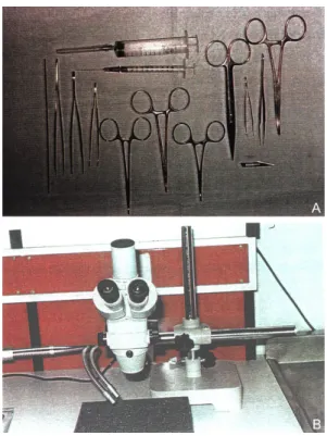
In this study 120 male Sprague-Dawley rats each weighing 180-220 g were used. Micro- surgical instruments (Figure 1A) and surgical microscope (Nikon; Figure 1B) were used. A vasospasm model called the “Rat Femoral Artery Vasospasm Model” proposed by Okada et al. was utilized [33].
Rats were divided into two groups. Vasospasm was induced in the right femoral arteries of rats, and these rats were treated with either Cyclosporine A, Bromocriptine, or a combina-tion therapy (Bromocriptine and Cyclosporine A). The left femoral arteries of the rats consti-tuted as control and surgical control groups (Table 1).
Group designations
Vasospasm Group (V) (n=30): Vasospasm was induced in the right femoral arteries.
Vasospasm+Cyclosporine A Group (VCyA) (n= 30): Vasospasm was induced in the right femo-ral arteries and Cyclosporin A was applied. Vasospasm+Bromocriptine Group (VBr) (n=30): Vasospasm was induced in the right femoral arteries, and Bromocriptine was applied.
Figure 1. A. Microsurgical instruments. B. Surgical microscope.
Vasospasm+Bromocriptine+Cyclosporine A Gr- oup (VBr+CyA) (n=30): Vasospasm was induced in the right femoral arteries, and Bromocri- ptine+Cyclosporine A was applied.
Control Group (K) (n=15): Left femoral arteries were left intact.
Surgical Control Group (CK) (n=15): A silastic sheath was wrapped around the left femoral arteries; blood samples were not obtained; and only physiologic saline was used.
Bromocriptine+Cyclosporine A Control Group (Br+CyAK) (n=15): Left femoral arteries were left intact and Bromocriptine+Cyclosporine A was applied.
Bromocriptine Control Group (BK) (n=15): Left femoral arteries were left intact, and Bromo- criptine was applied.
Bromocriptine Surgical Control Group (BCK) (n=15): A silastic sheath was wrapped around the left femoral arteries; blood samples were not given; and only physiologic saline and Bromocriptine were applied.
Cyclosporine A Control Group (CyAK) (n=15): Left femoral arteries were left intact, and only Cyclosporine A was applied.
Cyclosporine A Surgical Control Group (CyCK) (n=15): A silastic sheath was wrapped around the left femoral arteries. Instead of blood sam-ples, only physiologic saline and Cyclosporine A were applied.
Cyclosporine A+Bromocriptine Surgical Control Group (Br+CyACK) (n=15): A silastic sheath was wrapped around the left femoral arteries; instead of blood samples, only physiologic sa- line and Cyclosporine A+Bromocriptine were applied.
Rats were anesthesized with intraperitoneal 2 mg/kg ketamine HCI and laid supine on mush-room blocks.
The inguinal region of the rats were shaved and cleansed with a sterile povidone (PVD) iodine Table 1. Group designations
Vasospasm Group (V) (n=30) Vasospasm was induced in the right femoral arteries
Vasospasm+Cyclosporine A Group (VCyA) (n=30) Vasospasm was induced in the right femoral arteries and Cyclosporin A was applied
Vasospasm+Bromocriptine Group (VBr) (n=30) Vasospasm was induced in the right femoral arteries, and Bromocrip-tine was applied
Vasospasm+Bromocriptine+Cyclosporine A Group (VBr+CyA) (n=30) Vasospasm was induced in the right femoral arteries, and Bromocriptine+Cyclosporine A was applied
Control Group (K) (n=15) Left femoral arteries were left intact
Surgical Control Group (CK) (n=15) A silastic sheath was wrapped around the left femoral arteries; blood samples were not obtained; and only physiologic saline was used Bromocriptine+Cyclosporine A Control Group (Br+CyAK) (n=15) Left femoral arteries were left intact and Bromocriptine+Cyclosporine
A was applied
Bromocriptine Control Group (BK) (n=15) Left femoral arteries were left intact, and Bromocriptine was applied Bromocriptine Surgical Control Group (BCK) (n=15) A silastic sheath was wrapped around the left femoral arteries; blood
samples were not given; and only physiologic saline and Bromocrip-tine were applied
Cyclosporine A Control Group (CyAK) (n=15) Left femoral arteries were left intact, and only Cyclosporine A was applied
Cyclosporine A Surgical Control Group (CyCK) (n=15) A silastic sheath was wrapped around the left femoral arteries. Instead of blood samples, only physiologic saline and Cyclosporine A were applied
Cyclosporine A+Bromocriptine Surgical Control Group (Br+CyACK) (n=15) A silastic sheath was wrapped around the left femoral arteries; instead of blood samples, only physiologic saline and Cyclosporine A+Bromocriptine were applied
solution. Through a longitudinal 2 cm skin inci-sion (Figure 2), the femoral neurovascular bun-dle was explored under a surgical microscope. The femoral artery was dissected away from
the adjacent vein and nerve without traumatiz-ing the femoral artery (Figure 3A). A silastic sheath was wrapped and sutured around a 1.5 cm segment of the femoral artery (Figure 3B
Figure 3. A. The femoral neurovascular bundle. B. Silastic sheath and the femoral artery. C. Wrapping silastic sheat around the femoral artery. D. Blood injection into the silastic sheath wrapped around the femoral artery in order to create a subarachnoid hemorrhage model.
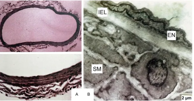
Figure 4. A. Control group, light microscopic examination. B. Control group, TEM examination the of femoral artery. (En: Endotelium, IEL: Internal elastic lamina, SM: Muscle).
and 3C). Blood drawn from inside the cardiac chambers was used as a source of whole blood. With an insulin injector, 0.1 cm3 of blood
sam-ples were percutaneously drawn from inside the cardiac chambers and injected into the silastic sheath wrapped around the femoral artery so as to create a subarachnoid hemor-rhage model (Figure 3D).
After hemostatic control, layers were closed in compliance with surgical principles. Rats were
awoken under normal room temperature in cages that each contained only one rat. The cages were warmed to an appropriate ambient temperature, and the rats were fed with stan-dard rat food for 7 days.
In each of the groups treated with only Bro- mocriptine or Cyclosporine A+Bromocriptine, a SAH model was constructed, and starting from the day of operation for 7 days through in- traperitoneal route, the VBr group received
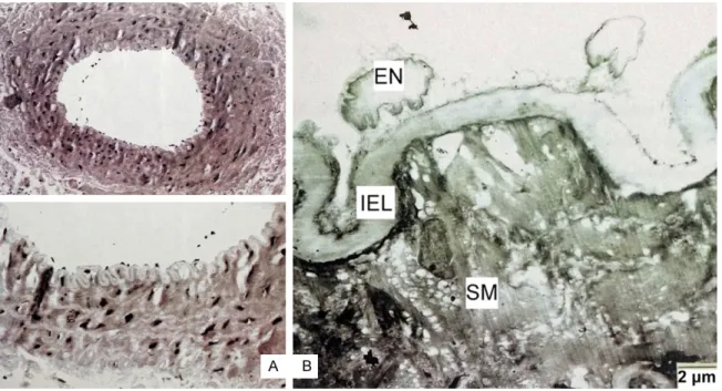
Figure 5. A. Vasospasm group, light microscopic examination. B. Vasospasm group TEM examination the of femoral artery. (En: Endotelium, IEL: Internal elastic lamina, SM: Muscle).
Figure 6. A. Bromocriptine+Cyclosporine A applied group, light microscopic examination. B. Bromocriptine+Cyclos- porine A applied group, TEM (EN: Endotelium, IEL: Internal elastic lamina, SM: Muscle).
Bromocriptine (2 mg/kg/d), the VCyA group received Cyclosporine A (10 mg/kg/d), and the VBr+CyA group received Bromocriptine (2 mg/ kg/d)+Cyclosporine (10 mg/kg/d).
The left femoral arteries of the rats in the con-trol group were left intact, while in the surgical control group the left femoral arteries of the
rats were wrapped with a silastic sheath, and only physiologic saline was instilled; blood sam-ples were not used. To assess the effect of Bromocriptine and Cyclosporine A on normal vessels and vessels wrapped only with a silas-tic sheath,control and surgical control groups received Bromocriptine (2 mg/kg/d)+Cyclos- porine A (10 mg/kg/d) through a
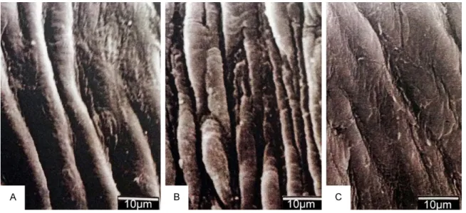
intraperitone-Figure 7. A. Control group, SEM examination the of femoral artery. B. Vasospasm group SEM examination the of femoral artery. C. Bromocriptine+Cyclosporine A applied group, SEM.
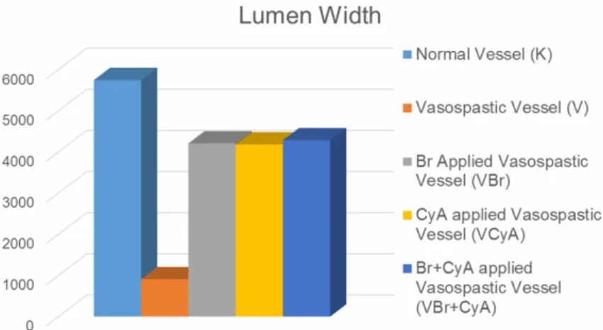
Table 2. Lumen widths
Average ± Std. Deviation P Vasospasm Group (V) 907.16 ± 124.81 P<0.001 Vasospasm+Bromocriptine Group (VBr) 4205.93 ± 124.81 P<0.001 Vasospasm+Cyclosporine A Group (VCyA) 4181.03 ± 625.98 P<0.001 Vasospasm+Bromocriptine+Cyclosporine A Group (VBr+CyA) 4283.83 ± 697.44 P<0.001 Control Group (K) 5744.00 ± 470.54 P>0.05 Bromocriptine+Cyclosporine A Control Group (Br+CyAK) 5773.33 ± 341.92 P>0.05 Surgical Control Group (CK) 5573.33 ± 306.65 P>0.05 Cyclosporine A+Bromocriptine Surgical Control Group (Br+CyACK) 5638.33 ± 376.42 P>0.05
Table 3. Vessel wall thicknesses
Average ± Std. Deviation P Vasospasm Group (V) 94.33 ± 13.28 P<0.001 Vasospasm+Bromocriptine Group (VBr) 49.47 ± 8.26 P<0.001 Vasospasm+Cyclosporine A Group (VCyA) 49.33 ± 8.13 P<0.001 Vasospasm+Bromocriptine+Cyclosporine A Group (VBr+CyA) 47.33 ± 8.26 P<0.001 Control Group (K) 36.06 ± 5.81 P>0.05 Bromocriptine+Cyclosporine A Control Group (Br+CyAK) 47.33 ± 8.26 P>0.05 Surgical Control Group (CK) 38.06 ± 4.85 P>0.05 Cyclosporine A+Bromocriptine Surgical Control Group (Br+CyACK) 37.06 ± 7.00 P>0.05
al route for 7 days. The rats were observed for 7 days. None of the rats died. Loss of weight and infection were not observed.
At the end of 7 days, the rats were anesthe-sized with percutaneously administered intra-peritoneal ketamine HCl (2 mg/kg/d) anesthe-sia and laid in the supine position on mushroom blocks. Previous incisions were opened, and the silastic sheath around the femoral artery was explored. Meanwhile, the skin over the sternum was shaved, and following cleansing of the surgical field with PVD iodine, the sternum was dissected away from its attachment to the ribs using dissection scissors, and the thora- cic cavity was explored. The pericardium was opened, and the left ventricular cavity was punctured. The serum set was connected to the puncture catheter. A mixture of 100 ml 0.03
Group) were applied, 1.5 cm segments of the right femoral arteries of the rats were excised to be subjected to light microscopic, TEM, and SEM examinations.
Light microscopic examinations were per-formed in the laboratories of the Department of Pathology, Marmara University, Faculty of Medicine. The femoral artery specimens were obtained and immersed in buffered 10% form-aldehyde solution. They were transferred to cassettes and placed in a tissue monitorization device. In this device, specimens were fixated with formol and dehydrated with a graded alco-hol solution. They were also treated with a xylene solution followed by paraffine. All these procedures lasted for 24 hours. After these processes were performed, tissue samples were embedded in paraffine blocks and frozen.
Figure 8. Comparision of lumen widths between normal vessel, vasospastic vessel and Br or CyA or both applied vasospastic vessel.
Figure 9. Comparision of vessel wall thicknesses between normal vessel, vasospastic vessel and Br or CyA or both applied vasospastic vessel.
M phosphate buffer (pH 7.4), 200 ml 4% paraformaldehyde and 1% glutaraldehyde solu-tion was injected under physi-ologic arterial pressure into the left ventricle. The injected solution was circulated throu- gh the entire vascular system and then drained from the opened right atrium. Then, a 1.5 cm segment of the inta- ct left femoral arteries was excised and harvested for light microscopic, TEM, and SEM examinations. In the sur-gical control groups, the left femoral arteries of the rats were wrapped with a silastic sheath, and physiologic saline was applied instead of blood. The 1.5 cm segments of the femoral arteries were excised to be subjected to light micro-scopic, TEM, and SEM exa- minations. In the vasospasm group, 1.5 cm segments of vasospasm-induced right fe- moral arteries were excised for further light microscopic, TEM, and SEM examinations. In vasospasm-induced grou- ps where only Bromocriptine (VBr Group), Cyclosporine A (VcyA Group), or Bromocrip- tine+Cyclosporine A (VBr+CyA
Five-micron sections were cut with a micro-tome, and these cut sections were deparaf-finized at 600°C in an incubator. Depara- ffinization was preceeded with xylene treat-ment that was repeated 3 times. Then the solu-tion was reacted with graded alcohol for rehy-dration, rinsed with water, and stained with hematoxylin-eosin. Prepared slides were exam-ined under a microscope (Olympus BX7, Japan) at 100X magnification, and their photos were taken for morphometric analyses. Luminal cross-sectional areas and the number of sq- uares within a lumen were calculated and taken as unit values. The same units were also used for the measurements of vascular wall thick-nesses. The morphometric analyses were per-formed, and the tissue samples were compared with regards to vascular lumen thickness and luminal cross-sectional areas.
For the TEM examinations, tissue samples were immersed in a buffered 2.5% glutaraldehyde solution and placed in a buffered 1% osmium tetraoxide solution and dehydrated with graded alcohol. Then the specimens were embedded in Epon 812 and examined under a JEOL 100C TEM (JEOL Ltd., Tokyo, Japan). For SEM exami-nations, tissue samples were immersed and left for 3 hours in 2.5% glutaraldehyde buffered with cacodylate. Then these cacodylate-buff-ered samples were post-fixated for one hour in 1% osmium tetraoxide and dehydrated in alco-hol and amyl acetate. The specimens were dried with CO2 at critical-point, coated with gold in the evaporator, and examined under a JEOL JSM 5200 SEM (JEOL Ltd., Tokyo, Japan).
Statistical evaluation
Statistical evaluation of the morphometric measurement data concerning vascular wall thickness and luminal cross-sectional areas of all groups were performed using Mann-Whitney U, Wilcoxon-signed rank, and Student-t tests.
Histological changes
In the control group, light microscopic examina-tion of the femoral artery vessels had a thin and smooth endothelium, a thin and unfolded internal elastic lamina, and concentrically arrayed smooth muscle cells (Figure 4A). In the surgical control group, the vascular structure did not demonstrate a significant difference when compared with the control group. In the
control group, during TEM examination the of femoral artery, development of normal organ-elles of endothelial cells were observed. Also observed was a continuum of endothelial ce- lls, a thin internal elastic lamina, and normal smooth muscle cells (Figure 4B). In the SEM examination, a flat and unfolded endothelium, a thin vessel wall, and a dilated lumen were observed (Figure 7A).
In the vasospasm group, in the light microscop-ic examination of the femoral artery, a promi-nent decrease in lumen diameter and a marked increase in the vascular wall thickness were detected. A disruption in the endothelial integ-rity was also observed as well as folding of the internal elastic lamina and vacuolization in the muscular layer (Figure 5A).
TEM examination of the vasospasm-induced group showed distortion of the endothelial cells, vacuolization, and a decrease in cytoplas-mic density. It also showed marked thickening and folding of the internal elastic lamina, sub-endothelial deposition, vacuolar degeneration, and myonecrosis (Figure 5B).
The SEM examination of the vasospasm-in- duced group showed increased and impaired endothelial foldings, craters, and a conspicu-ous thickening of the vascular wall (Figure 7B). During the light microscopic examination of the femoral artery in the vasospasm-induced group treated with Bromocriptine, just like in the con-trol group, the vessels had a thin and smooth endothelial layer, thin and patchy areas of fold-ed internal elastic lamina, and concentrically aligned smooth muscle cells.
In the vasospasm-induced group treated with Cyclosporine A, light microscopic examination of the femoral artery, similar findings to those seen in the control group were detected. In the vasospasm-induced group treated with Bromocriptine+Cyclosporine A, light microscop-ic analysis of the femoral artery did not reveal different findings compared to those of only Bromocriptine, or only Cyclosporine-A, applied groups (Figure 6A). The vascular structure in the surgical control group was not significantly different from that of the control group. TEM examination of the femoral artery in the vasospasm-induced Br+CyA Group showed
results similar to those seen in the control group, including physiologic development of the endothelial cell organelles, integrity and con-tinuum of the endothelial cells, thin internal elastic lamina, and normal smooth muscle cells (Figure 6B). During the SEM examination the following were observed: a flat and mildly fold-ed endothelium, a thin vessel wall, and enlargfold-ed lumens (Figure 7C).
Morphometric analysis
The mean values (± SD) of lumen diameters and vascular wall thicknesses were compared between the groups. Changes were not detect-ed in normal vessel walls and lumens after application of Bromocriptine or Cyclosporine A. Besides, the silastic sheath wrapped around the normal vessels did not lead to changes in the vessel wall or lumen. Also, after application of Cyclosporine A and Bromocriptine onto the silastic sheath wrapped vessel, the vessel wall and lumen did not demonstrate any alteration (P>0.05).
The control group and the vasospasm-induced groups were compared, and statistically signifi-cant decreases in lumen diameter increases in vascular wall thickness were detected (P<0.001). When the vasospasm group was compared with the VCyA, VBr and VBr+CyA groups, the vasospastic artery groups exposed to these drugs showed a decrease in vascular wall thickness and an increase in lumen diam-eters. The decreases in vascular wall thickness and increases in the lumen of vasospastic arteries in the Bromocriptine or Cyclosporine A groups did not significantly differ between these groups (P>0.05). A significant difference was not detected when both drugs were applied singly or in combination (Tables 2, 3); (Figures 8, 9).
Discussion
Inflammation and cellular and humoral immu-nity have been known to play a role in the pathogenesis of vasospasm developed after SAH. In 1981, Pellettieri et al. reported the presence of increased amounts of an immune complex circulating in the blood of patients with SAH and subsequently proposed the autoim-mune theory [34]. The autoimautoim-mune theory, which asserts that red blood cells released and fragmented during minor bleedings behave like
autoantigens and induce formation of immune complexes, was supported by Ostergaard et al., and the association between emerging immune complexes with vasospasm has been demon-strated [8].
Following the introduction of autoimmune theo-ry, studies investigating the use of agents with immunosuppressive activity in the prevention of vasospasm were performed.
Favourable outcomes on the vasospasm-inhibi-tory effects of Cyclosporine A were obtained in experimental studies and have been used in the prevention of graft rejection in organ trans-plantation and in the treatment of autoimmune diseases, including endogenous uveitis, psoria-sis, atopic dermatitis, and nephrotic syndro- me thanks to its potent immunosuppressive effects.
Cyclosporine A exerts its effects by blocking the development of cell-mediated reactions, forma-tion of T-cell dependent antibodies, producforma-tion and release of lymphokines (incl. interleukin-2), resting lymphocytes at G0 or G1 phases of the cell cycle, and inhibition of release of antigen-stimulated lymphokines from active T-cells [19, 21-24, 35].
The favourable effect of Cyclosporine A on vasospasm was firstly reported by Peterson et al. in the year 1990 and has been subsequently demonstrated by many researchers [6, 12, 23, 36, 37].
Cyclosporine A and Bromocriptine are used in combination in the treatment of autoimmune diseases and transplantation studies. Their combined use increases their immunosuppres-sive effects [25, 31, 32, 34].
We have not encountered any literature study that has investigated the vasospasm-preven-tive effect of Bromocriptine, which also has a potent immunosuppressive activity.
Bromocriptine demonstrates its immunosup-pressive effect by inhibiting the development of hypersensitivity, primary antibody response, T-lymphocyte dependent macrophage activa-tion, and proliferation of T and B lymphocytes. Bromocriptine (2-Bromo-x-ergocryptine) also has potent dopaminergic effects induced by the activation of dopamine receptors. D2 re-
ceptors that are affected by Bromocriptine exert their effects by activating the adenylate kinase enzyme. Because of its potent dopami-nergic effect, Bromocriptine inhibits the secre-tion of prolactine, and it is frequently used in the treatment of prolactinoma, galactorrhea, and amenorrhea. Still, due to its dopaminergtic effect, it is used as an adjunct to other treat-ment modalities in Parkison’s disease and acromegaly.
When used in combination with Cyclosporine A in the treatment of organ transplantation and autoimmune diseases, Bromocriptine reinforc-es the immunosupprreinforc-essive effect. Breinforc-esidreinforc-es, effectiveness of Bromocriptine against vaso-spasm has been demonstrated in many studies as well as in our investigation [6, 12, 23, 37]. Does the Bromocriptine+Cyclosporine A combi-nation also more effectively prevent the devel-opment of vasospasm?
We performed this study to find the answer to this question using the “Rat Femoral Artery Vasospasm Model” proposed by Okada et al. [33]. Rats are the most frequently preferred experimental animals in neuroanatomic, neuro-physiologic, and neuropharmacologic studies because they are cost-effective, easily avail-able, and they are easy to maintain. In the study where this model was developed, whole blood and washed red and white blood cells were applied singly around the femoral artery adven-titia, and the presence of maximum vasospasm was demonstrated at the end of the 7th day of their application. In the light and electron mic- roscopic examinations, morphologic changes were seen along the vascular wall that resem-bled changes occurring during the vasospasm of cerebral arteries following subarachnoidal bleeding [33].
Cerebral arteries differ from systemic arteries in many ways. These differences involve endo-thelial permeability, response to vasoactive agonists, and the nature of the adventitial matrix [38]. The response of cerebral arteries to various traumatic events (e.g., subarachnoi-dal bleeding) is never the same with the response elicited by systemic arteries. Clinical and experimental studies have demonstrated that vasospasm is dependent on the volume of blood in the subarachnoidal space and dura-tion of contact between blood and the vascular wall. Within this context, the tendency of
cere-bral vessels to develop vasospasm is simply associated with the persistence of blood around the vessel following subarachnoidal bleeding. This delayed arterial constriction rep-resents the typical individual response of vari-ous arteries to the periadventitial presence of blood [39-41]. Outcomes of subarachnoidal bleeding concerning vasospasm andthose of chronic constriction of rat femoral artery treat-ed with preiadventitial blood are comparable in degree, specificity, natural course, and histo-logical appearance [33].
For the investigation of pathologic mechanisms of vasospasm and its potential treatment modalities, this model appears to be simple and applicable.
In the study, a 0.1 ml autologous blood sample was used for a duration of 7 days. In many SAH models, the role of coagulated blood and the impact of its exposure time on ultrastructural changes of the vascular wall have been demon-strated [33, 40, 42-47]. A silastic sheath was used to prolong the exposure time between the whole blood sample and to increase the amount of whole blood coming in contact with the adventitia. Any effect of silastic material on the construction of the vasospasm model has not been reported in the literatüre [33, 40]. In our study, any statistically significant ultrastructur-al and morphologicultrastructur-al differences were not detected between control and surgical control groups.
The effects of Bromocriptine, and Cyclosporine A on vasospasm (when used at doses with maximum effectiveness) were investigated using ultrastructural and morphometric meth-ods. Since in the vasospasm model used, maxi-mum vasospasm does not become manifest before 7 days, in this study vascular wall sam-ples were obtained after 7 days of drug app- lication.
Among the basic criteria of vasospasm, includ-ing the cross-sectional area of the lumen and the vascular wall and wall thickness, only the first two parameters were used for morphomet-ric evaluation of outcomes. Statistical analysis was performed using the Student-t, Mann-Whitney U, and Wilcoxon signed-rank tests. Cyclosporine A, whose favourable effects in the prevention of vasospasm have been
demon-strated in previous studies, also markedly pre-cluded the development of chronic morphologic vasospasm in our study. We could not detect any change in the amplitude of this preventive effect when Bromocriptine, whose impact in the prevention of vasospasm has not been investigated so far, is used in combination with Cyclosporine A.
However most importantly, when we investigat-ed if Cyclosporine A+Bromocriptine might be more effective, an unexpected and pleasing development became manifest: In the preven-tion of a morphological vasospasm, Bromo- criptine demonstrated strong effects similar to Cyclosporine-A. Any difference was not detect-ed between Bromocriptine and cyclosporine A for the prevention of vasospasm. The mecha-nism of action of this effect is beyond the scope of this study. However potent immunosuppres-sive and dopaminergic effects of Bromocriptine have been already recognized. Its success in the prevention of vasospasm may be associat-ed with one or both of these mechanisms. Presence of dopamine-specific receptors in the vascular beds of cerebral vessels has been demonstrated in experimental and human studies using classical pharmacologic me- thods.
Bromocriptine is a semi-synthetic derivative of ergocriptine that is included in the ergotoxine fraction of ergot alkaloids. Vasodilatory effects of ergotoxine are already known. They demon-strate partial agonistic effectiveness on alpha-receptors via their blockage of vascular x-adren-ergic receptors. Their blocking efficacy beco- mes dominant in constricted vessels, and vaso-dilation occurs under these conditions. Our study is promising. Investigations concern-ing the strong effect of Bromocriptine on cere-bral vasospasm, which we revealed in this study, should be continued with experimental and clinical studies that also address its mech-anism of action. These investigations are nec-essary and inevitable so as to demonstrate the extent of beneficial effects of this drug against vasospasm in human beings.
Conclusion
Bromocriptine and Cyclosporine A markedly prevent the development of chronic morpho-logic vasospasm following SAH. With respect to
the prevention of vasospasm, Bromocriptine and Cyclosporine do not differ-both of them are equally effective. The combined use of both drugs does not change this preventive effect. Disclosure of conflict of interest
None.
Address correspondence to: Dr. Gökhan Canaz, Department of Neurosurgery, Haseki Research and Training Hospital, Fatih 34087, Istanbul. Tel: +90 212 529 44 00; E-mail: [email protected]
References
[1] Kassell NF, Torner JC, Haley EC Jr, Jane JA, Adams HP, Kongable GL. The International Cooperative Study on the Timing of Aneurysm Surgery. Part 1: Overall management results. J Neurosurg 1990; 73: 18-36.
[2] Chehrazi BB, Giri S, Joy RM. Prostaglandins and vasoactive amines in cerebral vasospasm after aneurysmal subarachnoid hemorrhage. Stroke 1989; 20: 217-24.
[3] Kwan AL, Lin CL, Wu CS, Chen EF, Howng SL, Kassell NF, Lee KS. Delayed administration of the K+ channel activator cromakalim attenu-ates cerebral vasospasm after experimental subarachnoid hemorrhage. Acta Neurochir (Wien) 2000; 142: 193-7.
[4] Takeuchi H, Tanabe M, Okamoto H, Yamazaki M. Effects of thromboxane synthetase inhibitor (RS-5186) on experimentally-induced cerebral vasospasm. Neurol Res 1999; 21: 513-6. [5] German JW, Lin CL, Wu CS, Chen EF, Howng
SL, Kassell NF, Lee KS. Systemic complement depletion inhibits experimental cerebral vaso-spasm. Neurosurgery 1996; 39: 141-5; discus-sion 145-6.
[6] Handa Y, Hayashi M, Takeuchi H, Kobayashi H, Kawano H, Kabuto M. Effect of cyclosporine on the development of cerebral vasospasm in a primate model. Neurosurgery 1991; 28: 380-5; discussion 385-6.
[7] Kasuya H and Shimizu T. Activated comple-ment components C3a and C4a in cerebrospi-nal fluid and plasma following subarachnoid hemorrhage. J Neurosurg 1989; 71: 741-6. [8] Ostergaard JR, Kristensen BO, Svehag SE,
Teisner B, Miletic T. Immune complexes and complement activation following rupture of in-tracranial saccular aneurysms. J Neurosurg 1987; 66: 891-7.
[9] Palestine AG, Muellenberg-Coulombre CG, Kim MK, Gelato MC, Nussenblatt RB. Bromocriptine and low dose cyclosporine in the treatment of experimental autoimmune uveitis in the rat. J Clin Invest 1987; 79: 1078-81.
[10] Pellettieri L, Nilsson B, Carlsson CA, Nilsson U. Serum immunocomplexes in patients with sub-arachnoid hemorrhage. Neurosurgery 1986; 19: 767-71.
[11] Peterson JW, Kwun BD, Hackett JD, Zervas NT. The role of inflammation in experimental cere-bral vasospasm. J Neurosurg 1990; 72: 767-74.
[12] Ryba M, Pastuszko M, Iwanska K, Bidzinski J, Dziewiecki C. Cyclosporine A prevents neuro-logical deterioration of patients with SAH--a preliminary report. Acta Neurochir (Wien) 1991; 112: 25-7.
[13] Tsementzis SA, Chao SW, Hitchcock ER, Gill JS, Beevers DG. Oligoclonal immunoglobulin G in acute subarachnoid hemorrhage and stroke. Neurology 1986; 36: 395-7.
[14] Bernton E, Bryant H, Holaday J, Dave J. Prolactin and prolactin secretagogues reverse immunosuppression in mice treated with cys-teamine, glucocorticoids, or cyclosporin-A. Bra- in Behav Immun 1992; 6: 394-408.
[15] Hiestand PC, Mekler P, Nordmann R, Grieder A, Permmongkol C. Prolactin as a modulator of lymphocyte responsiveness provides a possi-ble mechanism of action for cyclosporine. Proc Natl Acad Sci U S A 1986; 83: 2599-603. [16] Jara LJ, Lavalle C, Fraga A, Gómez-Sanchez
C, Silveira LH, Martínez-Osuna P, Germain BF, Espinoza LR. Prolactin, immunoregulation, and autoimmune diseases. Semin Arthritis Rheum 1991; 20: 273-84.
[17] Morkawa K, Oseko F and Morikawa S. Im- munosuppressive property of bromocriptine on human B lymphocyte function in vitro. Clin Exp Immunol 1993; 93: 200-5.
[18] Morikawa K, Oseko F and Morikawa S. Im- munosuppressive activity of bromocriptine on human T lymphocyte function in vitro. Clin Exp Immunol 1994; 95: 514-8.
[19] Cohen DJ, Loertscher R, Rubin MF, Tilney NL, Carpenter CB, Strom TB. Cyclosporine: a new immunosuppressive agent for organ trans-plantation. Ann Intern Med 1984; 101: 667-82.
[20] de la Monte SM, Bour C, Radhakrishnan VV, Jupiter JB, Smith RJ, Hedley-Whyte ET. Effects of cyclosporin A and predegeneration on sur-vival and regeneration of peripheral nerve al-lografts in rabbits. Surg Neurol 1988; 29: 95-100.
[21] Howard MA 3rd, Dacey, RG Jr and Winn HR. Brain xenografts: the effect of cyclosporin A on graft survival. J Neurosurg 1988; 69: 121-6. [22] Kahan BD. Individualization of cyclosporine
therapy using pharmacokinetic and pharmaco-dynamic parameters. Transplantation 1985; 40: 457-76.
[23] Manno EM, Gress DR, Ogilvy CS, Stone CM, Zervas NT. The safety and efficacy of cyclospo-rine A in the prevention of vasospasm in pa-tients with Fisher grade 3 subarachnoid hem-orrhages: a pilot study. Neurosurgery 1997; 40: 289-93.
[24] Nagata K, Sasaki T, Iwama J, Mori T, Iwamoto S, Nirei H, Hamada K, Kirino T. Failure of FK-506, a new immunosuppressant, to prevent cerebral vasospasm in a canine two-hemor-rhage model. J Neurosurg 1993; 79: 710-5. [25] Cardon SB, Larson DF and Russell DH. Rapid
elevation of rat serum prolactin concentration by cyclosporine, a novel immunosuppressive drug. Biochem Biophys Res Commun 1984; 120: 614-8.
[26] Carrier M, Wild J, Pelletier LC, Copeland JG. Bromocriptine as an adjuvant to cyclosporine immunosuppression after heart transplanta-tion. Ann Thorac Surg 1990; 49: 129-32. [27] Clodi M, Kotzmann H, Riedl M, Schmidt A,
Barnas U, Mühlbacher F, Mustafa G, Hörl WH, Waldhäusl W, Mayer G, Luger A. The long-act-ing dopamine agonist bromocriptine mesylate as additive immunosuppressive drug after kid-ney transplantation. Nephrol Dial Transplant 1997; 12: 748-52.
[28] Compton CC, Rizk I, Regauer S, Burd E, Holaday J, Kenner J. The effect of bromocryptine-in-duced hypoprolactinemia on xenogeneic and allogeneic skin graft survival in a mouse mod-el. J Burn Care Rehabil 1994; 15: 393-400. [29] Morinaga K, Hayashi S, Matsumoto Y, Omiya N,
Mikami J, Ueda M, Sato H, Inoue Y, Okawara S. [Hyponatremia and cerebral vasospasm in pa-tients with aneurysmal subarachnoid hemor-rhage]. No To Shinkei 1992; 44: 629-32. [30] Neidhart M. Synergism between long-acting
bromocryptine microcapsules and cyclospo-rine A in the prevention of various autoimmune diseases in rats. Experientia 1996; 52: 892-9. [31] Russell DH, Larson DF, Cardon SB, Copeland
JG. Cyclosporine inhibits prolactin induction of ornithine decarboxylase in rat tissues. Mol Cell Endocrinol 1984; 35: 159-66.
[32] Wilner ML, Ettenger RB, Koyle MA, Rosenthal JT. The effect of hypoprolactinemia alone and in combination with cyclosporine on allograft rejection. Transplantation 1990; 49: 264-7. [33] Okada T, Harada T, Bark DH, Mayberg MR.
A rat femoral artery model for vasospasm. Neurosurgery 1990; 27: 349-56.
[34] Pellettieri L, Carlson CA and Lindholm L. Is the vasospasm following subarachnoidal hemor-rhage an immunoreactive disease? Experientia 1981; 37: 1170-1.
[35] Cook DA. The pharmacology of cerebral vaso-spasm. Pharmacology 1984; 29: 1-16.
[36] Peterson JW, Nishizawa S, Hackett JD, Bun T, Teramura A, Zervas NT. Cyclosporine A reduces cerebral vasospasm after subarachnoid hem-orrhage in dogs. Stroke 1990; 21: 133-7. [37] Yanamoto H, Kikuchi H and Okamoto S. Effects
of protease inhibitor and immunosuppressant on cerebral vasospasm after subarachnoid hemorrhage in rabbits. Surg Neurol 1994; 42: 382-7.
[38] JA R. Architecture of the wall, in: SR G, editor. Handbook of physiology. Philadelphia: bethes-da; 1980. pp. 1-31.
[39] Clowes AW,Reidy MA and Clowes MM. Me- chanisms of stenosis after arterial injury. Lab Invest 1983; 49: 208-15.
[40] Mayberg MR, Okada T and Bark DH. The sig-nificance of morphological changes in cerebral arteries after subarachnoid hemorrhage. J Neurosurg 1990;72: 626-33.
[41] Ross R. George Lyman Duff Memorial Lecture. Atherosclerosis: a problem of the biology of ar-terial wall cells and their interactions with blood components. Arteriosclerosis 1981; 1: 293-311.
[42] Brismar J and Sundbarg G. Subarachnoid hemorrhage of unknown origin: prognosis and prognostic factors. J Neurosurg 1985; 63: 349-54.
[43] Findlay JM, Macdonald RL, Weir BK, Grace MG. Surgical manipulation of primate cerebral ar-teries in established vasospasm. J Neurosurg 1991; 75: 425-32.
[44] Kassell NF, Sasaki T, Colohan AR, Nazar G. Cerebral vasospasm following aneurysmal subarachnoid hemorrhage. Stroke 1985; 16: 562-72.
[45] Mayberg MR, Okada T and Bark DH. Mor- phologic changes in cerebral arteries after subarachnoid hemorrhage. Neurosurg Clin N Am 1990; 1: 417-32.
[46] Mayberg MR, Houser OW and Sundt TM Jr. Ultrastructural changes in feline arterial endo-thelium following subarachnoid hemorrhage. J Neurosurg 1978; 48: 49-57.
[47] Nakagomi T, Kassell NF, Sasaki T, Fujiwara S, Lehman RM, Johshita H, Nazar GB, Torner JC. Effect of subarachnoid hemorrhage on endo-thelium-dependent vasodilation. J Neurosurg 1987; 66: 915-23.