Turkish Journal of Vascular Surgery 2020;29(1):70-73 DOI: 10.9739/tjvs.2020.507
www.turkishjournalofvascularsurgery.org
Surgical Technique
A modified suture technique for peripheral arterial cardiopulmonary bypass
Emin Can Ata, Korhan ErkanlıReceived: June 19, 2019 Accepted: September 25, 2019 Published online: December 12, 2019
Correspondence: Emin Can Ata, MD. Medipol Mega Üniversitesi Hastanesi Kalp ve Damar Cerrahisi Kliniği, 34214 Bağcılar, İstanbul, Türkiye.
e-mail: [email protected]
Department of Cardiovascular Surgery, Medipol Mega University Hospital, Istanbul, Turkey
Citation:
Ata EC, Erkanlı K. A modified suture technique for peripheral arterial cardiopulmonary bypass. Turk J Vasc Surg 2020;29(1):70-73.
ABSTRACT
Peripheral cannulation for cardiopulmonary bypass is a frequently used technique in minimally invasive cardiac surgery and reoperation. In patients with a small arterial caliber, the purse suture on the femoral artery leads to marked stenosis after decannulation. In order to prevent this condition, we used double parallel U-sutures rather than purse sutures for arterial cannulation. Herein, we present this technique in the light of literature data.
Keywords: Cardiopulmonary bypass, peripheral cannulation, suture.
©2020 Turkish National Vascular and Endovascular Surgery Society. All rights reserved.
In minimally invasive surgery and redo cardiac surgery, percutaneous or open surgical methods are commonly used for peripheral cannulation through the femoral artery.[1] If the femoral artery diameter is
equal to or close to the cannula diameter, percutaneous placement may lead to catastrophic results due to the insertion force. In such cases, open access is more secure. In open access technique, after femoral artery exposed, the use of two purse sutures placed on the femoral artery is a conventional and standard technique. However, in this method, the femoral artery is subjected to visible narrowing after purse sutures are tied. Although this narrowing does not always cause consequent leg ischemia at once, long-term outcomes is often uncertain.
Herein, we present a modified suture technique in which we performed through femoral artery cannulation with double parallel U-sutures passed on the adventitia of the femoral artery.
Methods
Between April 2017 and October 2018, a total of 103 patients (65 males, 38 females; mean age 68.6±7.6 years; range, 42 to 78 years) who underwent peripheral cannulation in our clinic were included. Among these
patients, 90 (87%) underwent a redo operation, while 13 (13%) underwent a minimally invasive surgery. Double parallel U-suture technique was routinely used for femoral artery cannulation. The mean follow-up was 16.5±4 (range, 8 to 24) months. Femoral artery and veins were assessed through ultrasound at discharge, one month, and six months and at each outpatient visit, thereafter.
SuRgiCAl TeChnique
A through assessment of abdominal, iliac, and femoral arteries and veins were performed by physical examination and vascular ultrasound before the operation. Occlusion, stenosis or existence of plaque formation of these arteries were contraindicated for femoral cannulation. The patients with smaller calibration of femoral artery than the precalculated cannular size was also excluded. Computed tomography (CT) angiography was not routinely performed, unless highly suspicious of diffuse vascular disease.
The common femoral artery and vein were found with a 3 to 5-cm groin incision. After systemic heparinization, a single purse suture was placed on the femoral vein first and, then, two close and parallel
71 A simplified peripheral cannulation suture
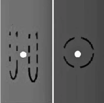
U-sutures were placed on the femoral artery adventitia. Each U-suture was created approximately 6 to 8 small jumping bites of the femoral artery adventitia horizontally. Both venous and arterial sutures were passed through tourniquets afterwards (Figure 1, 2).
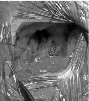
Venous cannulation was always placed first, due to deeper location than the artery. An 18-gauge Seldinger needle was used for both venous and arterial puncture. We do not prefer using surgical bistoury for cannula insertion. After the venous cannula was placed as usual fashion, the Seldinger needle was punctured through between the double parallel U-sutures (Figure 3). After the guidewire was introduced through the needle, its location was confirmed by transesophageal echocardiography whether it was within the descending aorta.
Figure 1. Femoral artery and vein cannulation sutures.
Figure 2. Schematic view of cannulation sutures (dashed line) and cannulation points (turquoise point).
Figure 3. Femoral artery puncture.
Turk J Vasc Surg 72
The proper size of the arterial cannula was, then, placed over the guidewire. No need to secure the tourniquets after placing the cannula (Figure 4). After weaning cardiopulmonary bypass (CPB), the venous cannula was removed first, and the purse suture was tied. Arterial cannula was removed, when half dose of protamine was administered and double parallel U-sutures were tied separately (Figure 5). Rare cases in which additional hemostasis suture was required, the third U-suture through the adventitia was placed and complete hemostasis was achieved. None of the patients had femoral artery complications through this technique.
DiSCuSSion
With the increase of cardiac reoperation and minimally invasive surgery, peripheral CPB has been increasingly widely used. Femoral artery and vein are the mostly preferred vasculatures both in percutaneous and open access.[1] Pseudoaneurysms formation,
retroperitoneal hemorrhage, and retrograde dissection were mostly encountered catastrophic complications of percutaneous method which requires an additional surgical intervention.[2] Direct vision insertion in open
access can highly preclude these kinds of adverse events. Two purse sutures to the femoral artery and vein have been used as a standard technique in open access to stop
bleeding after the cannula removal. In patients with a smaller femoral artery diameter than the cannula size, a larger purse suture is needed to achieve hemostasis after cannula removal. However, these purse sutures may cause femoral artery stenosis following the procedure, particularly in obese patients with a smaller femoral artery caliber. Furthermore, additional sutures are frequently needed in such cases and this may further narrow the femoral artery. In our technique, double parallel U-sutures pull the adventitia tissue horizontally and cover the cannulation site up, providing excellent hemostasis; therefore, no purse effect is expected in contrast with the standard technique.
Onan et al.[3] described a similar technique
and used multiple pledgeted sutures for arterial cannulation with excellent results. This technique was also applied to minimally and robotic cardiac surgery patients by Sen et al.,[4] and the mid-term results were
similar with former study. Based on our experiences, the pledgets are useless for femoral cannulation sutures and may complicate additional hemostasis suture placement, if needed. The adventitia tissue is a strong native pledget per se, and double parallel U-suture consisting of 6 to 8 small bites of adventitia can stop bleeding adequately. Another difference of our technique is that we use a single purse suture for the venous cannulation, and we believe the second purse suture is unnecessary. We examined this technique in a total of 103 patients, and no stenosis or occlusion was observed through both direct vision and postoperative ultrasound examination.
In conclusion, our technique is safe and easy to apply, which requires no drainage tube in the incision site. Using this technique, complications such as pseudoaneurysms, retrograde dissection, and retroperitoneal hematoma may be completely avoidable. However, long-term results are still needed.
Declaration of conflicting interests
The authors declared no conflicts of interest with respect to the authorship and/or publication of this article.
Funding
The authors received no financial support for the research and/or authorship of this article.
ReFeRenCeS
1. Nissen AP, Abreu J, Ornekian V, Nguyen S, Nguyen TC. Our preferred techniques for both direct and percutaneous femoral cannulation in minimally invasive mitral surgery. Asvide 2018;5:930.
73 A simplified peripheral cannulation suture
2. Lamelas J, Williams RF, Mawad M, LaPietra A. Complications associated with femoral cannulation during minimally invasive cardiac surgery. Ann Thorac Surg 2017;103:1927-32.
3. Onan B, Aydın Ü, Kadiroğulları E, Erkanlı K. An alternative peripheral arterial cannulation in minimally
invasive and robotic cardiac surgery. Cardiovasc Surg Int 2016;3:91-3
4. Sen O, Aydin U, Kadirogullari E, Bayram M, Karacalilar M, Kutluk E, et al. Mid-term results of peripheral cannulation after robotic cardiac surgery. Braz J Cardiovasc Surg 2018;33:443-7.