Volume 51, Number 11, 1997
0003-7028 / 97 / 5111-1741$2.00 / 0APPLIED SPECTROSCOPY
1741
q1997 Society for Applied SpectroscopyCharacterization of Denture Acrylic Resin Surfaces
Modi® ed by Glow Discharges
SËEFIK SU
È ZER,* NEHIR OÈZDEN, FUNDA AKALTAN, and GUÈNERI AKOVALI
Department of Chemistry, Bilkent University, 06533 Ankara, Turkey (SË .S.); Faculty of Dentistry, Ankara University, 06500 Ankara, Turkey (N.O., F.A.); Department of Chemistry, Middle East Technical University, 06531 Ankara, Turkey (G.A.)
Resin samples prepared by compression molding using a poly(methyl methacrylate) (PMMA) denture base material were ex-posed to radio-frequency (rf) glow discharges to improve the wett-ability of the material. Fourier transform infrared (FT-IR) re¯ ec-tance, X-ray photoelectron spectroscopy (XPS), and contact-angle measurements have been employed to characterize the changes in-troduced by the glow discharge plasma. FT-IR measurem ents can-not detect any modi® cation. XPS reveals an increase in the O/C atomic ratio. Contact angles of the plasma-treated samples are al-ways lower when compared with untreated ones. The increased O atomic concentration is attributed to formation of ± COH groups on the surface during plasma treatment. The O/C atomic ratio decreas-es upon heating the sampldecreas-es in vacuum to 1008C for 1± 2 min and exposing the samples to liquid CH2Cl2for 1± 2 min. Exposure to
distilled water for prolonged periods causes a slight decrease during the initial 1± 20 days but levels off to a constant value up to a period of 60 days. Plasma treatment seems to offer a durable increase in the wettability for these materials left in air or distilled water.
Index Headings: Denture base materials; PMMA; Plasma modi®
-cation; XPS; Contact angles.
INTRODUCTION
Poly(methyl methacrylate), PMMA, is the most com-monly used material for denture base. Therefore its ad-hesion to the underlying mucosa is very important and depends on various physicochemical factors. The basic requirement for good adhesion is intimate contact be-tween the adhesive and the adherent. The extent to which an adhesive will wet a surface depends, among other fac-tors, on the contact angle (u ) at which the adhesive meets the adherent surface. Since the tendency for the liquid to
spread increases as u decreases, the contact angle is a
useful inverse measure of spreadability or wettability.1
Kilani et al. measured contact angles between the syn-thetic saliva and distilled water on a selected number of base materials and reported that the contact angles be-tween saliva and distilled water were statistically
com-parable for a given denture base material.2The reported
contact angle values of water on PMMA varies between 60 and 808 depending on whether the advancing or re-ceding angle is measured. For the denture base materials that contain PMMA and other additives, the reported con-tact angle values are also in this range.
Surface modi® cation using various chemical and/or physical methods to improve its adhesion (assumed to be
related to wettability) has been reported.3± 6 Here, again,
improvement in the wettability was assessed by lowering the water contact angles after treatment. However, Mur-ray critically questioned the effectiveness of surface
treat-Received 11 November 1996; Accepted 23 May 1997. * Author to whom correspondence should be sent.
ment on PMMA when exposed in the mouth.6He treated
the PMMA surface using either (1) aqueous hydroxyl radicals generated by two chemical methods or (2) low-pressure electrical discharge. Using the contact angle measurements, he reported the treatment with aqueous hydroxyl radicals to be totally ineffective and the expo-sure to a low-presexpo-sure electrical discharge in water vapor and air to be initially effective in decreasing the value of the contact angle to near zero. He, however, reported that, with subsequent exposure to water vapor in a humidity chamber or immersion in water, a gradual increase in con-tact angles was observed, with an eventual return to their original values, and he concluded that there was no pros-pect for improvement by any surface treatment.
Here, we report Fourier transform infrared (FT-IR), X-ray photoelectron spectroscopy (XPS), and contact-an-gle study of the denture materials treated by glow-dis-charge plasma both to characterize the species formed on the surfaces and to assess the durability of such treat-ments. XPS is a surface-sensitive spectroscopic method and has been very successful in characterizing the phys-ical and chemphys-ical changes on the surfaces of
plasma-treated polymeric materials.7± 11In a recent study GroÈning
et al. reported an XPS investigation of the chemical mod-i® cations of the PMMA surfaces after plasma and
low-energy ion beam treatment.12 They found that reactive
atomic radicals or ions were responsible for incorporation of extra atoms (O) or groups (± COO) into the polymer surface. Use of XPS to study the adsorption of salivary
constituents on enamel has also been reported.13
EXPERIMENTAL
The conventional heat-compression mold technique was used to prepare acrylic resin test samples. The shal-low half of a ¯ ask was ® lled with type IV dental stone (Duralit-S, Degussa A.G., Frankfurt, Germany) and its surface smoothed in order to achieve ¯ at surfaces for the test samples. After a separating medium (Isolant, De Trey, London, U.K.) was applied to the stone surface, the upper half of the ¯ ask was ® lled with dental stone and the investment process completed. When the dental stone had set, the ¯ ask was opened and the separating medium applied to both dental stone surfaces. PMMA (Meliodent, Dental Bayer Limited Pharmaceuticals Div., Newburry, Berkshire, U.K.) was mixed according to the manufac-turer’s recommendations and cured by heat polymeriza-tion. The polymerized acrylic resin plaques were
sec-tioned to small ® lms with dimensions of 10 3 15 3 1
mm.
In a typical batch, 30 ® lms prepared under same con-ditions were sectioned into three groups, the ® rst of
1742
Volume 51, Number 11, 1997
TABLE I. Contact-angle measurements of untreated (C) and plas-ma-treated PMMA left in air (A) and in distilled water (D).
C (control) 0 DaysA 7 Days A D 14 Days A D 60 Days A D 51 54 53 48 46 42 53 45 62 54 58 49 44 41 55 43 61 51 48 49 47 44 54 48 71 47 54 38 54 45 52 50 70 ´´´ ´´´ ´´´ ´´´ 46 42 37 63 52 53 46 48 44 51 45
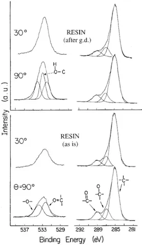
FIG. 1. O 1s and C 1s XPS of untreated PMMA at 908 and 308
take-off angles and after treatment with glow discharge. The enhanced O 1s signal is assigned to ± C± OH.
which was used as control samples; the second and the third groups were subjected to plasma treatment. The sec-ond group was left exposed to laboratory atmosphere, and the third group was placed in distilled water right after plasma treatment until analysis. In some cases, the ® lms were further subjected to in situ heating up to 100
8 C or liquid CH2Cl2 for periods of 1± 3 min before
anal-ysis.
Surface modi® cation was achieved inside a tubular
Py-rext reactor, which could be evacuated and/or ® lled with
various gases. Two copper electrodes on the Pyrext
re-actor were capacitatively coupled to a radio-frequency (rf) plasma system consisting of a ® xed-frequency (13.56-MHz) rf generator together with an rf power meter
and a matching network.14 In this work, only the samples
plasma-treated in evacuated reactor are reported. Since evacuation was achieved with a rotary pump only, the residual gases (; 1± 5 Torr) consisting mostly of air and water vapor were expected to take active part in the plas-ma processes. Two different power settings (5 and 10 W) for durations varying between 5 min to 1 h were applied. Contact-angle measurements were carried out with the
use of the entrapped air-bubble technique.15FT-IR
mea-surements were performed on a Bomem MD102 spec-trometer with a Harrick DRA-B03 diffuse re¯ ectance at-tachment. XPS measurements were performed on a
Kra-tos ES300 spectrometer with MgKa X-rays (1253.6 eV)
and a background pressure lower than 53 102 9Torr. All
measurements and treatments were repeated at least three times to ensure reproducibility.
RESULTS AND DISCUSSION
Surface Modi® cation. FT-IR measurements both in
absorbance/transmittance or in re¯ ectance mode could not detect any difference between the plasma-treated and untreated control samples, indicating that plasma treat-ment causes changes only in the very top layers. Contact-angle measurements, with the use of the entrapped-air method, on the plasma-treated samples yielded values
ranging between 48 and 548 , which were signi® cantly
lower than those of untreated control samples with an
average value of 638 . Samples left in distilled water gave
even lower values (44± 468 ). The results are summarized in Table I. Although several experiments using different power settings and/or durations were carried out, the val-ues reported in this table are for one set of experimental conditions only (5 W and 10 min in duration).
XPS spectra recorded at 908 and 308 take-off angles of
the control samples are given in Fig. 1. The general fea-tures are similar to the high-resolution spectra of PMMA
compiled by Beamson and Briggs.11 Although the
high-resolution spectrum of Ref. 11 assigns four different car-bons, we cannot resolve or can successfully differentiate between different hydrocarbon C 1s peaks using curve ® tting. Therefore, we ® tted the C 1s region into only three
peaks corresponding to CH, C± O, and C5 O groups. The
O 1s region was ® tted to two peaks corresponding to etheric and esteric oxygens. The main difference between our spectrum and that of pure PMMA reported by Ref. 11 is in the relative intensities of various C 1s peaks and also O/C atomic ratio. The O/C atomic ratio in our con-trol sample was 0.26, which deviates from 0.40 in pure PMMA. Similarly in the C 1s region, the ratio of both the etheric and esteric groups to total carbon intensity was around 0.14, which is less than 0.20 expected from stoi-chiometry. The ratio of the sum of etheric and esteric C 1s to total O 1s intensity is very close to the stoichio-metric ratio of 1.0. Therefore, the surface of our control samples seems to have additional CH groups, presumably stemming from the additives introduced through the var-ious processes used in preparation and molding. There is no distinguishable difference between the spectra record-ed at two different angles.
Spectra of the plasma-treated samples differ only in the O 1s region with a narrower and more intense peak that can be curve ® tted to three components. The extra peak in the O 1s region at 533.0 eV can be assigned to ± COH groups, in agreement with Refs. 7± 11 and 16± 18.
APPLIED SPECTROSCOPY
1743
TABLE II. XPS measurements of untreated (C) and plasma-treat-ed PMMA left in air (A) and in distillplasma-treat-ed water (D) for 14 days.
O 1s
C 1s
CH C± O C5 O
Resin B.E. (eV)a 532.5 285.0 286.7 288.9
(C) Intensitybat 908 (0.26) (0.73) (0.14) (0.14)
at 308 (0.27) (0.74) (0.14) (0.13)
Resin B.E. (eV)a 532.6 285.0 286.8 289.0
(A) Intensitybat 908 (0.42) (0.70) (0.17) (0.13)
at 308 (0.43) (0.67) (0.18) (0.15)
Resin B.E. (eV)a 532.6 285.0 286.8 288.8
(D) Intensitybat 908 (0.34) (0.70) (0.15) (0.15)
at 308 (0.35) (0.69) (0.15) (0.16)
aB.E.5 binding energy.
bCorrected for cross section and instrumental transmission function.
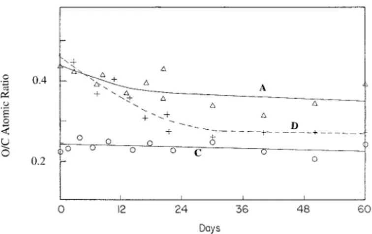
FIG. 2. Measured O/C atomic ratios of plasma-treated samples left in
a laboratory environment (A) and placed in distilled water (D) for a period of up to 60 days. Measurements in control samples not exposed to plasma (C) are also included.
A small increase in the relative intensity of the C± O com-ponent in the C 1s region is also consistent with the above assignment. Differences in the angle dependence of various components are slightly more pronounced after plasma treatment. Relevant XPS data are collected in Ta-ble II.
One of the main characteristics of cold plasma treat-ment is the formation of radicals in high concentration on polymer substrate surfaces, the chemical structure and half-life of which are known to depend on the structure
of the polymer and the plasma operational parameters.12,15
Besides the direct activation and functionalization with atomic oxygen of the gas phase, plasma radicals on the polymer surface can also react with molecular oxygen to form peroxides, which may lead to new species forma-tion, such as hydroperoxides through hydrogen addition
R´1 O2Ð ROO´Ð ROOH, which then can decompose to
form stable species such as esters alcohols, depending on plasma conditions.12,17,18
XPS data together with contact-angle measurements clearly reveal that the increased wettability must be re-lated to the chemical modi® cation of the surface com-position, especially to the increase in oxygen content. Since in XPS measurements one cannot detect hydrogen and can detect only relative changes in the surface com-position, an increase in the oxygen content must be in-terpreted as relative to carbon. Second, since there is not a strong angular dependence, the modi® cation is uniform up to several molecular layers into the bulk, greater at least than 5 nm, which is typically the XPS sampling
depth with the use of MgKa radiation,11 but de® nitely
less than 50 nm, which is an overestimated sampling depth observable by FT-IR.
Heating in vacuum at ; 100 8 C for 1± 2 min causes
approximately a 15% reduction in the observed O/C atomic ratio. Similarly, exposure of the plasma-treated
samples to 1± 2 min of liquid CH2Cl2 also decreases the
O/C atomic ratio by 20%. The decrease in the O/C atomic ratio as a result of (1) heating in vacuum, (2) exposure
to CH2Cl2, or (3) exposure to distilled water can be
ex-plained by either H2O elimination (possibly by ± H and
± OH recombination) or surface reconstruction.
Durability of the Modi® cation. To assess the
dura-bility of the plasma treatment, we followed up on the XPS O/C atomic ratios of the samples left in a laboratory environment (A) and the ones placed in distilled water
(D) for up to 60 days after plasma treatment (Fig. 2). Both in the samples left in air and those in distilled water, the O/C atomic ratios decrease within the ® rst two weeks but settle to 0.40 and 0.32, respectively, up to 60 days for air and water samples, as compared to 0.26 for un-treated control samples. The contact-angle measurements after 7, 14, and 60 days, however, reveal no signi® cant changes for the same samples left in air or water. This difference must be attributed to the different probing ca-pacities and/or sampling depths of the XPS and contact-angle methods. However, both methods indicate a durable (at least up to 60 days) modi® cation of the surfaces of the resin samples left in air or water, in contrast to
Mur-ray’s in vitro ® ndings.6 Differences between our results
and those of Murray could be due to the different plasma treatments, which are known to be very strongly depen-dent on various experimental parameters.12,14,16,17
CONCLUSION
Glow-discharge plasma alters the surfaces of the acryl-ic resin and increases the wettability, as shown both by XPS and contact-angle measurements. The increased wettability can be attributed to the presence of ± COH groups. Glow-discharge plasma treatment does seem to offer a durable (at least up to 60 days) wettability for these materials when left in air or distilled water.
1. A. W. Adamson, Physical Chemistry of Surfaces (Wiley, New York, 1990), 5th ed.
2. B. H. Z. Kilani, D. H. Retief, M. V. Guldag, D. J. Castleberry, and T. E. Fischer, J. Prosthet. Dent. 52, 288 (1984).
3. W. J. O’ Brien and G. Ryge, J. Prosthet. Dent. 15, 304 (1965). 4. H. D. Gesser and C. R. Castaldi, J. Prosthet. Dent. 25, 236 (1971). 5. R. E. Lindstrom, J. Pawelchak, A. Heyd, and W. J. Tarbet, J.
Pros-thet. Dent. 42, 371 (1979).
6. M. D. Murray, J. Prosthet. Dent. 59, 368 (1988).
7. D. T. Clark and H. R. Thomas, J. Polm. Sci. Polm. Chem. Ed. 16, 791 (1978).
8. J. D. Andrade, R. R. King, and D. E. Gregonis, J. Colloid Interface Sci. 72, 488 (1979).
9. T. J. Hook, J. A. Gardella, Jr., and L. Salvati, Jr., J. Mater. Res. 2, 117 (1987).
10. E. M. Lehockey and I. Reid, Surf. Interface Anal. 11, 302 (1988). 11. G. Beamson and D. Briggs, High Resolution XPS of Organic
1744
Volume 51, Number 11, 1997
12. P. GroÈning, O. M. KuÈttel, M. Collaud-Coen, G. Dietler, and L. Schlapbach, Appl. Surf. Sci. 89, 83 (1995).
13. Y. Kuboki, K. Teraoka, and S. Okada, J. Dent. Res. 66, 1016 (1987).
14. G. Akovali, J. Appl. Polym. Sci. 32, 4027 (1986).
15. A. N. OÈ zden, P. IÇmirzaliogÆlu, and M. Mutlu, Tr. J. Med. Sci. 23, 43 (1995).
16. G. P. Lopez, D. G. Castner, and B. D. Ratner, Surf. Interface Anal.
16, 267 (1991).
17. B. Y. Qu, Y. H. Xu, W. F. Shi, and B. Ranby, Macromolecules 25, 5215 (1992).