474
Journal of Neurological Sciences [Turkish] 31:(3)# 41; 474-488, 2014
http://www.jns.dergisi.org/text.php3?id=793 Research Article
Effects of the Glutamine on the Neuronal Cell Death in rat Ischemia-reperfusion Model
Omur KASİMCAN1, Bulent BAKAR2, Arzu KOSE3, Pergin ATİLLA4, Kamer KİLİNC5,
Sevda MUFTUOGLU4
1
Liv Hospital, Neurosurgery Department, Istanbul, Türkiye 2University of Kırıkkale School of Medicine, Neurosurgery Department, Kırıkkale, Türkiye 3Istanbul Medipol University, Anestesiology and Reanimation Department, Istanbul, Türkiye 4University of Hacettepe School of Medicine, Histology and Embriology Department, Ankara, Türkiye 5University of
Hacettepe School of Medicine, Biochemistry Department, Ankara, Türkiye Summary
Background: The aim of this study was to explore the effects of glutamine in brain
ischemia/reperfusion model in rat.
Methods: Right common carotid arteries of 24 Wistar albino rats were clamped for a duration
of 30 minutes. Two hours later, except CONTROL group, glutamine was infused into left femoral vein of rats in GLIV group; and glutamine and normal saline was administered into cisterna magna of rats in GLIS and SFIS groups, respectively. After 7 days, all animals were decapitated and each brain was divided into two hemispheres for histopathological and biochemical evaluation. The right hemisphere was called “Hypoxia/Reperfusion side (HRS)” and the left hemisphere was called “Toxicity side (TS)”.
Results: In TS and HRS, degenerated neuron counts of GLIV groups were significantly
higher than other groups' values. Degenerated neuron count values of TS were significantly lower than HRS values for GLIS, and SFIS groups, but the results of GLIV group in TS did not different from the GLIV group in HRS. LPO levels of TS and HRS of the groups was not statistically significant.
Conclusion: This study results showed that glutamine had no beneficial effect to the
hypoxia/reperfusion injury in rat model.
Key words: Glutamine, ischemia-reperfusion, intracisternal, toxicity
Ratlarda Beyin Hipoksi-reperfüzyon Modelinde Glutaminin Etkilerinin Araştırılması Özet
Amaç: Bu çalışmada glutamin adlı maddenin ratlardaki deneysel serebral hipoksi/reperfüzyon
hasarlanması üzerine etkileri araştırılmaya çalışıldı.
Yöntem ve Gereç: Yirmi dört adet Wistar albino ratın sağ ana karotis arterleri geçici
anevrizma klibi kullanılarak 30 dakika süre ile kapatıldı. İki saat sonra CONTROL grubundaki hayvanlar hariç glutamin adlı materyal GLIV grubundaki ratlara femoral venden; GLIS grubundakilere ise sisterna magnadan enjekte edildi. SFIS grubundaki ratlara sisterna magnadan serum fizyolojik verildi. Yedi gün sonra tüm hayvanların beyinleri çıkarılıp santral sulkustan ikiye bölündü; sağ hemisfere “hipoksi/reperfüzyon tarafı (HRS)”; karşı hemisfere “toksisite tarafı (TS)” ismi verildi ve dokular histopatolojik ve biyokimyasal analize tabi tutuldu.
Bulgular: TS ve HRS için GLIV grubunun dejenere nöron sayım sonuçları diğer gruplara
göre yüksek bulundu. Her bir grup için TS sayım sonuçları ile HRS sonuçları karşılaştırıldı ve toksisite tarafı SFIS ile GLIS gruplarının sayım sonuçlarının hipoksi/reperfüzyon tarafı
475
gruplarınkine göre anlamlı derecede düşük olduğu görüldü, ancak her iki tarafın GLIV gruplarının sonuçları arasında anlamlı fark saptanmadı. Her iki taraf doku lipid peroksidasyon (LPO) düzeyi sonuçları arasında istatistiksel anlamlı fark yoktu.
Sonuç: Bu çalışma sonunda glutamin adlı maddenin ratlarda oluşturulan hipoksi/reperfüzyon
yaralanması üzerinde yararlı etkilerinin bulunmadığı sonucuna ulaşıldı.
Anahtar Kelimeler: Glutamin, hipoksi/reperfüzyon, intrasisternal, toksisite INTRODUCTION
Ischemic brain injury arising from surgery, trauma, cerebrovascular occlusion or vasospasm related with subarachnoid haemorrhage is a common pathophysiological process observed in neurosurgical practice(3). Severe deficiency
of either oxygen or other substrates for normal function is likely to result in neuronal cell injury and death mediated by excitotoxic mechanisms(5). Moreover, the neuronal tissue damage may be accelerated by cell death when the blood supply is restored. This phenomenon is known as ischemia-reperfusion (IR) injury(13). This ischemic injury induces a molecular cascade that culminates in the formation of toxic proteases and free radicals. The superoxide radicals which have been identified as the primary reactive oxygen species (e.g. superoxide anion, chloride anion, hydroxyl radicals) could destroy the neuronal and axonal sheat by breakdown the lipoid component of the cell membranes as known lipid peroxidation (LPO) reaction(17). Although several pharmacological compounds were tested for protection of neuronal survival, preventing brain edema and keeping from oxidative stress in animal studies after ischemic brain injury, these studies results could not identified an exactly drug for treatment of this condition(7,10,13).
Glutamine, an antioxidant, is a precursor of glutathione that protects neurons from free radical damage during hypoglycemia, ischemia, and glutamate toxicity and its levels fall dramatically after major injury or infection(6,12). It has been demonstrated that glutamine protects against ischemia/reperfusion injury of the gut, heart, liver and skeletal muscle but the
mechanism of this protection has not been understood well(12). It is known that the glutamine can be used as an energy substrate by the mitochondria of the astrocytes or neurons after ischemia, which is important for neuronal survival under conditions when the utilization of pyruvate by cells in a post-ischemic period is restricted(2). It is also demonstrated that glutamine is a regulator of protein synthesis and degradation, and it plays an important role in regulating acid-base balance, promoting immune function, and improving adaptation to stress(13). The solution of the L-alanyl-L-glutamine is a highly stable dipeptide, and when infused intravenously it is promptly hydrolyzed to glutamine and alanin(12). The aim of this study was to explore the possible beneficial effects of intravenous or intracisternal application of L-alanyl-L-glutamine in brain ischemia-reperfusion model in rat.
MATERIAL AND METHODS Materials:
This study was performed in accordance with the guidelines for the use of laboratory animal subjects in research set by the Ethical Committee of Kırıkkale University (Number: 10/ 52; Date: 17.08.2010).
Twenty four male Wistar albino rats 250– 350 g body weight were used. The rats were placed in a temperature (21 ± 2 °C) and humidity (60 ± 5 %) controlled room for one week before the experiment, and 12 h light / 12 h dark cycle was maintained, and they were allowed free access to food and water before and after surgery. They were divided into five
476 groups each according to a table of a random numbers as follows:
- CONTROL group (consisted of the left hemisphere of the rats infused no chemical material; n: 6).
- SHAM group (consisted of the right hemisphere of the rats infused no chemical material; n: 6).
- SFIS group (infused normal saline solution intracisternally; n: 6).
- GLIS group (infused 20% L-alanyl-L-glutamine intracisternally; n: 6).
- GLIV group (infused 20% L-alanyl-L-glutamine intravenously; n: 6).
After decapitation procedures, each animal brain obtained from all groups described above were divided into two hemispheres called as:
1. Hypoxia/ reperfusion side (HRS) (consisted of the right cerebral hemisphere performed hypoxia/reperfusion injury) 2. Toxicity side (TS) (consisted of left cerebral hemisphere not performed hypoxia-reperfusion injury)
The right side brain tissues were used for histopathological and biochemical evaluation of the possible neuroprotective effects of 20% L-alanyl-L-glutamine. The remaining left side brain tissues were used to evaluate the possible neurotoxic effects of 20% L-alanyl-L-glutamine.
20% L-alanyl-L-glutamine with water for injection (Dipeptiven®, Fresenius-Kabi, Germany) was used in human subjects in dosage with 0.2-0.4 g/ kg/ day parenterally. Data sheet of the Dipeptiven® informs that its 100 ml bottle contains 20 g N(2)-L-alanyl-L-glutamine; and in rats, infusion of 50 mL/ kg body weight of N(2)-L-alanyl-L-glutamine over 4h/day led to tonic spasms, increased respiratory rate and exitus. However, its intracisternal administration has not been reported in medical literature yet. In present study, the dosage was settled as 0.15 g/ kg body weight of 20% L-alanyl-L-glutamine
(approximately 0.8 ml in volume) for the GLIV group intravenously; and because of the restricted area of the subarachnoid space, 0.075 g/ kg body weight (approximately 0.4 ml in volume) for the GLIS group intracisternally.
Anaesthesia was performed by using the intramuscular administration of ketamine HCl (Ketalar®; Pfizer Inc, USA) and xylazine HCl (Rompun® %2; Bayer HealthCare AG, Germany).
Methods:
All animals were placed under sedational anaesthesia with intramuscular 40 mg/kg ketamine HCl and 5 mg/kg xylazine HCl on spontaneous respiration at room temperature. Under an operating microscope, bifurcation of the right common carotid artery was exposed through a midline neck incision, and carefully separated from the surrounding vagus nerve. Then right common carotid artery was clamped with temporary microvascular aneurysm clip (Mizuho® Aneurysm Clip, Mizuho, Japan) for a duration of 30 minutes. After 30 minutes, the right carotid artery was unclamped. Two hours later from the removing of the microvascular clip, except those of CONTROL and SFIS groups, 0.15 g/kg 20% L-alanyl-L-glutamine was slowly infused into the left femoral vein of the rats in GLIV group; and in GLIS group, 0.075 g/kg (approximately 0.3 ml) 20% L-alanyl-L-glutamine was administered slowly into the cisterna magna of the rats by using a 26G needle when the free cerebrospinal fluid flow was seen. In SFIS group, 0.3 ml normal saline was slowly infused into the cisterna magna as described above.
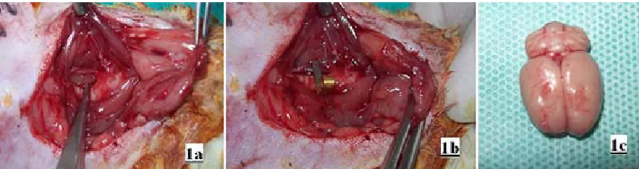
Seven days after the surgery; all animals were re-sedated with intramuscular 40 mg/kg ketamine HCl and 5 mg/kg xylazine HCl and their whole bloods were collected from inferior vena cava for sacrification. Then, all animals were decapitated (Figure 1) and first the brain tissues were divided into two hemispheres with a cut from sulcus centralis and then these half
477 hemispheres were also divided into two lobes with a cut from the hippocampal sulcus. The right side brain tissues were used for possible neuroprotective effects of 20% L-alanyl-L-glutamine. The right side brain tissues of the CONTROL group was used as “SHAM group”. The remaining left side brain tissues were used to evaluate of the possible neurotoxic effects of 20% L-alanyl-L-glutamine to the brain tissues. Then those brain tissues were immediately harvested for future biochemical and histopathological examinations. For histopathological examination, the neural tissue specimens that those of the posterior half side of the brain hemisphere which contained hippocampus were stored in 10% buffered formaldehyde solution at room temperature. For biochemical examination, the neural tissue specimens obtained from the anterior half side of the cerebral hemisphere were stored at −30°C at dry air.
Histopathological evaluation:
For histopathological examination, all tissue samples were processed according to the routine light microscopic tissue processing technique. Serial sections of 5
μm thickness stained with haematoxylene– eosin (H&E) were examined and photographed by a microscope (Olympus BX-50). The number of the degenerated neurons (called pycnotic neurons or “red neurons”) in each group was counted and calculated separately in three areas per section of the hippocampal CA and DG region as an average per rat. The investigators who performed these measurements (P.A and F.S.M) were blind to the study groups and experimental materials.
Biochemical analysis
LPO level was assayed by measuring malondialdehyde (MDA) as thiobarbituric acid-reactive substances (TBARS) levels using the thiobarbituric acid method(9). Biochemical analyses were performed by thiobarbituric acid application; and then 532 nm, spectrophotometry (Shimadzu1 UV-120-02 Spectrophotometer) was used for measuring the MDA levels in nanomoles per gram of wet tissue. The investigator who performed these measurements (K.K) was blind to the study groups and experimental materials.
Figure 1: During surgical procedures (1a) first the right common carotid artery was exposed and (1b) then
clamped with temporary microvascular clip. (1c) After decapitation the macroscopic appearance of the cerebral cortex following ischemia/ reperfusion injury.
478
Statistical analysis
Histopathological cell count results of the TS and HRS and the tissue LPO levels of the TS were not normally distributed; and the variation was not homogenous between all groups. So, these results were statistically analyzed by the Krusskal– Wallis test, and p values lower than 0.05 were considered to be significant. Mann– Whitney U test and Bonferroni multiple comparisons test (post hoc evaluation) were performed to determine the statistical differences between the groups, and p values lower than 0.0083 for TS groups and p values lower than 0.005 for HRS groups were considered to be significant. The tissue LPO levels were normally distributed and the variation was homogenous between all HRS groups. Therefore, they were statistically analyzed by the One-Way Analysis of Variance (One-Way ANOVA) test, and p values lower than 0.05 were considered to be significant.
Furthermore, the Wilcoxon signed-rank test (i.e. the nonparametric analog of the “paired samples t” test) was performed to determine the statistical differences between the results of the cell counts of the TS and HRS for each group. p values lower than 0.05 were considered to be significant(8).
RESULTS
Light microscopy
Toxicity side (TS):
Specimens of the CONTROL and SFIS group showed degenerated and vacuolized neurons localized in the tip of the DG region of the hippocampus. Marked edema located in the neuropil and vascular stasis with perivascular edema was observed in the DG and CA3 regions of the hippocampus, except SFIS group specimens.
Degenerated neurons in GLIV group were observed in the tip of DG region of the hippocampus. Marked edema located in the neuropil and vascular stasis was also observed in DG and CA regions, but perivascular edema was not seen.
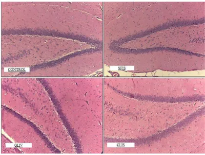
Specimens of the GLIS group showed much more degenerated neurons located in the DG region of the hippocampus than GLIV group' s. However, stromal edema was lesser than other groups (Figure 2) Hypoxia -reperfusion side (HRS):
Specimens of the SHAM group showed large edematous areas with vascular stasis and perivascular edema located in the neuropil. Additionally degenerated neurons were also observed in DG and CA regions of the hippocampus. Those histopathological observations were less common seen in SFIS group.
Specimens of the GLIV group showed advanced degree of numerous pycnotic neurons located in the CA and tip of the DG regions. Marked stromal and perivascular edema with vascular stasis was prominent in the neuropil and due to the edema, stromal layer was almost separated from the pycnotic neurons.
Degenerated neurons in GLIS group were lesser than GLIV group. Stromal edema with vascular stasis localized in the DG region of the hippocampus; and perivascular edema was not seen in any specimen (Figure 3).
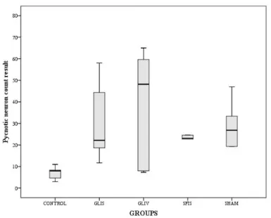
Degenerated neuronal cell count results:
The variation of degenerated neuron counts of TS among all groups was statistically
significant (X2=13.463, p=0.004),
determined by the Kruskal-Wallis test ( Table 1, Table 2).
The Mann Whitney U test results that were applied to red neuron counts of TS described statistically significant differences between GLIV-SFIS (Z=-2.892, p=0.004), and GLIV-CONTROL
479 groups (Z=-2.892, p<0.004) (Table 3, Figure 4).
The variation of degenerated neuron counts of HRS among all groups was also statistically significant (X2=11.110, p=0.025), determined by the Kruskal-Wallis test (Table 1, Table 2).
The Mann Whitney U test results that were applied to red neuron counts of HRS described statistically significant differences between SHAM-CONTROL (Z=-2.887, p=0.004), and GLIS-CONTROL groups (Z=-2.887, p<0.004) (Table 4, Figure 5).
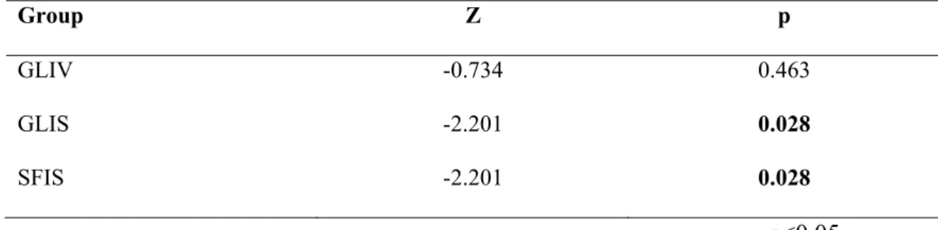
Moreover, the mean values of degenerated cell count results of TS were compared to HRS for SFIS, GLIV, and GLIS groups by the Wilcoxon Signed Ranks test. Except the GLIV group, the results explained that the cell count values of the TS of the GLIS 2.201, p=0.028), and SFIS group
(Z=-2.201, p=0.028) were significantly different to the HRS values (Table 5)
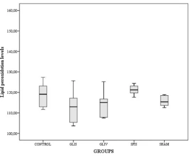
Biochemical Analysis:
The variation of MDA levels of TS among all groups was not statistically significant (X2=1.499, p=0.682), determined by the Kruskal-Wallis test (Table 1, Table 2, Figure 6). In addition, the variation from the mean values of the MDA levels of the HRS of the groups was not statistically significant (F=2.052, p=0.118), determined by the One Way-ANOVA test (Table 1, Table 6, Figure 7).
Furthermore, the tissue MDA level results of TS were compared with HRS for GLIV, GLIS, SFIS groups by the Wilcoxon Signed Ranks test. The test results explained that there was no statistical difference between the TS and the HRS values (p> 0.05).
Figure 2: Toxicity side specimens of the CONTROL, SFIS, GLIV and GLIS group show the marked edema,
480
Figure 3: Hypoxia/ reperfusion side specimens of the SHAM, SFIS, GLIV and GLIS group show the marked
edema, degenerated and vacuolized neurons localized in the hippocampus (HEx200).
Figure 4: The variation of the pycnotic neuron counts for toxicity side. Each error bar shows the minimum and
481
Table 1: Descriptive table of the all groups. (LPO:lipid peroxidation; SD: standart deviation)
Side Group Variable Minimum Maximum Mean SD
Toxicity side CONTROL LPO 111.69 127.35 118.89 6.46
Cell count 3.00 11.00 7.17 2.87 GLIV LPO 107.04 117.19 114.44 3.87 Cell count 14.00 52.00 28.61 16.80 GLIS LPO 103.23 155.27 119.03 18.52 Cell count 2.00 17.33 8.61 6.36 SFIS LPO 108.31 118.88 115.99 3.99 Cell count 2.00 4.33 3.22 0.87
Hypoxia/ Reperfusion side SHAM LPO 112.54 118.88 115.71 2.55
Cell count 19.33 47.00 28.78 10.39 GLIV LPO 107.46 125.23 114.58 6.55 Cell count 7.33 65.00 39.39 25.95 GLIS LPO 103.65 125.65 112.96 8.09 Cell count 11.67 58.00 29.50 17.76 SFIS LPO 117.62 124.38 121.21 2.50 Cell count 8.33 44.33 24.37 11.51 CONTROL LPO 111.69 127.35 118.89 6.46 Cell count 3.00 11.00 7.17 2.87
482
Figure 5: The variation of the pycnotic neuron counts for hypoxia/ reperfusion side. Each error bar represents
the minimum and maximum values of the red neuron count.
Figure 6: The mean values of the tissue lipid peroxidation level of the toxicity side groups. Each error bar shows
483
Table 2: This table demonstrates that there were statistically significant differences between all groups for comparison of degenerated neuron cell count values of the toxicity side and hypoxia-reperfusion side. However, tissue lipid peroxidation levels were not different among the groups in the toxicity side. Krusskal-Wallis test, p<0.05.(df: degrees of freedom; LPO: lipid peroxidation; X2:
chi-square)
Side Variable X2 df p
Toxicity side LPO 1.499 3 0.682
Cell count 13.463 3 0.004
Hypoxia/reperfusion side Cell count 11.110 4 0.025
p<0.05 Figure 7: The mean values of the tissue lipid peroxidation level of the hypoxia/ reperfusion side groups. Each
484
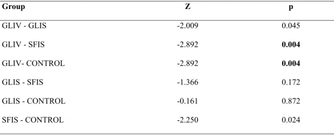
Table 3: Apart from the CONTROL, SFIS and GLIS groups, GLIV group showed a more pronounced pycnotic neuron cells in toxicity side. Mann Whitney U test and Bonferroni Multiple Comparisons test,
p< 0.0083. (Z: Z score) Group Z p GLIV - GLIS -2.009 0.045 GLIV - SFIS -2.892 0.004 GLIV- CONTROL -2.892 0.004 GLIS - SFIS -1.366 0.172 GLIS - CONTROL -0.161 0.872 SFIS - CONTROL -2.250 0.024 p<0.0083
Table 4: Pycnotic neuron cell counts did not different in GLIV, GLIS, SHAM and SFIS groups of the hypoxia/ reperfusion side. Mann Whitney U test and Bonferroni Multiple Comparisons test, p< 0.005. (Z: Z score) Group Z p SHAM - GLIV -0.801 0.423 SHAM - GLIS -0.480 0.631 SHAM - SFIS -0.801 0.423 SHAM - CONTROL -2.887 0.004 GLIV - GLIS -0.480 0.631 GLIV - SFIS -0.801 0.423 GLIV - CONTROL -1.764 0.078 GLIS - SFIS -0.080 0.936 GLIS - CONTROL -2.887 0.004 SFIS - CONTROL -2.580 0.010 p< 0.005
485
Table 5: Comparisons of the degenerated neuron count values of toxicity side to hypoxia- reperfusion side for each of SFIS, GLIV, and GLIS groups demonstrated that the pycnotic neuron cell count values of the SFIS, and GLIS groups were much higher at the hypoxia-reperfusion side than toxicity side. Wilcoxon Signed Ranks test, p < 0.05. (Z: Z score)
Group Z p
GLIV -0.734 0.463
GLIS -2.201 0.028
SFIS -2.201 0.028
p<0.05
Table 6: The tissue lipid peroxidation levels were not different among the groups in the hypoxia-reperfusion side. One Way-ANOVA test, p<0.05 (F: F factor; LPO: lipid peroxidation)
Variable F p
LPO 2.052 0.118
p<0.05
DISCUSSION
Glutamine is synthesized primarily in skeletal muscle, lungs, and adipose tissue. Within the body, glutamine comprises more than 60% of the free amino acids(1). Recently, it has been shown that despite large repositories of glutamine, its stores may become depleted particularly in the course of a catabolic insult such as injury(16). In literature, the authors have suggested that hypoxia and ischemia could increase the production of glutamate from glutamine. It is also known that glutamate is an important pathogenic factor for neuronal cell death in cerebral ischemia(3,5). On the other hand, some studies have demonstrated that neurons can also use this product as an alternative energy substrate after ischemia/ reperfusion, and this promotes neuronal survival(11,12,14). Because of clarifying the confused data mentioned above and
evaluating the possible beneficial effects of glutamine in the hypoxia/ reperfusion injury in brain this study was constructed as a stroke model; and two hours later from the performing of the hypoxia/ reperfusion injury intravenous or intracisternal 20% L-alanyl-L-glutamine were administered to the rats. In present study, the pycnotic neuron count results of the SHAM, SFIS, GLIV and GLIS groups were almost equivalent in HRS. Although the statistical analyses results were just significant between the CONTROL-SHAM and CONTROL-GLIS group, it could be said that the mean values of the CONTROL group was lower than other groups' values (especially lower than GLIV group's values) significantly (see Table 1). These results may suggested that intravenous or intracisternal 20% L-alanyl-L-glutamine administration could not ameliorate the hypoxia/ reperfusion injury in rat brain. On
486 the other hand, pycnotic neuron count results of the GLIV group were much more than CONTROL, SFIS and GLIS group values in TS. This result suggested that intravenous 20% L-alanyl-L-glutamine administration may induce the neuronal cell degeneration in normal neural tissue in rat. Additionally, except the CONTROL, SHAM and GLIV groups, pycnotic neuron counts of the HRS were much more than TS for all groups. So, it could be said according to these results that our stroke model has been effective to describe the hypoxia-reperfusion model; and intravenous administration of the 20% L-alanyl-L-glutamine may induce the neuronal cell degeneration both ischemic and normal neural tissue in rat.
If the ischemic condition of the neural tissue is prolonged, it would depress the antioxidants levels by overproduction of the superoxide anions(1,13,14). Lipoproteins
located in the neural cell membrane are susceptible to LPO triggered by reactive oxygen and reactive nitrogen species produced by inflammatory cells and glial cells(2). Mondello et al showed in their study that the treatment with glutamine could decrease the degree of LPO in injured organs in mice(4). However, Stelmashook et al recommended in their study that glutamine stimulates glutamate-dependent neuronal damage in cerebellar tissue cultures when mitochondrial respiration is impaired(15). In present study, it was found that there was no statistical difference between the LPO levels of the TS and HRS among all groups. Additionally, there was no statistical difference between the LPO levels when LPO values of the TS were compared to the HRS values for all groups. It can be said according to these results that glutamine infused through intracisternal or intravenous route could not decrease LPO levels in the ischemic neural tissue as an antioxidant. Furthermore it may have negative effects on the normal brain tissues in rat (See Table 1, Table 2, and Table 5). Actually, numerous studies have indicated
that the extracellular concentration of excitatory amino acids such as glutamate and aspartate is abnormally increased after brain ischemia. These excitatory amino acids can interact with their receptors, resulting in Na+ and Ca+2 influx as well as the release of intracellular Ca+2, which results in Na+ and Ca+2 overload leading to neuronal death(3). Finally, because we
could not observe antioxidant effects of the glutamine on the ischemic neural tissues sufficiently, we may speculate that the neuronal damage may be developed by the glutaminergic receptor over expression induced by glutamine in present study,.
Study limitations
This study has some limitations. First, this study could not performed in an optimal laboratory conditions (such as thermoregulatory heating unit, continuous blood pressure monitoring, arterial blood gas monitoring, etc) because of some technical problems. Second, because of some financial limitations present study could not contain more specific
histopathological analysis (e.g.
immunohistochemical and electron microscopic findings which can show ultrastructural details of the inflammatory response, neuronal necrosis and oedema) which can identify other mechanisms of the hypoxia-reperfusion injury. Third, because of the same financial limitations this study could not be supported by using more specific biochemical analyses for other inflammatory pathways of the hypoxia-reperfusion injury (such as apoptotic pathways, reduced glutathione level, nitrite/ nitrate level, and xhantine oxidase activity level measurements). Fourth, the authors reported in literature that rats have a anastomotic vessels like Willis' polygon around their skull base and hypoxia/reperfusion model developed by the temporary compression of the common carotid artery could not be effective to produce stroke in rat brain. They have recommended to use the “mongolian gerbils” in experimental stroke models(12).
487 However, our study results showed that our hypoxia/ reperfusion model is sufficiently effective in rats.
CONCLUSION
This study demonstrated that
1. Intravenous 20% L-alanyl-L-glutamine may induce the neuronal cell degeneration both ischemic and normal neural tissue in rat
2. Neither intravenous nor intracisternal 20% L-alanyl-L-glutamine could reduce the tissue lipid peroxidation levels.
In conclusion, it can be said that 20% L-alanyl-L-glutamine had not any beneficial effect to the hypoxia/ reperfusion injury in rat, and it may promote the neuronal cell degeneration in the ischemia/ reperfusion injury in rat.
Conflict of interest and funding
The authors have not disclosed any financial relationship with any company whose product might be affected by the research described or with any company that makes or markets a competing product.
Correspondence to:
Bulent Bakar
E-mail: [email protected]
Received by: 03 January 2014 Revised by: 09 July 2014 Accepted: 21 July 2014
The Online Journal of Neurological Sciences (Turkish) 1984-2014
This e-journal is run by Ege University Faculty of Medicine,
Dept. of Neurological Surgery, Bornova, Izmir-35100TR
as part of the Ege Neurological Surgery World Wide Web service.
Comments and feedback: E-mail: [email protected] URL: http://www.jns.dergisi.org
Journal of Neurological Sciences (Turkish) Abbr: J. Neurol. Sci.[Turk]
ISSNe 1302-1664
REFERENCES
1. Curthoys NP, Watford M. Regulation of glutaminase activity and glutamine metabolism. Annu Rev Nutr. 1995; 15: 133-159.
2. Ferretti G, Bacchetti T. Peroxidation of lipoproteins in multiple sclerosis. J Neurol Sci. 2011; 311: 92-97.
3. Harukuni I, Bhardwaj A. Mechanisms of brain injury after global cerebral ischemia. Neurol Clin. 2006; 24: 1-21.
4. Jang HJ, Kwak JH, Cho EY, We YM, Lee YH, Kim SC, Han DJ. Glutamine induces heat-shock protein-70 and glutathione expression and attenuates ischemic damage in rat islets. Transplantation Proceedings 2008; 4: 2581– 2584.
5. Lee A, Lingwood BE, Bjorkman ST, Miller SM, Poronnik P, Barnett NL, Colditz P, Pow DV. Rapid loss of glutamine synthetase from astrocytes in response to hypoxia: implications for excitotoxicity. J Chem Neuroanat. 2010; 39: 211-220.
6. Mondello S, Galuppo M, Mazzon E, Italiano D, Mondello P, Aloisi C, Cuzzocrea S. Glutamine treatment attenuates the development of organ injury induced by zymosan administration in mice. Eur J Pharmacol. 2011; 658: 28-40. 7. Nagahiro S, Uno M, Sato K, Goto S, Morioka
M, Ushio Y. Pathophysiology and treatment of cerebral ischemia J Med Invest. 1998; 45: 57-70.
8. Nie NH, Hull CH, Jenkins JG. SPSS. Statistical Package For Social Science. New York, Mc Graw Hill Inc, 1975.
9. Ohkawa H, Ohishi N, Yagi K. Assay for lipid peroxides in animal tissues by thiobarbituric acid reaction. Anal Biochem 1979; 95: 351– 358.
10. Park JY, Byeon JH, Park SW, Eun SH, Chae KY, Eun BL. Neuroprotective effect of human placental extract on hypoxic-ischemic brain injury in neonatal rats. Brain Dev. 2012; 35: 68-74.
488
11. Pascual JM, Carceller F, Roda JM, Cerdn S. Glutamate, glutamine, and GABA as substrates for the neuronal and glial compartments after focal cerebral ischemia in rats, Stroke 1998; 29: 1048–1056.
12. Pires VL, Souza JR, Guimarães SB, Silva Filho AR, Garcia JH, Vasconcelos PR. Preconditioning with L-alanyl-L-glutamine in a Mongolian Gerbil model of acute cerebral ischemia/reperfusion injury. Acta Cirúrgica Brasileira 2011; 26 (Suppl. 1): 14-20.
13. Saleem S, Zhuang H, Biswal S, Christen Y, Doré S. Ginkgo biloba extract neuroprotective action is dependent on heme oxygenase 1 in ischemic reperfusion brain injury. Stroke 2008; 39: 3389-3396.
14. Stelmashook EV, Isaev NK, Lozier ER, Goryacheva ES, Khaspekov LG. Role of glutamine in neuronal survival and death during brain ischemia and hypoglycemia. Int J Neurosci. 2011; 121: 415-422.
15. Stelmashook EV, Lozier ER, Goryacheva ES, Mergenthaler P, Novikova SV, Zorov DB, Isaev NK. Glutamine-mediated protection from neuronal cell death depends on mitochondrial activityNeuroscience Letters 2010; 482: 151– 155.
16. Tapiero H, Mathé G, Couvreur P, Tew KD. II. Glutamine and glutamate. Biomed Pharmacother. 2002; 56: 446-457.
17. Yang Y, Rosenberg GA. Blood-brain barrier breakdown in acute and chronic cerebrovascular disease. Stroke. 2011; 42: 3323-3328.