Curr Urol 2014;8:189–193 DOI: 10.1159/000365715
Key Words
Abstract Copyright © 2014 S. Karger AG, Basel
Urolithiasis • Bone mineral density •
Oxalobacter formigenes • Oxalate degradation
Introduction: Lower bone mineral density (BMD) and re-duced Oxalobacter formigenes colonization are common findings in urolithiasis patients. But none of the studies con-ducted investigated the relationship between decreased bone mineral density and reduced Oxalobacter coloniza-tion. Here we evaluated the relation between BMD and O. formigenes colonization in urolithiasis patients. Materials and Methods: 50 stone formers (48.9 ± 11.9 years) and 50 control (47.2 ± 13.4 years) adult male subjects were included in the study. Alterations in O. formigenes colonization were determined as absolute O. formigenes count from fecal sam-ples by real time polymerase chain reaction using species specific primers. BMD was evaluated from t- and z- scores calculated by using dual energy absorptiometry in the total femoral neck and lumbar spine (L2–L4). Results: Low BMD was observed in 18 (36%) urinary stone forming patients and in 7 (14%) control subjects in the lumbar area (p < 0.05). The mean O. formigenes count in stone formers and control sub-jects were 19,257 (5,791± 1,117.93) and 143,850 (2,815,725
Low Oxalobacter Formigenes Colonization
is Associated with Reduced Bone Mineral
Density in Urinary Stone Forming Patients
Sadrettin Pence
aIbrahim Ikizceli
bEmin Ozbek
cNecip Ozan Tiryakioglu
a, dHilal Eren
a, e, hEmre Can Polat
fHalime Hanım Pence
gaDepartment of Molecular Medicine, Institute for Experimental Medical Research, Istanbul University; bDepartment of Emergency
Medicine, Cerrahpasa Medical School, Istanbul University; cDepartment of Urology, Okmeydani Training and Research Hospital; dDepartment of Molecular Biology and Genetics, Halic University; eDepartment of Histology and Embryology, Faculty of Medicine,
Medipol University, Istanbul; fDepartment of Urology, Balikligol State Hospital, Sanliurfa; gZeytinburnu District Directorate of Health; hREMER, Regenerative and Restorative Medicine Research Center, Medipol University, Istanbul , Turkey.
Original Paper
Received: January 21, 2015 Accepted: April 9, 2015
Published online: November 10, 2015
± 3,946,044.7) (p < 0.05) respectively. We observed a cor-relation between decreased lumbar BMD and O. formigenes colonization and testosterone levels in stone formers. Our results indicated that diminished O. formigenes colonization in the gut of urinary stone forming subjects was associated with reduced BMD.
Introduction
Urolithiasis is one of the most common benign urolog-ical diseases. Epidemiologurolog-ical data indicate an increase in the incidence and prevalence of urolithiasis [1]. The lifetime prevalence of kidney stone disease is estimated to range between 1% and 15%. Adult males are affected 2 or 3 times more frequently than adult females, but the gender gap has been declining in recent years. Occur-rence of urolithiasis is relatively rare before age 20, and peaks in the fourth and sixth decades of life. Accumu-lated evidence from studies showed that the prevalence
and incidence of urinary stone disease are in direct cor-relation with the patient’s body mass index (BMI) in both sexes [2].
Urinary calculi are composed of several elements with calcium being the most prevalent constituent. Calcium oxalate stones make up about 60% of mixed calcium oxalate stones while hydroxyapatite constitutes 20% of all urinary stones [3]. Studies show that urinary oxalate excretion is a significant factor in stone formation [4, 5]. Recent studies investigating oxalate urolithiasis mainly focused on the role of the colonic gram negative-anaer-obic bacterium Oxalobacter formigenes. Several studies
support the role of O. formigenes in regulation of serum
oxalate levels while reporting an inverse correlation be-tween O. formigenes colonization and stone formation
and recurrence [6, 7]. This bacterium uses oxalate as its main carbon and energy source. As a result of this, colo-nization of the colon by O. formigenes decreases the
in-testinal oxalate concentrations and oxalate hypoabsorp-tion resulting in reduced risk of calcium oxalate stone formation [8].
Several studies have shown that bone mineral density (BMD) is reduced in calcium oxalate urolithiasis [9–11]. Despite the fact that previous studies have shown that both decreased BMD and O. formigenes colonization are
associated, the relation between O. formigenes
coloniza-tion and BMD in urinary stone forming patients has not been investigated. Therefore we decided to explore this possible association with a real time polymerase chain reaction (PCR) based system using a species specific primer pair.
Material and Methods
Patients and Controls
Fifty male calcium oxalate urolithiasis patients and 50 non-stone forming control subjects were included in the study. Follow-ing the approval of the local ethics committee, necessary informed consent forms were obtained from all participants. All control subjects were non-stone forming adults and in good health, as evidenced by their medical history and their normal profile on a complete metabolic serum panel. Participant who used any anti-biotics in the last 3 months were excluded from the study since the use of antibiotics could decrease or in some cases completely abolish O. formigenes colonization [12]. Since menopausal status
also affects BMD, both female stone formers and controls were excluded from the study in addition to any individual with sys-temic or metabolic disease which may affect bone mineral den-sity. To minimize the uncontrolled impact on testosterone levels, patients undergoing anti-androgen therapy or those who were taking testosterone preparations due to androgen deficiency and 5-α reductase inhibitors for benign prostate hyperplasia were also
excluded from the study. Total testosterone levels were measured from morning samples to adjust for the circadian rhythm in tes-tosterone release.
Urinary Calcium and Oxalate Levels
Urinary and serum calcium levels and serum testosterone lev-els were measured using an automated analyzer (Model 7170; Hitachi High-Technologies, Tokyo, Japan). Urinary oxalate ex-cretion levels were determined by using a commercial enzyme kit (Sigma Diagnostics Inc., MO, USA) from urine specimens ob-tained via 24-hour collection. Baseline urinary calcium and ox-alate levels were compared with previously reported normal val-ues (calcium 70–180 mg/24 h, oxalate 0.08–0.49 mmol/24 h for men). O. formigenes counts were quantified from stool samples
which were stored at -80°C until analysis. All subjects provided at
least 3 stool samples.
Quantitative Analysis of O. formigenes
Bacterial DNA was extracted from each sample at least three times using 1g stool per sample with the i-genomic Stool DNA Extraction Kit (iNtRON Biotechnology) according to the man-ufacturer’s specifications. The exact amount of O. formigenes
in each sample was determined by single color real time PCR with previously described species-specific primer pairs. We used Power SYBR Green Master Mix (Life Technologies) for real time PCR and all cycling conditions were modified accordingly except annealing temperatures which were previously reported by Kumar
et al. [13]. In addition to 45 cycles of amplification, we performed
a disassociation curve analysis in order to confirm amplification specificity. A single melting point of 85°Cwas considered to in-dicate amplicon specificity. Primers were checked for specificity using Primer BLAST [14] and were shown to be specific for the Oxc gene of O. formigenes. Each reaction was run against a set of
dilutions containing pure O. formigenes DNA (1 ng DNA = 4.82
× 105 cells) in order to obtain a standard curve. The lower limit
of detection was determined as 5 × 10³. When a sample yielded a
quantity at the lower limit we performed conventional multiplex PCR both with the aforementioned primer pair and a universal bacterial primer pair. Universal primers were used to check if the absence of an amplicon was due to an unsuccessful PCR or to the actual absence/scarcity of O. formigenes. Multiplex reactions
were run on agarose gels and the existence of a single band of expected size for universal primers indicated the success of the reaction and absence of bacteria. None of the multiplex reactions indicated an unsuccessful PCR.
Determination of Bone Mineral Density
BMD (g/cm2), bone area (cm2), and bone mineral content (g),
were measured for the femur neck, and total lumbar vertebra (L2– L4) using dual-energy X-ray absorptiometry (DEXA, Hologic Inc, Waltham, QDR Elite W 4500) in both groups. Obtained data were corrected for age, height, and the BMI. These measurements were used to calculate T and Z-scores. The Z score was obtained by comparing a subject’s bone density to those with the same gen-der and age, while the T score was obtained by comparing the bone density to 30 year old subjects with the same ethnicity and gender. The obtained values were used to express the decrease in BMD. All scores were evaluated according to the classification criteria set by the World Health Organization and expressed as the mean standard deviation [15].
Statistics
We used the Pearson Chi-square test for statistical analysis of lumbar and femoral BDM values and the Independent Samples T-test for the evaluation of other measurements except O. formi-genes counts. Since the Shapiro-Wilk test showed that the number
of O. formigenes counts was not normally distributed among
pa-tients and healthy controls (p < 0.05), the Mann-Whitney U test was used to assess significant differences in O. formigenes counts
among subjects. Linear regression was used to determine the asso-ciation between BDM in the lumbar region and age, BMI, testos-terone levels, and O. formigenes counts. All statistical tests were
two-tailed, and statistical significance was defined as p < 0.05. All analyses were conducted using SPSS version 15.0 (SPSS Inc., Chicago, Illinois, USA).
Results
Median O. formigenes counts in patients and healthy
subjects were 19,257 (mean 5,791 ± 111.93) and 143,850 (mean 2,815,725 ± 3,946,044.7) respectively. Our results indicated a strong association between the number of
O. formigenes counts and calcium oxalate kidney stone
formation (Mann-Whitney U = 661, n1 = n2 = 50, p < 0.001). Urinary calcium levels were higher in patients but the difference was not statistically significant. Serum tes-tosterone and urinary citrate concentrations were lower in patients when compared with control subjects while urinary oxalate levels were higher in patients. The differ-ences in serum testosterone levels, and oxalate and citrate concentrations were found to be statistically significant. BMD measurements in the lumbar region yielded signifi-cantly lower values in patients (p < 0.05). BMD measure-ments in the femoral neck area also yielded lower values
but the difference was not statistically significant (p > 0.05). We observed lower O. formigenes counts in stone
formers in comparison to non-stone formers (table 1). We also evaluated the association between BMD, age, BMI, testosterone levels, and O. formigenes counts using
linear regression. In urinary stone forming patients, we observed a significant correlation between lower BMD and testosterone levels (p = 0.011, Beta = 0.34) and de-creased O. formigenes counts (p = 0.02, Beta = 0.32).
In addition, we also observed a negative correlation be-tween BMD and age but this correlation was not statisti-cally significant (table 2).
When the same comparison was performed for BMD at the femoral neck region both for healthy subjects and stone formers no significant association was detected. Upon observing a significant difference in testosterone
Table 1. Comparison of clinical findings and other characteristics in patients and controls
Age (year) BMI (kg/m2)
Low total testosterone2
Urine calcium (mg/24h) Urine citrate (mg/24h) Urine oxalate (mg/24h) Lumbar T score < -1 Femoral T score < -1 Median OF count 48.9 ± 11.9 26.7 ± 4.5 18 (36%) 267.2 ± 103,8 260.1 ± 121.2 39.9 ± 16.4 19 (38%) 10 (20%) 19,257 47.2 ± 13.4 26.1 ± 3.3 7 (14%) 229.4 ± 104.5 533.4 ± 263.9 29.5 ± 15.8 8 (16%) 6 (12%) 143,850 0.066 0.47 0.008 0.073 < 0.001 0.002 0.013 0.275 < 0.001
Characteristics Patients Controls p1
OF = Oxalobacter formigenes; BMI = body mass index. 1p values were separately calculated for comparison of each parameter. 2 A
total testosterone concentration of < 285ng/ml is considered low.
Table 2. BMD in the lumbar region is significantly correlated with testosterone levels and O. formigenes colonization in urinary
stone forming patients
r Lumbar BMD p Age (years) BMI (kg/m2) Testosterone (ng/ml) O. formigenes count 0.07 0.17 0.34 0.32 0.625 0.243 0.011 0.02 BMD = Bone mineral density; BMI = body mass index.
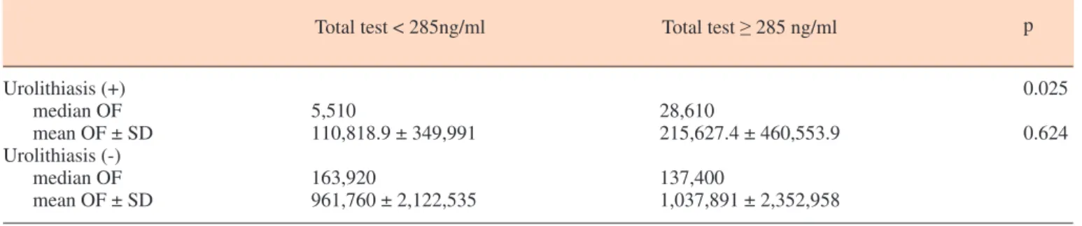
levels we categorized patients into 2 groups as the low testosterone (< 285 ng/ml) and normal testosterone (≥ 285 ng/ml) groups and compared O. formigenes counts.
This analysis showed a significant association between decreased O. formigenes colonization and low
testoster-one (table 3). Discussion
Urinary oxalate excretion has a crucial role in the formation of calcium oxalate stones. Even normal con-centrations of urinary oxalate have been shown to affect calcium oxalate stone formation in terms of recurrence and severity [7]. O. formigenes colonization has been
suggested to have an impact on calcium oxalate urolith-iasis since this bacterium uses oxalate as its main energy source. Batislam et al. [16] reported that O. formigenes
colonization is significantly lower in stone formers and subjects with significantly diminished colonization have higher rates for the coexistence of hyperoxaluria and hypercalciuria. Other studies corroborated these results, reporting reduced urinary oxalate concentrations in cal-cium oxalate stone forming patients colonized with O. formigenes in comparison to non-colonized stone
for-mers [17]. Our results concerning O. formigenes
coloni-zation are in accordance with previous findings. Urolith-iasis not only affects the urinary system, but also other systems including the skeletal system. Stone formers have been reported to have a lower BMD than non-stone formers [9–11]. To the best of our knowledge our study is the first one to investigate the association between bone mineral density and O. formigenes colonization. Due to
the lack of an established threshold value for O. formi-genes counts in the gut we used absolute quantification
to compare O. formigenes counts in both groups [8, 19].
We detected significantly higher O. formigenes counts in
control subjects in comparison to calcium oxalate stone formers. In addition, we observed decreased O. formi-genes colonization in patients with a lower BMD in the
lumbar region than in those with a normal BMD. How-ever we could not confirm these findings in the femo-ral neck region. In non-stone former males there was no significant difference in O. formigenes colonization
re-gardless of the osteoporotic status which indicated that
O. formigenes has little to no effect on BMD in non-stone
forming men. The marked decrease in BDM in the pa-tient group may be a consequence of the impaired cal-cium absorption due to the formation of non-absorbable calcium-oxalate complexes [20]. Such complexes are more likely to occur when reduced O. formigenes
con-centrations result in higher oxalate concon-centrations in the gut. In addition to urinary calcium oxalate excretion, BMD is also affected by testosterone levels in men [21, 22]. Therefore we measured serum testosterone concen-trations in patients and control subjects and determined significantly lower testosterone concentrations in litho-genic men. This finding implies that reduced BMD may be attributed to the decreased total testosterone levels in lithogenic men when other parameters do not show a significant difference including O. formigenes counts.
Interestingly, previous studies investigating the role of endogenous steroids in urolithiasis have produced con-tradictory results. Naghii et al. [23] reported significantly higher testosterone levels in stone forming men while 2 other studies conducted by Zhao et al. [24] and Watson et al. [25] showed no correlation between testosterone levels and urolithiasis. In contrast with these studies we observed an inverse correlation between calcium-ox-alate stone formation and testosterone levels. When
Table 3. Comparison of O. formigenes counts and total testosterone levels
Urolithiasis (+) median OF mean OF ± SD Urolithiasis (-) median OF mean OF ± SD 5,510 110,818.9 ± 349,991 163,920 961,760 ± 2,122,535 28,610 215,627.4 ± 460,553.9 137,400 1,037,891 ± 2,352,958
Total test < 285ng/ml Total test ≥ 285 ng/ml p
OF = Oxalobacter formigenes; SD = standard deviation.
0.025 0.624
we investigated the relation between the O. formigenes
concentration and low testosterone levels, we observed a marked decrease in the O. formigenes colonization in
patients with low testosterone levels. While it is possible for plasma oxalate concentrations to have an impact on the testosterone levels via an unknown mechanism, this discrepancy is probably a result of the limitations of this study. Limitations of this study include small sample size and the lack of dietary control. The effect of diet on ox-alate concentrations could have been compensated for by using a food questionnaire. In conclusion, based on our
findings we suggest that O. formigenes may play an
es-sential role in calcium oxalate metabolism, affecting the bone mineral density in oxalate stone formers.
Acknowledgements
The authors declare no conflict of interest. This proj-ect was supported by Istanbul University Research Fund (Project No: 24949). Finally, we would like to express our gratitude to Dr. John Knight for providing the Ox-alobacter formigenes strain and associated data.
References
Turney BW, Reynard JM, Noble JG, Ke-oghane SR: Trends in urological stone dis-ease. BJU Int 2012;109:1082–1087. Taylor EN, Stampfer MJ, Curhan GC: Obe-sity, weight gain, and the risk of kidney stones. JAMA 2005;293:455–462.
Wilson DM: Clinical and laboratory ap-proaches for evaluation of nephrolithiasis. J Urol 1989;141:770–774.
Batz M, Kohlbecker G: Significance of se-rum and urinary oxalate in calcium stone for-mers; in Rose GA, Robertson WG, Watts RW (eds): Oxalate in Human Biochemistry and Clinical Pathology. London, The Wellcome Foundation Ltd, 1979, pp181–185.
Yriberri J, Posen S: A semi-automatic en-zymic method for estimating urinary oxalate. Clin Chem 1980;26:881–884.
Jiang J, Knight J, Easter LH, Neiberg R, Holmes RP, Assimos DG: Impact of dietary calcium and oxalate, and Oxalobacter formi-genes colonization on urinary oxalate excre-tion. J Urol 2011;186:135–139.
Siener R, Bangen U, Sidhu H, Honow R, von Unruh G, Hesse A: The role of Oxalobacter formigenes colonization in calcium oxalate stone disease. Kidney Int 2013;83:1144– 1149.
Kaufman DW, Kelly JP, Curhan GC, Ander-son TE, Dretler SP, Preminger GM, Cave DR: Oxalobacter formigenes may reduce the risk of calcium oxalate kidney stones. J Am Soc Nephrol 2008;19:1197–1203.
Tugcu V, Ozbek E, Aras B, Ozbay B, Islim F, Tasci AI: Bone mineral density measurement in patients with recurrent normocalciuric cal-cium stone disease. Urol Res 2007;35:29–34.
Arrabal-Polo MA, Arrabal-Martin M, Arias-Santiago S: Bone and metabolic markers in women with recurrent calcium stones. Ko-rean J Urol 2013;54:177–182.
Arrabal-Polo MA, Arrabal-Martin M, de Haro-Munoz T, Lopez-Leon VM, Merino-Sa-las S, Ochoa-Hortal MA, Garrido-Gomez J, Lahoz-Garcia C, Zuluaga-Gomez A: Mineral density and bone remodelling markers in pa-tients with calcium lithiasis. BJU Int 2011; 108:1903–1908.
Sidhu H, Hoppe B, Hesse A, Tenbrock K, Brömme S, Rietschel E, Peck AB: Absence of Oxalobacter formigenes in cystic fibrosis patients: a risk factor for hyperoxaluria. Lan-cet 1998;352:1026–1029.
Kumar R, Ghoshal UC, Singh G, Mittal RD: Infrequency of colonization with Oxalobac-ter formigenes in inflammatory bowel dis-ease: possible role in renal stone formation. J Gastroenterol Hepatol 2004;19:1403–1409. Ye J, Coulouris G, Zaretskaya I, Cutcutache I, Rozen S, Madden TL: Primer-BLAST: a tool to design target-specific primers for polymerase chain reaction. BMC Bioinfor-matics 2012;13:134.
Orimo H, Hayashi Y, Fukunaga M, Sone T, Fujiwara S, Shiraki M, Kushida K, Miyamoto S, Soen S, Nishimura J, Oh-Hashi Y, Hosoi T, Gorai I, Tanaka H, Igai T, Kishimoto H: Diagnostic criteria for primary osteoporo-sis: year 2000 revision. J Bone Miner Metab 2001;19:331–337.
Batislam E, Yilmaz E, Yuvanc E, Kisa O, Kisa U: Quantitative analysis of colonization with real-time PCR to identify the role of Ox-alobacter formigenes in calcium oxalate uro-lithiasis. Urol Res 2012;40:455–460.
Troxel SA, Sidhu H, Kaul P, Low RK: Intes-tinal Oxalobacter formigenes colonization in calcium oxalate stone formers and its relation to urinary oxalate. J Endourol 2003;17:173– 176.
Prokopovich S, Knight J, Assimos DG, Hol-mes RP: Variability of Oxalobacter formi-genes and oxalate in stool samples. J Urol 2007;178:2186–2190.
Kwak C, Kim HK, Kim EC, Choi MS, Kim HH: Urinary oxalate levels and the enteric bacterium Oxalobacter formigenes in pa-tients with calcium oxalate urolithiasis. Eur Urol 2003;44:475–481.
Borghi L, Nouvenne A, Meschi T: Probiotics and dietary manipulations in calcium oxalate nephrolithiasis: two sides of the same coin? Kidney Int 2010;78:1063–1065.
Chin KY, Ima-Nirwana S: Sex steroids and bone health status in men. Int J Endocrinol 2012;2012:208719.
Gielen E, Vanderschueren D, Callewaert F, Boonen S: Osteoporosis in men. Best Pract Res Clin Endocrinol Metab 2011;25:321– 335.
Naghii MR, Babaei M, Hedayati M: Andro-gens involvement in the pathogenesis of renal stones formation. PloS One 2014;9:e93790. Zhao Z, Mai Z, Ou L, Duan X, Zeng G: Se-rum estradiol and testosterone levels in kid-ney stones disease with and without calcium oxalate components in naturally postmeno-pausal women. PLoS One 2013;8:e75513. Watson JM, Shrewsberry AB, Taghechian S, Goodman M, Pattaras JG, Ritenour CW, Ogan K: Serum testosterone may be associ-ated with calcium oxalate urolithogenesis. J Endourol 2010;24:1183–1187. 1 2 3 4 5 6 7 8 9 10 11 12 13 14 15 16 17 18 19 20 21 22 23 24 25