Effect of Tooth Whitening (Bleaching) Agent on Dentin
Microhardness
H. Miray UYAN
1Sema YILDIRIM
2Abstract
Objectives: The aim of this study is to investigate the effect of whitening agents on dentin micro hardness.
Materials and methods: 60-incisor maxillary (20 per group) tooth were applied root chanel treatment: one is control group and the others are applied to whitening products namely, whiteness super endo (Dentscare LTDA;37% Carbamide peroxide) and opalescence endo(Ultradent,USA;35% Hydrogen peroxide). Whitening agents were applied in every four days for 12 days. Hardness assessed by using Vickers test, after the end of treatment. Scanning electron microscopy (SEM) analyses performed after the end of bleaching treatment.
Results: The results show that the micro hardness decreased for both agents; where as, the micro hardness of whiteness superendo is less than opalescence endo.
Keywords: dentin micro hardness, intracoronal bleaching, whitening agent
Diş Beyazlatma Ürünlerinin Dentin Mikrosertliğine Etkileri Özet
Amaç: Bu çalışmanın amacı diş beyazlatıcı ürünlerin dentin mikrosertleşmeleri üzerine etkilerini incelemektir.
Materyel ve Yöntemler: 60-kesici maksiller (grup başına 20 adet) dişe kanal tedavisi uygulanmıştır. Bir tanesi kontrol olmak üzere, diğerlerine sırasıyla şu beyazlatıcı ürünler uygulanmıştır. whiteness super endo (Dentscare LTDA;37% Karbamid peroksit) ve opalescence endo (Ultradent,USA;35% Hidrojen peroksit). Beyazlatıcı ürünler 12 gün süreyle, her 4 günde bir kez uygulanmıştır. Tedavinin sonunda sertleşme Vickers testi ile değerlendirilmiştir. Beyazlatıcı tedavinin sonunda Taramalı elektron mikroskobu ile (TEM) değerlendirmeler yapılmıştır.
Sonuçlar: Sonuçlar, her iki ürünün de mikro sertleşmeleri azalttığını göstermekle birlikte; whiteness superendo’ nun mikro sertleşmeleri opalescence endoya göre daha azdır.
1. Introduction
The whiteness of the teeth is always an essential element of health and esthetics (1). Since the importance of esthetic considerations increasing nowadays, giving the natural appearance of the tooth to the patient by restoration has been one of the most important issues of modern dentistry (2).
Various factors can be the cause of coloration of the teeth. This situation occur direct discoloration of enamel dentin or reflection of discoloration of dentin from semi-transparent enamel (3). There are wide variety of chemical agent were used for many years to treat the discoloration of enamel dentin (4).
The bleaching agents, a more conservative method, has become a good alternative for restoration the natural color of stained teeth, instead of prosthetic applications, such as; crown and veneer. Since often usage of tooth whitening applications, there are many clinical studies has made in order to evaluate the efficacy, reliability and potential effect this method (5).
Whitening treatment is a method that aimed to reaction of the free oxygen, which emerge from whitening agents with colored molecules in order to return the natural color of the teeth(6). The most common materials used for bleaching; hydrogen peroxide, sodium perborate, and carbamide peroxide which is usually used for extra coronary bleaching where as; sodium perborate is also used for the intracoronary bleaching.
It is believed that the bleaching also causes increased brittleness of the coronal tooth structure because of desiccation of the dentin and enamel(7). However, this has not been proven conclusively because little is known about the effect of bleaching on the biomechanical properties of teeth. Since dentin constitutes a major part of the tooth structure, any change in the biomechanical properties of dentin after bleaching is likely to have an impact on the overall strength of the tooth (8).
Many studies have looked at the various biomechanical properties of dentin and some of more commonly studied properties include micro hardness and strength properties, such as tensile strength and shear strength(9). A number of recent studies have compared the biomechanical properties of dentin from vital teeth. The results of these studies indicated that endodontically treated teeth were not weaker than vital teeth. In contrast, the number of studies that examined the biomechanical properties of bleached dentin is very limited. Lewinstein et al. examined the effect of hydrogen peroxide and sodium perborate on the micro hardness of human dentin. Intact teeth were sectioned longitudinally and bleaching agents were applied to the polished dentin surfaces for up to 30 min. It was found that 30% hydrogen peroxide reduced the micro hardness of dentin after 5 min, but treatment with sodium perborate mixed with hydrogen peroxide did not alter the micro hardness of dentin at the of the observation period(10).
This study examined the effects of different bleaching agents on dentin and it is aimed to compare the results with each other.
2. Materials and Methods
Sixty intact human maxillary incisors extracted for orthodontic reasons were used. The teeth were placed in distilled water and the soft tissue attached to the root surface was removed with a scalpel. Endodontic access cavities were prepared using a diamond bur in a high-speed hand piece. The pulp tissue was removed ana a no.15 h type file was inserted into the root canal until the tip of the file was seen at the apical foramen. The working length was determined by subtract 1 mm from the length of the file. Cleaning and shaping was carried out using 1% sodium hypochlorite as the irrigant. The root canal was dried with paper points and filling with AH Plus and 15-40 gutha percha points. Cavit was packed into the root canal.
The teeth were randomly distributed into 3 groups, and applied different whitening agents, (Table1) Group 1, opalescense endo, 35% hydrogen peroxide -containing; Group 2, Whiteness super endo %37 carbamide peroxide-containing; Group 3, cotton pellet soaked with distilled water. Each tooth was stored in an individually labeled, capped plastic vial containing distilled water. The plastic vials were stored at 37 C for 12 days. After 12 days, the teeth were sectioned and dentine from the teeth was subjected to microhardness testing.
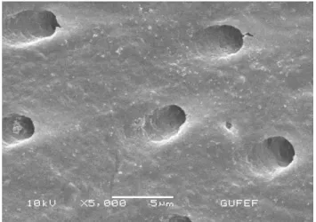
Table 1. Bleaching Agents
Product Name Opalescence Endo Whiteness Super Endo
Manufacturer Ultradent Products Dentscare LTDA
Manufacturer
Contact Info 505 West 10200 South, South Jordan, Utah 84095, USA Av.Edgar Nelson Meister,474,Districo Industrial 89219-501-Joinville-SC
Active Substance Hydrogen Peroxide Carbamide peroxide, glycol, deionized
water
Concentration (Approx) % 35 %37
The root of each was embedded in a block of acrylic resin, 2mm apical to the CEJ to facilitate subsequent sectioning and testing of the specimens. Each specimen was blotted dry and a vickers hardness test was performed using a digital variance was conducted on the data obtained. Data comparisons were conducted using Mann Whitney U tests. All statistical analyses were performed at the 0,05 level of significance.
During the experimental procedures, bleaching products were removed by suction and washed thoroughly with distilled, de-ionized water. After the bleaching procedures, samples were fixed with 2% glutaraldehyde, washed in distilled, de-ionized water, critical point dried and sputter coated with gold (120 s, 70 nm) for SEM analysis. Photomicrographs (3500-x magnification, 15 kV) were obtained from the samples and compared with group 3 by a single assessor, evaluating
3. Results
According to the statistical analysis, the microhardness of the control group was higher than both Opalescence Endo and Whiteness Super endo, where as; the maximum reduction in microhardness has noticed in the Whiteness Super Endo group.
The result, which is obtained by evaluation of dentin microhardness, is the proof of mineral loss or earnings on the hard tissue of tooth. There is a positive correlation between microhardness and mineral structure of teeth. Although reduction in the microhardness could decrease the fragility, when it comes to fragility of teeth there are many other factors, which should take in to consideration. Reduction in microhardness due to mineral loss in dentin, can cause weaken teeth and increase the tendency to tooth decay which is the major cause of increased the fragility of the teeth.
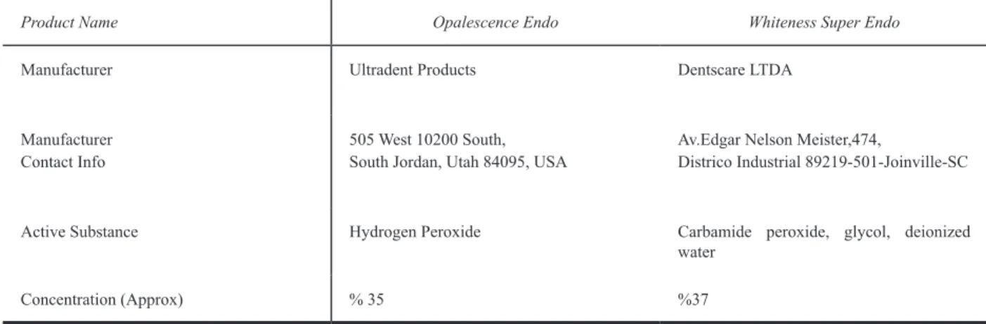
Figure 1. Control Group
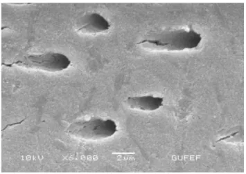
Figure 3. Whiteness Super endo Group
It is observed that, a deep erosion area formed on the dentine tubule, due to the effect of bleaching agents, which could be the main reason for mineral loss of dentin and also reduction of microhardness which cause increase of the fragility and tendency of tooth decay.
4. Discussion
Various studies have shown that hydrogen peroxide, especially in high concentration, affects the biomechanical properties of dentine even after exposure for a short period. Saleh & Ettman found that irrigation with 3% hydrogen peroxide and 5% sodium hypochlorite used alternatively and left for 60 s significantly reduced the microhardness of dentine(11).
Lewinstein et al. showed that the reduction in microhardness of dentine after treatment with 30% hydrogen peroxide was time-related. In their study, the specimens were assessed at 5, 15 and 30 min after application of hydrogen peroxide and significant, progressive reduction in microhardness was observed (12).
Hydrogen peroxide and Carbamide peroxide can cause a decrease in dentin microhardness during intracoronal bleaching. In this case, the degradation of morphological structure of the dentin and the deterioration of inorganic structure is an expected result.
Although the effect of whitening agents on the dentin are not fully understood, some studies reveal that, hydrogen peroxide cause the dissolution of the inorganic material of dentin, thus affect decrease in the calcium phosphor ratio, and loss of mineral on the organic matrix of dentin due to protein denaturalization. The hardness of dentin affected from the Ph of the whitening agents, buffer capacity of the dentin, and also the density difference of dentin tubules. Some researchers investigate the effect of whitening agents and conjunction use of these agents with materials that known to be effect on dentin microhardness.
Daniel pinto de Oliveira et al. studied on the differentiation of dentin microhardness when applied whitening agents alone and conjunction with chlorhexidine gel and find out that chlorhexidine jel has no effect on dentin microhardness. However, they find out that chlorhexidine gel can be used for an antimicrobial agent in cavity during intracoronal bleaching(13). Chng et al studied the effect of the different whitening agents with different concentration on the dentin microhardness, hydrogen peroxide, sodium perborate and carbamide peroxide with different concentration was used in this study, and results show that the highest rate of reduction on dentin microhardness observed with hydrogen peroxide 35 %, carbamide peroxide 35 %(14). Zalkind et al. had reported that there is a close relationship between the enamel dentin change and bleaching effect. Studies show that, whitening agent can change the mineral composition and also micro morphology of the enamel dentin thus reduction on dentin microhardness.
The linear relationship between the calcium and phosphor loss and reduction on dentin microhardness show that hardness measurements can be used as an indicator for the relationship between mineralization degree of enamel dentin and caries enamel. Mineral loss induced by bleaching agents, occurs under the enamel surface similar to initial caries lesions. This loss of mineral content is seen as an increase in range of enamel prisms, increase in surface roughness and increase of the adhesion of Streptococcus mutants. Hosaya et al reported that streptococcus mutants colonies increased on the whitened enamel dentin, after repeated bleaching sessions bacterial adhesion growth occurred, and the maximum number of bacterial colonies formed after five bleaching sessions followed by etching(15). Caries lesions affecting dentine enamel easily move toward tissues and can cause the deeper cavities. In addition to this, caries lesions formed on the whitened enamel tissue, due to extra coronal bleaching method, can also occur on the dentin tissue during intracoronal bleaching. Intracoronal bleaching application for the teeth, which were root canal treatment, progression of caries lesions is expected without causing pain. This situation would adversely affect restorations made after.
In order to achieve optimal results in the bleaching treatment, the structure and the concentration and also preventable side effect profile of whitening agent is also important. Although has no clinical symptoms, bleaching agents can cause a chemical and micro structural changes which affect the surface properties of teeth, the degree of mineralization, and probably the development of caries-like lesions should not be forgotten.
It is observed that different bleaching agents used in this study decrease the microhardness of dentin tissue. On the contrary, the hardness has been observed higher for none bleached teeth. Although decrease in the microhardness mainly reduces the fragility, in the case of teeth, other factors must be take in to consideration.
Teeth whitening procedure damage the tooth structure, which is irreversible. The fragility of the tooth, is related with the amount of water contained in dentin, mineralization, and change of the collagen and non-collagen proteins. Especially the bleaching agents, that contain peroxide, due to dissolution and decay in the structure of dentin collagen, that weakens the tooth structure, increase the tendency to caries, and thus increases the fragility.
REFERENCES
[1] Hasson H,Ismail AI,Neıva G.Home -based chemically induced whitening of teeth in adults The Cochrane Collaboration 2007.
[2] Goldstein RE,Garber DA.Complete Dental Bleaching Quintessence Pub Co,Chicago,1995. [3] Rotstein I,Friedman S.pH variation among materials used for ıntracoronal bleaching.J Endod 17,376,1991.
[4] Costas FL,Wong M,Intracoronal ısolatin barriers:Effect of location on root leakage and effective ness of bleaching agents.J Endod 17,8,1991.
[5] Cherry DV, Bowers DE,Thomas Jr L,Redmond AF.Acute toxicological effects of ingested tooth whitens in female rats.J Dent Res 1993,72:1298-1303
[6] Freccia WF,et al.An ın vıtro comparison of non vital bleaching techniques in the discolored tooth.J Endod 8,70,1982.
[7] Rotstein I,et al. Histochemical analysis of dental hard tissues following bleaching .J Endod 22,23,1996.
[8] Martin dunitz,greenwall linda.Bleaching technıques in restorative dentistry.2001.20-24 [9] Goldstein RE, Garber DA. Complete dental bleaching.Chıcago: quıntessence
Publishing;1995.165
[10] Lewinstein I, Hirschfeld Z, Stabholz A, Rotstein I. Effect of hidrogen peroxide and sodium perborate on the microhardness of human enamel and dentin. J Endod 20:61-3,1994. [11] Kwong K, Mohammed S, McMillan MD, Stokes AN. Evaluation of a 10 percent carbamide peroxide gel vital bleaching agent. N Z Dent J 1993;89:18-22.
[12] Lewinstein I, Hirschfeld Z, Stabholz A, Rotstein I. Effect of hidrogen peroxide and sodium perborate on the microhardness of human enamel and dentin. J Endod 20:61-3,1994. [13] In vitro assessment of a gel base containing 2% chlorhexidine as a sodium perborate ‘s vehicle for ıntracoronal bleaching of discolored teeth. 2006,32(7),672-674.
[14] Lım MY et all.An in vitro comparison of bleaching efficiacy
[15] Singleton LS; Wagner MJ. Peroxide tooth whitener concentration. Versus composite resin etching. J Dent Res, 71: .281-286, 1992.