ISSN 1996-0808 ©2012 Academic Journals
Full Length Research Paper
Active constituents of some Satureja L. species and
their biological activities
Tulin Askun*, Gulendam Tumen, Fatih Satıl and Didem Karaarslan
Department of Biology, Faculty of Science and Art, Balikesir University, 10145, Balıkesir, Turkey.
Accepted 20 May, 2011
Methanol extracts from the aerial parts of three Satureja L. spp. belonging to the family Lamiaceae were
studied. S. cilicica is endemic to Turkey; S. icarica and S. coerulea extend to West Anatolia; S. icarica
extends to the Turkish-Greek border and S. coerulea extends to the Turkish-Bulgarian border. S.
icarica, S. coerulea, and S. cilicica extracts had strong fungicidal effects at high concentrations and
fungistatic effects at lower concentrations. Plant methanol extracts were investigated for their
antibacterial, antifungal and antimycobacterial activity. To our knowledge, this is the first report of S.
cilicica mycobactericidal activity against M. tuberculosis. Phenolic constituents were detected using
high performance liquid chromatography (HPLC) analyses; the effective constituents of methanol
extracts of these plants are presented, and their activities and their ethnobotanical uses are discussed.
Key words: Antibacterial, antifungal, antimycobacterial, Lamiaceae, Satureja icarica, Satureja coerulea,
Satureja cilicica.
INTRODUCTION
Satureja L. is a genus of common aromatic plants
belonging to the family Lamiaceae. Satureja is
represented by 15 species in Turkey, which is an
important biodiversity hotspot for the Lamiaceae family
(Davis, 1982; Tumen et al., 2000). The endemism ratio of
this genus is 33% in Turkey; Satureja species grow
mainly in south and west Anatolia. Satureja species are
economically and medicinally important because of their
high content of essential oils. Previous studies on the
essential oils of Satureja species found in Turkey have
been reported (Tumen and Baser, 1996; Tumen et al.,
1997, Tumen et al., 1998; Baser, et al., 2000).
Members of the Satureja genus are called kekik in
Turkish, and some species are exported as the herb
thyme (Satil et al., 2008). Traditional Turkish folk
remedies use S. cuneifolia and S. thymbra, which are
collected from the wild, and S. hortensis, which is known
locally as “Cipriska” or “Koc Otu” (Sahin et al., 2003) and
is cultivated for use as a diuretic and digestive aid in
various regions of Turkey (Baytop, 1999; Sahin et al.,
*Corresponding author. E-mail: [email protected]. Tel: 0090 2666121278. Fax: 0090 2666121215.
2003). The species S. hortensis is also native to southern
Europe and has been naturalised to regions of North
America. In Europe, summer savoury (Satureja hortensis
L.) and winter savoury (Satureja montana L.) are the
most important Satureja species for cultivation. S.
hortensis has a sweeter and more delicate aroma and
fragrance than S. montana (Skocibusic and Bezic, 2004;
Bezic et al., 2005). Both summer and winter savoury are
used to flavour food (Bowles, 2004). In previous studies,
essential oils and extracts from S. hortensis
demon-strated a variety of useful properties: antibacterial activity;
antifungal activity, particularly against Aspergillus flavus
under in vitro conditions (Dikbas et al., 2008; Gulluce et
al., 2003; Sahin et al., 2003; Boyraz and Ozcan, 2006);
antioxidant activity; antispasmodic activity; and
anti-diarrheal as well as sedative properties (Deans and
Svoboda, 1989; Gulluce et al., 2003; Hajhashemi et al.,
2000; Madsen et al., 1996; Dorman and Hiltunen, 2004).
Carvacrol and γ-terpinene were identified as the primary
phenolic constituents of S. hortensis (Ryu et al., 2004). S.
montana L., showed effective antibacterial activity against
E. coli, methicillin-resistant Staphylococcus aureus and
Candida albicans (Skocibusic et al., 2006). De Oliveira et
al. (2011) reported that the antimicrobial effect of S.
4624 Afr. J. Microbiol. Res.
Table 1. Herbarium data of plants such as locality, altitude, collection time and herbarium number of species.
Genus species authority (Labiatae) Locality Altitude (m) Collection
time
Herbarium number
Satureja icarica P. H. Davis Gökçeada, Çanakkale 250 09/22/2002 FS1024
Satureja coerulea Janka in Velen Demirköy, Kırklareli 600 10/29/2001 FS1006
Satureja cilicica (endemic) P. H. Davis Andıran, Geben kasabası, Kahraman Maraş 1400 08/27/2000 FS1180
sausages and Zavatti et al. (2011) showed that S.
montana is a medicinal plant used to treat male sexual
dysfunctions in rats.
Ozkan et al. (2007) determined antioxidant activities of
Satureja cilicica essential oil in butter and in vitro. The
essential oil of S. cilicica exhibited a strong antioxidant
activity in butter. Carvacrol (59.2%) was the main
component in the oils of S. icarica. The oil of S. coerulea
contained caryophyllene (10.6%) and caryophyllene
oxide (8.0%) as main constituents. The main component
of S. cilicica are carvacrol, p-cymene,
-terpinene (Kirimer
et al. 1993). The main component is carvacrol
(52.04-55.97%) in S. icarica, caryophyllene (10.3-12.2%) in S.
coerulea (Tümen et al., 1998 a, c).
Satureja spp. are widely distributed across Turkey and
neighbouring regions: S. cilicica is endemic to Turkey; S.
icarica and S. coerulea extend to West Anatolia; S.
icarica extends to the Turkish-Greek border; and S.
coerulea extends to the Turkish-Bulgarian border (Davis,
1982). However, despite the ubiquity of these species,
the antibacterial, antifungal and antimycobacterial activity
of their methanol extracts has not been studied
previously. The goal of this study was to identify the
major phenolic constituents in methanolic extracts of
Satureja icarica P. H. Davis, Satureja coerulea Janka in
Velen and Satureja cilicica (endemic) P. H. Davis using
HPLC, and to determine the antibacterial, antifungal and
antimycobacterial activities of these constituents.
MATERIALS AND METHODS Plant materials
Aerial parts (herbs in the flowering stage) of plants were collected from different parts of Turkey between 2000 and 2002. The plants were identified by Assoc. Prof. Dr. F. Satil at Balıkesir University, Turkey. Voucher specimens were deposited in the herbarium of Department of Biology, Balikesir University. Herbarium plant data, such as locality, altitude, and collection time and identification number of species are given in Table 1.
Preparation of plant extracts
The plants [S. icarica (23 g), S. coerulea (25 g), and S. cilicica (29 g), (endemic)], were air-dried at room temperature. Extracts were prepared using 1 L of methanol (98%) at room temperature over a period of ten days according to the method of Seshadri (1962). The methanol extracts were filtered through filter paper concentrated
using a rotary evaporator and dried in vacuo at 40°C. The total yields from S. icarica, S. coerulea and S. cilicica were 1.10, 1.25 and 1.27 g, respectively. All stocks were stored at -20°C. To conduct antimicrobial activity tests, samples were dissolved in dimethyl sulphoxide (DMSO) and prepared at a concentration of 100 mg/ml. All samples were sterilised using syringe membrane filters.
High performance liquid chromatography (HPLC) conditions HPLC was performed using a Shimadzu HPLC device according to published techniques for the preparation of phenolic constituents (Caponio et al., 1999). A DAD (diode array detector) detector (Imax = 278) and SIL-10ADvp auto sampler were used for reverse-phase gradient system chromatography. An SCL-10Avp system controller, a LC-10ADvp pump and a DGU- 14A degasser were used. A CTO-10Avp oven was used with an Agilent Zorbax Eclipse XDB-C18 (250 × 4.60 mm) 5 µm column. The A and B mobile phase components were 3% acetic acid and methanol, respectively, and the flow rate was 0.8 ml/min. The column temperature was 30°C and the injection volume was 20 µL. Gallic acid, catechin, caffeic acid, epicatechin, p-coumaric acid, ferulic acid, vitexin, rutin, naringin, hesperidin, apigenin-7-glucoside, rosmarinic acid, eriodictyol, quercetin, naringenin, luteolin, apigenin, and carvacrol were used as chromatography standards.
Preparation of microorganisms and inocula
A total of seven microorganisms were used for antimicrobial activity studies: Staphylococcus aureus (ATCC 6538P), Klebsiella
pneumoniae (CCM 2318), Escherichia coli (ATCC 11230), Pseudomonas aeruginosa (ATCC 27853), Proteus vulgaris (ATCC
6897), Bacillus cereus (CCM 99) and Candida albicans (ATCC 10239). Methanol extracts from the three plants were examined against the following fungi: Aspergillus niger van Tiegh (TA 47-3),
Aspergillus flavus Link (TA 41-17), Aspergillus ochraceus K. Wilh.
(MUCL 39534), and Fusarium proliferatum (Matsushima) Nirenberg (TA 18-2). The fungi were subcultured on Czapek-Dox Agar (Oxoid CM 97), Malt Extract Agar (Oxoid CM 59), Sabouraud 2% Dextrose Agar (Merck) and Sabouraud 2% Dextrose Broth (Merck), respectively. Mycobacterium tuberculosis strain H37Ra from American Type Culture Collection (ATCC 25177) was used for the antimycobacterial bioassay.
Preparation of bacterial and fungal inocula
Day-old cultures of bacteria grown on nutrient agar (NA) plates were suspended in sterile saline solution until the turbidity was equal to a 0.5 McFarland standard of 106 colony forming units (CFU) per ml (Koneman et al., 1997). The plates were inoculated using the bacterial suspensions (15 μL per well) and were incubated overnight at 37°C. All tests were performed in triplicate.
For fungi, the isolates were subcultured on potato dextrose agar and incubated at 35°C for 7-14 days. Fungal suspensions were standardised at a spectrophotometric absorbance of 0.600 at 450 nm.
Antibacterial and antifungal activity tests
Stock solutions of all extracts were prepared in DMSO. The extracts were screened for antimicrobial activity using the agar diffusion technique. Sulphamethoxazole/trimethoprim (Oxoid) was used as a standard drug for bacteria (25 µg/disk) and amphotericin B (BioChemica) was used for fungi (30 µg/disk).
Petri dishes were inoculated with the bacterial/fungal suspensions (100 μL per dish). Filter paper disks (Whatman No. 1; 6 mm diameter) were soaked with 15 µL of each extract (100 and 50 mg/ml) and the disks were applied to the agar plate surfaces. The plates were incubated at 37°C overnight for bacteria and 27°C for two nights for fungi. Determination of the disc diffusion results for each extract was performed according to National Committee for Clinical Laboratory Standards Guidelines (NCCLS, 2006a).
Inhibition zone diameters were measured three times at different angles, and the means and standard deviations were calculated. Fungal colony inhibition (I%) was calculated as a percentage according to the following equation, where Dc is the diameter of the control zone (mm), and Ds is the diameter of the sample zone (mm).
I% = (Dc − Ds)/Dc*100
Minimal inhibitory concentrations (MICs) were determined using a modified microdilution method according to the National Committee of Clinical Laboratory Standard guidelines (NCCLS, 2006b) for bacteria and for fungi (NCCLS, 2008). Sterile 96-well microplates were used for the assay (0.2 ml volume, Fisher Scientific).
Samples were diluted to twice the desired initial test concentration using nutrient broth (NB) (Oxoid), and all microplate wells were filled with NB (100 μL). Each test sample (100 μL) was added to the first well, and serial two-fold dilutions were performed to obtain final concentrations within the range of 12.5–0.4 mg/ml. The lowest concentrations that were determined, after macroscopic evaluation, to inhibit the growth of the organisms tested were determined to be the MICs.
Preparation of Mycobacterium tuberculosis Inocula
Bacterial suspensions of M. tuberculosis were prepared either from Lowenstein–Jensen slants or from complete 7H9 broth cultures. To prepare an inoculum that was less than 15 days old from a culture grown on Lowenstein-Jensen medium, a suspension was prepared in Middlebrook 7H9 broth. The turbidity of the suspension was adjusted to a 1.0 McFarland standard. The suspension was vortexed for several minutes and was allowed to stand for 20 min for the initial settling of larger particles. The supernatant was transferred to an empty sterile tube and was allowed to stand for an additional 15 min. After being transferred to a new sterile tube, the suspension was adjusted to a 0.5 McFarland turbidity standard by visual comparison. One ml of the adjusted suspension was diluted in 4 ml of sterile saline solution.
To prepare M. tuberculosis inoculum using a BACTEC MGIT tube with positive growth, the positive tubes were used beginning from the day after the sample first became positive (day-1 positive), up to and including the fifth day (day-5 positive). The positive tubes that were older than five days were subcultured into fresh growth medium. Tubes that were day-1 and day-2 positive were used in the inoculation procedure for the susceptibility tests. The tubes that were between days 3 and 5 positive were diluted using 1 ml of the
positive broth and 4 ml of sterile saline solution; the 5 ml diluted suspension samples were used for the inoculation procedures.
Antimycobacterial activity tests
Antimycobacterial bioassays were performed using the microplate Alamar blue assay (MABA) (Collins and Franzblau, 1997). The methanol extracts were sterilised by filtration using 13 mm diameter (0.22 µm pore size) filters (Millipore, Bedford, MA).
An oleic acid, albumin, dextrose and catalase (OADC) mixed supplement (0.5 ml) was added to Middlebrook 7H9 broth. The broth mixture was vortexed and 1.75 μL was added to the first microplate well. The remaining wells were filled with Middlebrook 7H9 Broth (100 μL). Extract (25 μL) was added to the first well, and the final extract concentration in the first well was 12.5 mg/ml. After mixing by pipetting several times, two-fold dilutions were performed from the first well to the next well (100 μL), excluding the positive and negative control wells. Final extract concentrations in the wells were between 12.5 mg/ml and 0.024 mg/ml. Streptomycin (STR), ethambutol (EMB), and isoniazid (INH) were used as standard drugs. Concentrations of STR, EMB and INH in the two-fold dilution series ranged from 41.5 to 0.040 µg/ml, 83 to 0.16 µg/ml, and 2.07 to 0.004 µg/ml, respectively.
Determination of minimal inhibitory concentrations (MICs) for mycobacterium tuberculosis
Microplates were inoculated with the bacterial suspension (20 μL per well except for the negative control wells) and incubated at 37 °C for 6 days. Alamar blue (15 μL, Trek Diagnostic system) was then added to the bacterial growth control wells (without extract) and the microplates were incubated at 37°C for an additional 24 h. If the dye turned from blue to pink (indicating positive bacterial growth); then Alamar blue solution was added to the other wells to determine the MIC values. All tests were performed in triplicate.
Determination of mycobactericidal activity
The plant extracts described above were used in mycobactericidal activity tests. Two-fold dilution series in triplicate sets of parallel microplate wells were used for each extract. To determine the minimum bactericide concentrations (MBCs), fresh Middlebrook 7H9 culture broth (185 μL) was transferred to each well, and 15 μL of an mycobacterial suspension, from MIC concentration and higher concentration wells obtained from the MIC test described above was added to each well, in order to determine the minimum bactericide concentration (MBC).
Two microplate wells were used as positive and negative controls, and each test was repeated in triplicate. For the negative controls, 200 ml of fresh broth (Middlebrook 7H9 culture medium and OADC) was used. For positive controls, including 185 μL and inoculums from former positive control wells (15 μL) was used. After 24 h of incubation and colour development using the Alamar blue solution, MBCs were recorded as the minimum extract concentration that did not cause any colour change in cultures when reincubated in fresh medium.
RESULTS
Standard compounds for HPLC chromatograms were
gallic acid, catechin, caffeic acid, epicatechin, p-coumaric
acid, ferulic acid, vitexin, rutin, naringin, hesperidin,
apigenin- 7 -glucoside, rosmarinic acid, eriodictiol,
4626 Afr. J. Microbiol. Res.
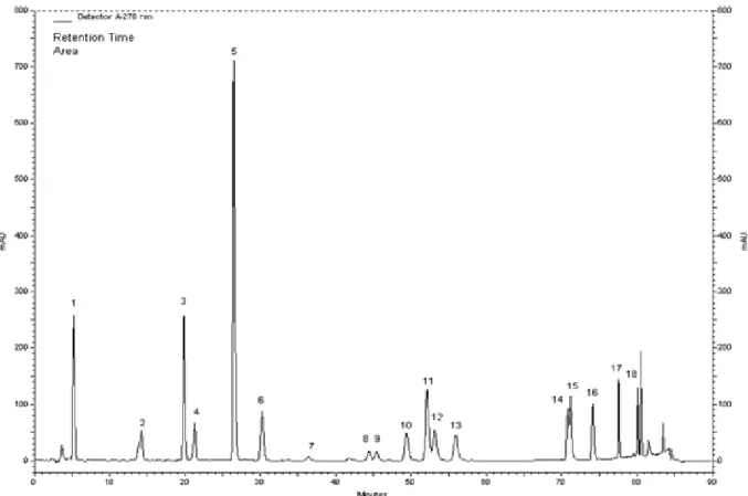
Figure 1. HPLC chromatogram for the standard compounds: (1) gallic acid, (2) catechin, (3) caffeic acid, (4) epicatechin, (5) p-coumaric acid, (6) ferulic acid, (7) vitexin, (8) rutin, (9) naringin, (10) hesperidin, (11) apigenin-7-glucoside, (12) rosmarinic acid, (13) eriodictyol, (14) quercetin, (15) naringenin, (16) luteolin, and (17) apigenin, (18) karvakrol.
quercetin, naringenin, luteolin, apigenin, and carvacrol.
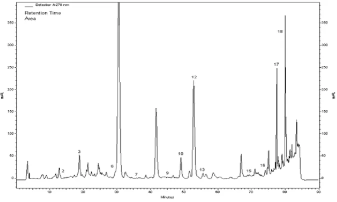
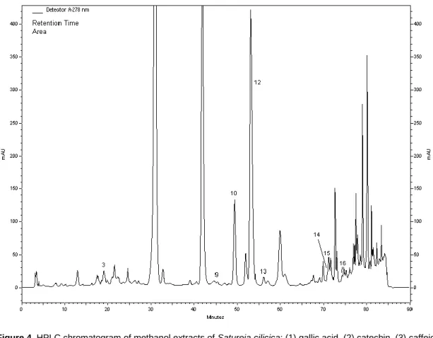
The major phenolic constituents determined by HPLC
analyses of the methanol extracts were carvacrol,
hesperidin and apigenin for S. icarica; rosmarinic acid,
carvacrol and caffeic acid for S. coerulea; and rosmarinic
acid, hesperidin and quercetin for S. cilicica (Figures 1, 2,
3 and 4; Table 2).
Inhibition of bacteria and fungi by methanol extracts of
Satureja spices was tested by measuring the sizes of
inhibition zones (mm). The microbial colony inhibition
efficacy of the extracts was different in some cases,
depending on which inhibition zone diameters were larger
than the standard drug inhibition zones for bacteria and
fungi (Table 3).
The extracts were tested for antibacterial activity
against P. vulgaris, K. pneumoniae, B. cereus, P.
aeruginosa, E. coli, and S. aureus and antiyeast activity
against C. albicans (Table 4). M. tuberculosis was used
for the antimycobacterial activity test (Table 5). Bacteria
were tested for susceptibility
to the reference drug
Sulphamethoxazole/Trimethoprim (Oxoid). STR, EMB
and INH (BD) were used as standard drugs for M.
tuberculosis.
The S. icarica methanol extract was the most effective
against E. coli (MIC of 0.8 mg/ml). Other bacteria (B.
cereus, P. vulgaris, P. aeruginosa, K. pneumoniae, S.
aureus) and yeast (C. albicans) showed inhibition
between MICs of 1.6 to 6.3 mg/ml. The S. icarica extract
exhibited bacteriostatic activity (MIC > 12.5 mg/ml)
against other bacteria.
The S. coerulea methanol extract was the most
effective against B. cereus (MIC of 6.3 mg/ml). The
efficacy of inhibition against other bacteria (P. vulgaris, P.
aeruginosa, K. pneumonia, S. aureus, E. coli) and yeast
(C. albicans) was moderate (MIC of 12.5 mg/ml). S.
coerulea extract showed bacteriostatic/fungistatic activity
at the same concentration (MBC/MFC of 12.5 mg/ml)
against S. aureus, P. vulgaris and C. albicans.
The methanol extract from S. cilicica was shown to be
most effective against P. vulgaris and S. aureus (MIC of
1.6 mg/ml). MIC values for other bacteria varied between
3.1 and 6.3 mg/ml. S. cilicica extract showed bactericidal
activity (MBC of 3.1 mg/ml) against S. aureus and
bacteriostatic activity (MIC > 12.5 mg/ml) against other
bacteria.
The filamentous fungi showed various sensitivities to
the extracts that were tested. Strong activity was
recorded for methanol extracts of S. icarica, S. coerulea,
and S. cilicica, which completely inhibited fungi in a MIC
range of 6.3 to 12.5 mg/ml. The fungus F. proliferatum
Figure 2. HPLC chromatogram of methanol extracts of Satureja icerica: (1) gallic acid, (2) catechin, (3) caffeic acid, (4) epicatechin, (5) p-coumaric acid, (6) ferulic acid, (7) vitexin, (8) rutin, (9) naringin, (10) hesperidin, (11) apigenin-7-glucoside, (12) rosmarinic acid, (13) eriodictyol, (14) quercetin, (15) naringenin, (16) luteolin, and (17) apigenin and (18) karvakrol.
Figure 3. HPLC chromatogram of methanol extracts of Satureja coerulea: (1) gallic acid, (2) catechin, (3) caffeic acid, (4) epicatechin, (5) p-coumaric acid, (6) ferulic acid, (7) vitexin, (8) rutin, (9) naringin, (10) hesperidin, (11) apigenin-7-glucoside, (12) rosmarinic acid, (13) eriodictyol, (14) quercetin, (15) naringenin, (16) luteolin, and (17) apigenin and (18) karvakrol.
4628 Afr. J. Microbiol. Res.
Figure 4. HPLC chromatogram of methanol extracts of Satureja cilicica: (1) gallic acid, (2) catechin, (3) caffeic acid, (4) epicatechin, (5) p-coumaric acid, (6) ferulic acid, (7) vitexin, (8) rutin, (9) naringin, (10) hesperidin, (11) apigenin-7-glucoside, (12) rosmarinic acid, (13) eriodictyol, (14) quercetin, (15) naringenin, (16) luteolin, and (17) apigenin and (18) karvakrol.
Table 2. Chemical concentrations in the methanol extracts of Satureja species.
Compound Satureja icarica Satureja coerulea Satureja cilicica
gallic acid n.a n.a n.a
catechin 322.7 298.3 n.a
caffeic acid 622.2 505.1 214.2
epicatechin n.a n.a n.a
p-coumaric acid n.a n.a n.a
ferulic acid 81.9 138.8 n.a
vitexin 95.2 134.0 n.a
rutin n.a n.a n.a
naringin 127.8 n.a 239.1
hesperidin 2318.1 74.0 5663.2
apigenin-7-glucoside n.a n.a n.a
rosmarinic acid 9716.0 13662 20358
eriodictyol 231.8 13.0 275.9
quercetin n.a n.a 179.3
naringenin 151.7 56.6 394.8
luteolin 165.3 117.0 241.5
apigenin 2256.0 n.a 973.5
carvacrol 13786.0 1219 n.a
Table 3. Disk susceptibility testing for bacteria and fungi.
Microorganism Statistics
Mean zone of inhibition (mm)
Satureja icarica Satureja coerulea Satureja cilicica
Concentration (mg/disc)
1.5 0.75 Sd drug 1.5 0.75 Sd drug 1.5 0.75 Sd drug
P. vulgaris Mean± SD 15.5±0.57 8±0 36±3.46 6±0 6±0 38.75±2.21 12.25±1.5 9.5±1 10.87±4.5 R% 56.94 77.77 84.51 84.51 -12.64 12.64 K. pneumonia Mean± SD 13±0.81 8.5±0.57 30±0 6±0 6±0 30±0 12.75±1.25 8±0 30±0 R% 56.66 71.66 80 80 57.5 73.33 B. cereus Mean± SD 10±0 9.5±0.57 6±0 7.5±0.57 6±0 6±0 16.25±3.30 15±1.41 6±0 R% -66.66 -58.33 -25 0 -170.83 -150 P. aeruginosa Mean± SD 14.5±0.57 6±0 6±0 6±0 6±0 6±0 13±1.41 10.5±0.57 6±0 R% -141.66 0 0 0 -116.66 -75 E. coli Mean± SD 6±0 6±0 30±0 6±0 6±0 30±0 6±0 6±0 30±0 R% 80 80 80 80 80 80 S. aureus Mean± SD 15.25±0.5 8±0 33±0 6±0 6±0 33±0 16.25±0.5 11.25±0.5 33±0 R% 53.78 75.75 81.81 81.81 50.75 65.90 C. albicans Mean± SD 6±0 6±0 6±0 6±0 6±0 6±0 22±0 11±0 6±0 R% 0 0 0 0 -266.66 -83.33 A. ochraceus Mean± SD 6±0 6±0 6±0 6±0 6±0 6±0 7.5±0.57 6±0 7±0.81 R% 0 0 0 0 0 0 -7.14 14.28 A. niger Mean± SD 6±0 6±0 13.75±2.98 9±0.81 6±0 13±1.41 6±0 6±0 10.75±0.95 R% 56.36 56.36 30.76 53.84615 44.18 44.18 A. flavus Mean± SD 37.5±6.45 32.5±2.08 6.75±0.5 6±0 6±0 6±0 6±0 6±0 7.25±0.95 R% -455.55 -381.48 0 0 0 20.83 20.83 F. proliferatum Mean± SD 6±0 6±0 8.5±2.38 6±0 6±0 6±0 12.75±2.21 8±0.81 7±0.81 R% 29.41 29.41 0 0 -82.14 -14.28
4630 Afr. J. Microbiol. Res.
Table 4. Antibacterial and antifungal activities of methanol extracts from the plants. Results are shown as MICs and MBCs/MFCs.
Microorganism Satureja icerica Satureja coerulea Satureja cilicica
A B A B A B P. vulgaris 1.6 >12.5 12.5 12.5 1.6 >12.5 K. pneumonia 3.1 >12.5 12.5 >12.5 6.3 >12.5 B. cereus 1.6 >12.5 6.3 >12.5 3.1 >12.5 P. aeruginosa 1.6 >12.5 12.5 >12.5 6.3 >12.5 E. coli 0.8 >12.5 12.5 >12.5 3.1 >12.5 S. aureus 6.3 >12.5 12.5 12.5 1.6 3.1 C. albicans 1.6 >12.5 12.5 12.5 6.3 >12.5 A. ochraceus 6.3 >12.5 6.3 12.5 12.5 >12.5 A. niger 6.3 >12.5 12.5 12.5 6.3 >12.5 A. flavus 12.5 >12.5 12.5 >12.5 6.3 >12.5 F. proliferatum 6.3 12.5 6.3 12.5 6.3 12.5 A: MIC (mg/ml); B: MBC (mg/ml).
Table 5. Antimycobacterial activity results for the methanol extracts against M. tuberculosis H37Ra (ATCC 25177) determined by MABA.
Plant S. icarica S. coerulea S. cilicica Standard drug
STR EMB INH
M. tuberculosis A B A B A B A B A B A B
n.a. n.a. n.a. n.a. 0.8 0.8 0.16 0.16 1.2 1.29 0.01 0.016 A: MIC (mg/ml); B: MBC (mg/ml).
was the most sensitive to inhibition by the three plant
extracts; the extracts also exhibited fungicidal activity
against F. proliferatum (MFC of 12.5 mg/ml). S. icarica
and S. coerulea extracts showed fungistatic activity
against the other fungi that were tested (MICs of 6.3–12.5
mg/ml). S. coerulea extracts also showed fungicidal
activity against A. ochraceus and A. niger (MIC of 12.5
mg/ml) and fungistatic activity against A. flavus.
According to the MABA test results for the activity of
the three plant extracts against M. tuberculosis, S. cilicica
extracts showed the highest activity (MIC and MBC of 0.8
mg/ml). The remaining extracts, S. icarica and S.
coerulea, showed higher MBC values (1.6 mg/ml) than
those of S. cilicica.
DISCUSSION
This study demonstrated that the addition of crude
methanol extracts of S. icarica, S. coerulea, and S.
cilicica to growth medium inhibited the growth of the
mycotoxigenic filamentous fungi, F. proliferatum, A.
flavus, A. ochraceus and A. niger. This property may
provide a safe and effective method of protecting food
from mycotoxigenic fungi. The results of this study
showed that the activities of methanol extracts were
concentration dependent: S. icarica, S. coerulea, and S.
cilicica extracts had strong fungicidal effects at high
concentrations
and
fungistatic
effects
at
lower
concentrations.
According to the results presented here, S. icarica had
fungicidal activity against F. proliferatum and fungistatic
activity against the other fungi and bacteria that were
tested. S. coerulea also showed fungicidal activity against
A. ochraceus and A. niger and showed bactericidal
activity against P. vulgaris, S. aureus and C. albicans.
Our previous research on the effects of plant-derived
methanol extracts (including T. spicata and O.
minutiflorum) against fungi (Askun et al., 2008) and
bacteria (Askun et al., 2009) formed the basis for
developing research on antimicrobial activity. S. cilicica
extract showed bactericidal activity against S. aureus and
fungicidal activity against F. proliferatum. S. cilicica
extract also showed fungistatic and bacteriostatic activity
against the other fungi and bacteria tested.
Methanol extracts of Satureja spp. were evaluated for
their in vitro antimycobacterial activity against M.
tuberculosis using the Alamar blue susceptibility test. The
methanol extract of S. cilicica showed antimycobacterial
activity at a concentration of 0.8 mg/ml. Although S.
icarica extract showed the same MIC value (0.8 mg/ml)
Similarly, the MIC value for S. corulea extract was 0.4
mg/ml, whereas the MBC was 1.6 mg/ml. However, all
plant methanol extracts killed M. tuberculosis. MIC/MBC
values for the standard drugs were 0.16/0.16 µg/ml,
1.29/1.29 µg/ml and 0.016/0.016 µg/ml for STR, EMB and
INH, respectively. To our knowledge, this study is the first
to report the mycobactericidal activity of S. cilicica against
M. tuberculosis.
The use of MABA has several advantages when
compared with other methods, such as the ability to use
small quantities of extract in 200 μL wells (Molina-Salinas
et al., 2006) and a shorter time requirement. The use of
Middlebrook 7H9 Broth and OADC supplement in
multi-well plates reduces the cultivation time from 3-4 weeks
(using solid medium such as Lowenstein-Jensen) to 7–10
days (Sethi et al., 2004). A reduction in cultivation time is
beneficial when conducting research, especially when
working with rare species, as the impact on natural
environments and the potential for over-collecting species
is minimised.
Plant methanol extracts contain many chemicals such
as alkaloids, amino acids, flavonoids, glycosides,
phytosterols, saponins, steroids, tannins and triterpenoids
(Kumar et al., 2009). Methanol extracts may therefore
yield a spectrum of antibacterial components that is
different from those previously described. In a review of
literature on the antimicrobial activity of different plant
extracts, Parekh et al. (2005) noted that methanol
extracts were more active than aqueous extracts.
The HPLC results demonstrated that none of the
Saturea
species
extracts
contained
gallic
acid,
epicatechin, p-coumaric acid, rutin, or
apigenin-7-glucoside. Compared to the other species, the major
phenolic constituents in S. icarica were carvacrol,
hesperidin and apigenin. While, the concentration of
carvacrol in S. icarica was 11.3 times higher than that S.
coerulea; the concentration of apigenin was 2.3 times
higher than that S. cilicica. Although, caffeic acid and
catechin were found in the extract of S. icarica but there
was no quercetin present.
Although rosmarinic acid, carvacrol and caffeic acid
were the major phenolic constituents of S. coerulea, the
amounts of ferulic acid and vitexin were higher than in the
other two species. In contrast, naringin, quercetin, and
apigenin were not detected.
Rosmarinic acid, hesperidin and quercetin were the
major phenolic constituents in the S. cilicica methanol
extract. Although the amounts of naringin, hesperidin,
rosmarinic acid, eriodictyol, naringenin, and luteolin were
high relative to the other two species, the S. cilicica
extract did not contain catechin, ferulic acid, vitexin, or
carvacrol.
Extracts of natural products are a common starting
point in the search for new antimycobacterial agents. As
shown previously, rosmarinic acid might be responsible
for the antimycobacterial activity observed in S. cilicica
(Askun et al., 2009). The results of this study support the
findings of our previous research. In addition, Mandalari
et al. (2007) reported that paired combinations of
eriodictyol, naringenin and hesperidin showed both
synergistic and antagonistic interactions that were
dependent on the test indicator organism and their cell
wall structure. In addition to these interactions, the results
presented here showed synergistic interactions, which
supports the suggestion that rosmarinic acid and
hesperidin may be the main bioactive antimicrobial
constituents present in S. cilicica extract.
Rosmarinic acid is a natural phenolic compound with
two phenolic rings (Petersen and Simmonds, 2003).
Hesperidin is produced in high concentrations by
members of the Rutaceae and Lamiaceae famillies, and it
has been shown that dietary hesperidin deficiency has
been linked to abnormal capillary leakiness (Martínez et
al., 2011). Hesperidin is also well known for having
antioxidant, analgesic and anticarcinogenic properties
(Hirata et al., 2005) and sedative effects (Fernández et
al., 2005). Naringin is present in grapefruit and citrus, and
the antioxidant (Haenen et al., 1997), antimicrobial (Han
and You, 1988) and anticancer (So et al., 1996)
properties of naringin have been studied. Naringenin also
occurs in citrus fruits. Renugadevi and Prabu (2009)
showed that the nephroprotective potential of naringenin
against Cd toxicity might be due to its antioxidant and
metal chelating properties. In addition, naringenin has
antimutagenic (Choi et al., 1994) and anticancer (So et
al., 1997) properties. Rosmarinic acid and chlorogenic
acid are caffeic acid esters that are common in
Lamiaceae plants (Petersen et al., 2009). The
biosynthesis of rosmarinic acid starts with the amino
acids L-phenylalanine and L-tyrosine. Rosmarinic acid
occurs in plants that are thought to have health benefits,
and it has antiviral, antibacterial, anti-inflammatory and
antioxidant properties. In plants, the role of rosmarinic
acid is thought to be related to defence (Petersen et al.,
2003). Carvacrol activity results from the interaction of
the carvacrol hydroxyl group with the cytoplasmic
membrane, which changes the membrane permeability
with respect to protons and potassium ions (Ultee et al.,
2002). Carvacrol has many biological properties, such as
antibacterial (Lambert et al., 2001) and antifungal
(Manohar et al., 2001) activity.
Arunasree (2010) identified the mechanism by which
carvacrol induces cell death in human metastatic breast
cancer cells and demonstrated that carvacrol induces
apoptosis in breast cancer (MDA-MB 231) cells.
Furthermore, studies of bio-based food packaging have
emphasised a need for the involving of natural active
agents (for example, antioxidants or antimicrobial
constituents). Recently, the antimicrobial packaging
industry has shown a great interest in natural active
agents for inhibiting or delaying the growth of pathogenic
bacteria and fungi on foods (Padgett et al., 1998; Dorman
and Deans, 2000).
4632 Afr. J. Microbiol. Res.
accounts of the effects of plant extracts on
M.tuberculosis (Adeniy et al., 2004; Rojas et al., 2006;
Askun et al., 2009).
These results provide a basis for the selection of
candidate plant species for further phytochemical and
pharmacological
investigation.
From
a
practical
perspective, S. cilicica extract may be a suitable
candidate
for
the
development
of
plant-based
pharmaceutical products for use against tuberculosis.
After determining that some of the methanol extracts
had high antibacterial, antifungal and antimycobacterial
activities, further research should involve the isolation
and purification of the effective constituents from the
extracts.
ACKNOWLEDGEMENTS
The authors are grateful to the Scientific and
Technological Research Council of Turkey (TUBITAK).
This research was supported by a grant from TUBITAK
and TBAG (Research grant no.104T336).
REFERENCES
Adeniy BA, Groves MJ, Gangadharam PRJ (2004). In vitro
anti-Mycobacterial activities of three species of Cola plant extracts
(Sterculiaceae). Phytother. Res., 18(5): 414 - 418.
Arunasree KM (2010). Anti-proliferative effects of carvacrol on a human metastatic breast cancer cell line, MDA-MB 231. Phytomedicine, 17: 581-588.
Askun T, Tumen G, Satil F, Ates M (2008). Characterization of the phenolic composition of five plant methanol extracts and their antimicrobial activities. Pharm. Biol., 46(10–11): 688–694.
Askun T, Tumen G, Satil, F, Ates M (2009). In vitro activity of methanol extracts of plants used as spices against Mycobacterium tuberculosis and other bacteria. Food Chem., 116 (1): 289-294.
Baser KHC, Tumen G, Satil F, Kirimer N (2000). Comparative morphological and chemical studies on Satureja L. species from West Anatolia. In: Ozhatay N (ed), Proceeding of the Second Balkan Botanical Congress (SBBC), 14-18: 129-132.
Baytop T (1999). Therapy with medicinal plants in Turkey: Past and Present. Publications of the İstanbul University: No:3255 (First edition), p. 332.
Bezic N, Skocibusic M, Dunkic V (2005). Phytochemical composition and antimicrobial activity of Satureja montana L. and Satureja
cuneifolia Ten. essential oils. Acta. Bot. Croat., 64(2): 13–322.
Bowles EJ (2004). The chemistry of aromatherapeutic oils. 3rd ed. Allen & Unwin Academic. Crows Nest, NSW, Australia, pp. 21-38.
Boyraz N, Özcan M (2006). Inhibition of phytopathogenic fungi by essential oil, hydrosol, ground material and extract of summer savory (Satureja hortensis L.) growing wild in Turkey. Int. J. Food Microbiol., 107(3): 238-242.
Caponio F, Alloggio V, Gomes T (1999). Phenolic compounds of virgin olive oil: influence of paste preparation techniques. Food Chem., 64(2): 203-209.
Choi JS, Park KY, Moon SH, Rhee SH, Young HS (1994). Antimutagenic effect of plant flavonoids in the Salmonella assay system. Arch. Pharm. Res., 17: 71-75.
Collins LA, Franzblau SG (1997). Microplate alamar blue verses BACTEC460 system for high-throughput screening of compounds against Mycobacterium tuberculosis and Mycobacterium avium. Antimicrob. Agents Chemother., 41: 1004-1009.
Davis PH (1982). Flora of Turkey and The East Aegean Islands. Edinburgh University Press: Edinburgh, 7: 314-323.
De Oliveira TL, de Araújo Soares R, Ramos EM, das Graças Cardoso M, Alves E, Piccoli RH (2011). Antimicrobial activity of Satureja
montana L. essential oil against Clostridium perfringens type A
inoculated in mortadella-type sausages formulated with different levels of sodium nitrite. Int. J. Food Microbiol., 144: 546-555. Deans SG, Svoboda KP (1989). Antibacterial activity of summer savory
(Satureja hortensis L.) essential oil. J. Hort. Sci., 64(2): 205–210. Dikbas N, Kotan R, Dadasoglu F, Sahin F (2008). Control of Aspergillus
flavus with essential oil and methanol extract of Satureja hortensis. Int. J. Food Microbiol., 124(2): 179–182.
Dorman HJD, Deans SG (2000). Antimicrobial agents from plants: antibacterial activity of plant volatile oils. J. Appl. Microbiol., 88: 308-316.
Dorman HJD, Hiltunen R (2004). Fe (III) reductive and free radical-scavenging properties of summer savory (Satureja hortensis L.) extract and subfractions. Food Chem., 88(2): 193–199.
Fernández SP, Wasowski C, Paladini AC, Marder M (2005). Synergistic interaction between hesperidin, a natural flavonoid and diazepam. Eur. J. Pharmacol., 512: 189-198.
Gulluce M, Sokmen M, Daferera D, Agar G, Ozkan H, Kartal N, Polissiou M, Sokmen A, Sahin F (2003). In vitro antibacterial, antifungal and antioxidant activities of the essential oil and methanol extracts of herbal parts and callus cultures of Satureja hortensis L. J.
Agric. Food. Chem., 51(14): 3958-3965.
Haenen GRMM, Paquay JBG, Korthouwer REM, Bast A (1997). Peroxynitrite scavenging by flavonoids. Biochem. Bioph. Res. Co., 236: 591-593.
Hajhashemi V, Sadraei H, Ghannadi AR, Mohseni M (2000). Antispasmodic and antidiarrheal effect of Satureja hortensis L. essential oil. J. Ethnopharmacol., 71(1–2): 187–192.
Han SS, You IJ (1988). Studies on antimicrobial activities and safety of natural naringin in Korea. Korean J. Mycol., 16: 33-40.
Hirata A, Murakami Y, Shoji M, Kadoma Y, Fujisawa S (2005). Kinetics of radical-scavenging activity of hesperetin and hesperidin and their inhibitory activity on COX-2 expression. Anticancer Res., 25: 3367-3374.
Kirimer N, Tümen G, Özek T, Baser KHC (1993). The Essential Oil of
Satureja cilicica P.H. Davis. J. Essent. Oil Res., 5: 547-548.
Koneman E, Allen SD, Janda M, Shreckenberger PC, Winn WC (1997). Guidelines for the collection, transport, processing, analysis and reporting of cultures from specific specimen sources. In: Colour Atlas and Textbook of Diagnostic Microbiology. Philadelphia 5. Ed. J.B Lippincott Co., pp. 121-170.
Kumar A, Ilavarasan R, Jayachandran T, Decaraman M, Aravindhan P, Padmanabhan N, Krishnan MRV (2009). Phytochemicals investigation on a tropical plant, Syzygium cumini from Kattuppalayam, Erode District, Tamil Nadu, South India, Pak. J. Nutr., 8(1): 83-85.
Lambert R, Skandamis PN, Coote PJ, Nychas GJE (2001). A study of the minimum inhibitory concentration and mode of action of oregano essential oil, thymol and carvacrol. J. Appl. Microbiol., 91: 453–462. Madsen HL, Andersen L, Christiansen L, Brockhoff P, Bertelsen G
(1996). Antioxidative activity of summer savory (Satureja hortensis L.) and rosemary (Rosmarinus officinalis L.) in minced, cooked pork meat. Food Res. Technol., 203(4): 333–338.
Mandalari G, Bennett RN, Bisignano G, Trombetta D, Saija A, Faulds CB, Gasson MJ, Narbad A (2007). Antimicrobial activity of flavonoids extracted from bergamot (Citrus bergamia Risso) peel, a byproduct of the essential oil industry. J. Appl. Microbiol., 103(6): 2056-2064. Manohar V, Ingram C, Gray J, Talpur NA, Echard BW, Bagchi D,
Preuss HG (2001). Antifungal activities of Origanum oil against
Candida albicans. Mol. Cell. Biochem., 228: 111-117.
Martínez AL, González-Trujano ME, Chávez M, Pellicer F, Moreno J, López-Muñoz FJ (2011). Hesperidin produces antinociceptive response and synergistic interaction with ketorolac in an arthritic gout-type pain in rats. Pharmacol. Biochem. Be., 97: 683-689. Molina-Salinas GM, Ramos-Guerra MC, Vargas-Villarreal J,
Mata-Cárdenas BD, Becerril-Montes P, Said-Fernández S (2006). Bactericidal Activity of Organic Extracts from Flourensia cernua DC against Strains of Mycobacterium tuberculosis. Arch. Med. Res., 37(1): 45-49.
(2006a). National committee for clinical laboratory standards.performance standards for antimicrobial disk susceptibility tests; approved standard. Seventh Edition. NCCLS document M2-A9, Wayne, Pennsylvania, 26(1): 21-28.
National Committee for Clinical Laboratory Standards (NCCLS) (2006b). National Committee for Clinical Laboratory Standards. Methods for dilution antimicrobial susceptibility tests for bacteria that grow edition aerobically; Approved Standard. Seventh Edition NCCLS Document M7-A7, Wayne, Pennsylvania, 26(2): 1-16. National Committee for Clinical Laboratory Standards (NCCLS) (2008).
Reference methods for broth dilution antifungal susceptibility testing of filamentous fungi. Approved Standard. Second Edition NCCLS document M38-A2. Wayne, Pennsylvania, 28(16): 1-29.
Ozkan G, ªimºek B. Kuleasan H (2007). Antioxidant activities of Satureja
cilicica essential oil in butter and in vitro. J. Food Eng., 79:
1391-1396.
Padgett T, Han IY, Dawson PL (1998). Incorporation of foodgrade antimicrobial compounds into biodegradable packaging films. J. Food Prot., 61: 1330–1335.
Parekh J, Jadeja D, Chanda S (2005). Efficacy of aqueous and methanol extracts of some medicinal plants for potential antibacterial activity. Turk. J. Biol., 29: 203-210.
Petersen M, Simmonds MSJ (2003). Rosmarinic acid. Phytochemistry, 62: 121-125.
Petersen M, Simmonds MSJ, Abdullah Y, Benner J, Eberle D, Gehlen K, Hücherig S, Janiak V, Kim KH, Sander M, Weitzel C, Wolters S (2009). Evolution of rosmarinic acid biosynthesis. Phytochemistry, 70: 1663-1679.
Renugadevi J, Prabu SM (2009). Naringenin protects against cadmium-induced oxidative renal dysfunction in rats. Toxicology, 256: 128-134. Rojas R, Bustamante B, Ventosýlla P, Fernádez I, Cavýedes L, Gýlman
RG, Lock O, Hammond GB (2006). Larvicidal, antimycobacterial and antifungal compounds from the bark of the Peruvian plant Swartzia
polyphylla DC. Chem. Pharm. Bull., 54(2): 278-279.
Ryu SD, Park CS, Baek HM, Baek SH, Hwang SY, Chung WG (2004). Anti-diarrheal and spasmolytic activities and acute toxicity study of Soonkijangquebo, a herbal anti-diarrheal formula. J. Ethnopharmacol., 91(1): 75-80.
Sahin F, Karaman I, Gulluce M, Ogutcu H, Sengul M, Adiguzel A, Ozturk S, Kotan R (2003). Evaluation of antimicrobial activities of
Satureja hortensis L. J. Ethnopharmacol., 87(1): 61-65.
Satıl F, Dirmenci T, Tümen G, Turan Y (2008). Commercial and Ethnic Use of Satureja (Sivri Kekik) Species in Turkey. Ekoloji, 17(67): 1-7. Seshadri TR (1962). Isolation of Flavonoid Compounds from Plant
Materials. In: Geissman, T.A., The Chemistry of Flavonoid Compounds. Oxford: Pergamon, pp. 184-186.
Sethi S, Sharma S, Sharma SK, Meharwal SK, Jindal SK, Sharma M (2004). Drug susceptibility of Mycobacterium tuberculosis to primary antitubercular drugs by nitrate reductase assay. Indian J. Med. Res., 120: 468-471.
Skocibusic M, Bezic N (2004). Phytochemical analysis and in vitro antimicrobial activity of two Satureja species essential oils. Phytother. Res., 18(1): 967–970.
Skocibusic M, Bezic N, Dunkic V (2006). Phytochemical composition and antimicrobial activities of the essential oils from Satureja
subspicata Vis. growing in Croatia. Food Chem., 96(1): 20–28.
So FV, Guthrie N, Chambers AF, Carroll KK (1997). Inhibition of proliferation of estrogen receptor-positive MCF-7 human. Cancer Lett., 112: 127-133.
So FV, Guthrie N, Chambers AF, Moussa M, Carroll KK (1996). Inhibition of human breast cancer cell proliferation and delay of mammary tumorigenesis by flavonoids and citrus juices. Nutr. Cancer, 26: 167-181.
Tumen G, Baser KHC (1996). Essential oil of Satureja spicigera (C. Koch) Boiss from Turkey. J. Essent. Oil Res., 8: 57–58.
Tumen G, Kýrýmer N, Ermin N, Baser KHC (1998). The essential oil of
Satureja cuneifolia, Planta Med., 64(1): 81-83.
Tumen G, Kirimer N, Baser KHC (1997). The essential oils of Satureja L. occurring in Turkey. In Proceeding of the 27th International Symposium on essential oils. Vienna, Austria: Allured Publishing Corporation, pp. 250–254.
Tumen G, Satýl F, Duman H, Baser KHC (2000). Two New Records for the Flora of Turkey: Satureja icarica P.H. Davis, S.pilosa Velen. Turk. J. Bot., 24(1): 211-214.
Ultee A, Bennik MHJ, Moezelaar R (2002). The phenolic hydroxyl group of carvacrol is essential for action against the food-borne pathogen
Bacillus cereus. Appl. Environ. Microb., 68: 1561–1568.
Zavatti M, Zanoli P, Benelli A, Rivasi M, Baraldi C, Baraldi M (2011). Experimental study on Satureja montana as a treatment for premature ejaculation. J. Ethnopharmacol., 133: 629-633.