LIPOTOXIC ENDOPLASMIC RETICULUM
STRESS-ASSOCIATED INFLAMMATION:
MOLECULAR MECHANISMS AND
MODIFICATION BY A BIOACTIVE LIPOKINE
A THESIS
SUBMITTED TO THE DEPARTMENT OF MOLECULAR
BIOLOGY AND GENETICS AND THE GRADUATE
SCHOOL OF ENGINEERING AND SCIENCE OF
BILKENT UNIVERSITY IN PARTIAL FULFILLMENT OF
THE REQUIREMENTS FOR THE DEGREE OF MASTER
OF SCIENCE
BY
ŞEYMA DEMIRSOY
SEPTEMBER 2012
ii
I certify that I have read this thesis and that in my opinion it is fully adequate, in scope and in quality, as a thesis for the degree of Master of Science.
Assist.Prof.Dr. Ebru ERBAY
I certify that I have read this thesis and that in my opinion it is fully adequate, in scope and in quality, as a thesis for the degree of Master of Science.
Assist. Prof. Dr. Uygar TAZEBAY
I certify that I have read this thesis and that in my opinion it is fully adequate, in scope and in quality, as a thesis for the degree of Master of Science.
Assist. Prof. Dr. Çağdaş D. SON
Approved for the Graduate School of Engineering and Science
Prof. Dr. Levent Onural Director of the Graduate School
iii
ABSTRACT
Lipotoxic endoplasmic reticulum stress-associated inflammation:
molecular mechanisms and modification by a bioactive lipokine
Şeyma Demirsoy
M.Sc.in Molecular Biology and Genetics
Supervisor Assist. Prof. Dr. Ebru Erbay
September 2012, 127 Pages
Physiologic or pathologic processes that disturb protein folding in the endoplasmic reticulum (ER) activate a signaling pathway named the unfolded protein response (UPR). UPR promotes cell survival by reducing misfolded protein levels. The three proximal stress sensors of the UPR are known as PKR-resemble like ER kinase (PERK), inositol-requiring enzyme-1 (IRE1) and activating transcription factor 6 (ATF6), which monitor the quality of protein folding in the ER membrane and relay that information to the rest of the cell. If ER homeostasis can not be restored, prolonged UPR signaling can lead to cell death.
Recent studies have shown metabolic overload, particularly high levels of fatty acids and cholesterol can induce ER stress and activate UPR signaling. These studies also demonstrated ER stress is a central mechanism that underlies the pathogenesis of metabolic diseases including obesity, type 2 diabetes, insulin resistance, atherosclerosis and hepatosteatosis. Understanding how nutrient excess activates the UPR and its novel molecular mechanisms of operation during metabolic stress could facilitate the development of novel and effective future therapeutics aiming to restore ER homeostasis.
iv
The molecular mechanisms of lipid induced activation of UPR and how the three proximal UPR stress sensors are linked to lipid metabolism and inflammation is not well understood. One of the UPR stress sensors, PERK, is a trans-membrane serine/threonine kinase with only two known downstream substrates, the eukaryotic translation initiation factor (eIF2) that controls translation initiation, and an anti-oxidant transcription factor, Nuclear factor eryhthroid-2-related factor-2 (Nrf2), that keeps redox homeostasis. One of the existing road blocks in studying PERK signaling has been the lack of molecular or chemical tools to regulate its activity. For my thesis studies, I developed a chemical-genetic approach to specifically modify PERK’s kinase activity. In this approach, the ATP binding pocket of a particular kinase is altered via site-directed mutagenesis in order to accommodate a bulky ATP analog that is not an effective substrate for the wild type kinase. Thus, only the mutated kinase can be targeted by the activatory or inhibitory bulky ATP analogs and this form of the kinase is referred to as ATP analog sensitive kinase (ASKA). Furthermore, I identified specific siRNA sequences that can be efficiently delivered to mouse macrophages and significantly reduce PERK expression. Both of these methods can be applied to study the direct impact of PERK activity on lipotoxic ER stress- associated inflammation. The results of the siRNA mediated PERK expression silencing experiments showed that PERK has a direct contribution to lipid-induced pro-inflammatory response in macrophages.
Finally, I examined whether palmitoleate, a bioactive monounsaturated fatty acid previously shown to reduce lipid-induced ER stress and death, could also modify lipotoxic ER stress-associated inflammation. Based on the results from my experiments,
v
palmitoleate is highly effective in preventing lipid induce inflammation. Unexpectedly, I also observed that palmitoleate could significantly block LPS-induced inflammation, too.
In summary, during my thesis study I generated several useful tools including siRNA mediated knock-down of PERK and a novel chemical-genetic tool to directly and specifically modify PERK kinase activity. The findings from my studies demonstrate that PERK plays a significant role in lipid-induced inflammation, suggesting modification of PERK activity or its direct pro-inflammatory substrates could become desirable approaches to inhibit obesity-induced inflammation that contributes to the pathogenesis of diabetes and atherosclerosis. The outcome of my studies also showed that palmitoleate can significantly reduce lipotoxic-ER stress associated inflammation, which may explain its beneficial impact on both insulin resistance and atherosclerosis. Furthermore, the ATP-analog sensitive PERK mutant developed in my thesis can be coupled with proteomics to identify the full repertoire of PERK substrates during metabolic stress. In conclusion, the findings and tools developed in my thesis studies can form the basis of future studies to identify the molecular details of PERK’s involvement in lipid induced inflammation, the identification of novel PERK substrates during metabolic stress and the development of new therapeutic strategies against metabolically induced inflammation in obesity, diabetes and atherosclerosis.
vi
ÖZET
Lipotoksik endoplazmik retikulum stresine bağlı inflamasyon: Bir
biyoaktif lipokine tarafından gerçekleştirilen moleküler mekanizmalar
ve modifikasyonlar
Şeyma Demirsoy
Moleküler Biyoloji ve Genetik Yüksek Lisansı
Tez Yöneticisi: Yrd.Doç. Dr. Ebru Erbay
Eylül 2012, 127 Sayfa
Endoplazmik retikulumdaki (ER) protein katlanmasını bozan fizyolojik ya da patolojik süreçler katlanmamış protein yanıtı (KPY) adı verilen bir sinyal yolağını aktifleştirir. KPY yanlış katlanmış protein seviyelerini düşürerek hücre kurtuluşunu sağlar. KPY’nin 3 yakınsal sensörü PKR’yi anımsatan ER kinazı (PERK), inositol-gerektiren enzim-1 (IRE1) ve aktifleştiren transkripsiyon faktör 6’dır. Bunlar ER zarındaki protein katlanmasının kalitesini gözlemler ve bu bilgiyi hücrenin geri kalanına yayar. Eğer ER dengesi yeniden sağlanamazsa, uzun süre devam eden KPY hücre ölümüne neden olabilir.
Son zamanlarda yapılan çalışmalar göstermiştir ki metabolik aşırı yüklenme, özellikle yüksek seviyede bulunan yağ asitleri ve kolesterol, ER stresi tetikleyebilir ve KPY sinyal yolağını aktifleştirebilir. Bu çalışmalar aynı zamanda ER stresinin obezite, tip 2 diyabet, insülin direnci, aterozkleroz ve karaciğer yağlanması da dâhil olmak üzere metabolik hastalıkların patolojilerinin temelinde yatan merkezi bir mekanizma olduğunu da göstermiştir. Besin maddelerinin fazlasının KPY’yi nasıl aktifleştirdiğini ve buna
vii
bağlı özgün stres mekanizmalarının metabolik stres sürecinde nasıl çalıştığını anlamak, gelecekte ER dengesini yeniden sağlamaya yönelik özgün ve etkili tedavi yöntemlerinin gelişimini kolaylaştıracaktır. Yağ fazlasının, bu stres sensörlerini nasıl aktive ettiği ve özellikle de yağ metabolizması ve inflamasyon olmak üzere değişmiş hücresel yanıtlarla nasıl ilişkilendirileceği konusu halen araştırılmayı gerektirmektedir.
KPY’nin yağlarla aktifleştirilen moleküler mekanizması, ve KPY’nin üç yakınsal sensörünün yağ metabolizması ve inflamasyonla olan bağlantısı henüz anlaşılamamıştır. KPY’nin sensörlerinden biri olan PERK, translasyon mekanizmasının başlayışını kontrol eden ökaryotik translasyon başlatan faktör α (eIF2α) ve redoks dengesini sağlayan bir antioksidan transkripsiyon faktörü olan çekirdeksel faktör eritroid-2-ilgili faktör-2 (Nrf2) olmak üzere yalnızca iki bilinen substrata sahip hücre zarına gömülü serine/threonin kinazdır. PERK’den kaynaklanan sinyal yolağını çalışmadaki var olan en büyük engellerden biri onun aktivitesini düzenleyen kimyasal ve moleküler bir tekniğin eksikliği olmuştur. Bu çalışmada PERK’ün özellikle kinaz aktivitesini modifiye eden kimyasal-genetik bir yaklaşım geliştirdim. Bu yaklaşıma göre belirli bir kinazın ATP bağlayan paketi, mutasyona uğratılmamış doğal kinaz için etkin bir substrat olmayan iri hacimli ATP analoğuna uyum sağlayabilmesi için mutasyona uğratılır. Böylece sadece mutasyona uğramış kinaz, aktive edici veya engelleyici özelliğe sahip iri hacimli ATP analogları tarafından hedeflenebilir ve buna da ATP analoğuna duyarlı kinaz (AZKA) denilmektedir. Bunun dışında fare makrofajlarına etkili bir şekilde ulaştırılabilecek ve PERK ifadesini önemli ölçüde azaltabilecek özel siRNA dizileri tespit ettim. Bu iki metot da PERK aktivitesinin, lipotoksik ER stresine-bağlı inflamasyon üzerindeki doğrudan etkisinin araştırılması için kullanılabilir. PERK
viii
ifadesini azaltmaya yönelik siRNA deney sonuçları, makrofajlarda palmitat uyarısına bağlı pro-inflamatuvar yanıta PERK’ün doğrudan bir katkısının olduğunu göstermiştir.
Son olarak yağlara bağlı ER stresi ve ölümünü azaltmak için daha önceden de gösterilen bir biyoaktif tekli doymamış yağ asidi olan palmitoleatın, lipotoksik ER stresine-bağlı inflamasyonu modifiye edip edemeyeceğini de inceledim. Yapmış olduğum deneylerin sonuçlarına göre palmitoleat, yağa bağlı inflamasyonun engellenmesinde oldukça etkilidir. Beklenmeyen bir şekilde palmitoleatın LPS’e bağlı inflamasyonu da önemli ölçüde engellediği gözlemlenmiştir.
Özet olarak tez çalışmam boyunca PERK kinaz aktivitesini doğrudan ve özel olarak modifiye etmek için etkin bir kimyasal-genetik mekanizması ve PERK ifadesini azaltabilen siRNA dizileri gibi yararlı araçlar geliştirdim. Yapmış olduğum çalışmanın sonuçları PERK’ün yağa bağlı inflamasyonda önemli bir rol oynadığını göstermiştir. Bu da PERK aktivitesinde veya PERK’ün doğrudan pro-inflamatuvar hedeflerinde yapılacak modifikasyonun, diyabet ve aterozkleroz patogenezine katkıda bulunan obeziteye bağlı inflamasyonun engellenmesinde arzu edilen yaklaşımlardan biri olabileceği anlamına gelmektedir. Bu sonuçlar aynı zamanda palmitoleatın lipotoksik ER stresine bağlı inflamasyonu azaltabileceğini göstermiştir ve bu da hem insülin direncini hem de aterozkleroz üzerinde yarattığı yararlı etkiyi açıklayabilir. Ayrıca, geliştirmiş olduğum bu ATP-analoglarına duyarlı PERK mutantı, metabolik stres sırasında PERK’ün hedeflerinin tümünü tespit etmek için proteomik çalışmaları ile birleştirilebilir. Sonuç olarak, tez çalışmamda geliştirdiğim bulgular ve araçlar, yağa bağlı inflamasyonda PERK’ün rolünün moleküler mekanizmasının tanımlanması, metabolik stress sırasında PERK’e özgün hedeflerinin tespiti ve obezite, diyabet ve
ix
aterozkleroz hastalıklarında metabolik inflamasyona karşı yeni tedavi yöntemlerinin geliştirilmesi yönünde yapılacak olan çalışmalara temel oluşturabilir.
x
ACKNOWLEDGEMENT
I am deeply indebted to my supervisor Assit.Prof.Dr.Ebru Erbay whose help, stimulating suggestions and encouragement helped me during my research and writing of this thesis. I learned a lot from her and it felt proud for working with her.
I want to thank Assist.Prof.Dr.Uygar Tazebay and Assist.Prof.Dr.Çağdaş D.Son for being members of my thesis committee and helpful suggestions.
I would like to give my special thanks to the former members of our group, Aylin Göke, Gülhan Saraçaydın and Arda Mızrak for helping the first step of this master thesis. Additionally, I can not find words to express my gratitude to the current members of our group, Özlem Tufanlı and Büşra Yağabasan and our new member and a former senior student, İnci Şimşek for their friendships, patience and support during my studies. I have furthermore to thank to Elif Senem Kayalı and Hakan Köksal for assisting me during my experiments.
I would like to express my deepest appreciation to Bilkent MBG doctoral program student, Verda Bitirim who was always willing to help me as a valuable friend during my experiments and writing of thesis. Many thanks to Defne Bayık, Gözde Güçlüer, Aysegül Örs and Gülesin Eren for their friendships and moral support. It would be a lonely lab without them.
This thesis would not have been possible without Assoc.Prof. İhsan Gürsel, Assist.Prof.Uygar Tazebay and Prof.Dr.Mehmet Öztürk and their groups allowing me to
xi
use their instruments and materials. I would like to express my gratitude to all those who gave me the possibility to complete this thesis.
I would like to thank all past and present MBG family for providing me an enjoyable work place. Without them the journey in my study would be challenge to me.
I want to give my special thanks to TUBITAK for supporting me with a scholarship during my thesis study.
I wish to thank my very close friends Esra Yalçın, Reyhan Mutlu, İlke Ilgaz, Tuğçe Başer, Özge Kalman and Nihan Dilşat Dağtaş for their friendships and moral support. Furthermore, I would like to thank to my very close friend Emrah Ataş for friendship as well as helping me with grammar corrections for my thesis. All helped me to be who I am.
I would like to express my special thanks to Seda Yağabasan and Mehmet Yağabasan for cheering me up and standing by me through the good times and bad during my thesis study.
Last but not the least, many thanks and gratitude to my family for giving me the strength to plod on despite my constitution wanting to give up and throw in the towel.
xii
TABLE OF CONTENTS
ABSRACT ... iii
ÖZET ... vi
ACKNOWLEDGEMENT ... x
TABLE OF CONTENTS ... xii
LIST OF FIGURES ... xvii
LIST OF TABLES ... xix
1. Introduction ... 1
-1.1 Endoplasmic Reticulum Function And Biogenesis ... 1
-1.2 Endoplasmic Reticulum Stress ... 5
-1.2.1 Endoplasmic Reticulum Stress : The Causes And Consequences ... 5
-1.2.2 The Unfolded Protein Response ... 7
-1.2.3 The Adaptive And Destructive Outcomes Of The Unfolded Protein Response ……… 14
-1.3 The Interface Between Endoplasmic Reticulum Stress And Inflammation ... 16
-1.4 Endoplesmic Reticulum Stress And Inflammation In Cardiometabolic Syndrome ………...………..- 20 -
1.5 Restoring ER Stress ... 23
-1.6 Chemical Genetics And Its Applications ... 25
-2. OBJECTIVES AND RATIONALES ... 31
-xiii
3.1 MATERIALS... 34
-3.1.1 General Laboratory Reagents ... 34
-3.1.2 Tissue Culture Materials And Reagents ... 34
-3.1.3 Bacterial Strains ... 35
-3.1.4 Enzymes ... 35
-3.1.5 Nucleic Acids ... 35
-3.1.6 Oligonucleotides ... 36
-3.1.7 Electrophoresis, Photography And Spectrophotometry ... 37
-3.1.8 Electroporation ... 37
-3.1.9 Antibodies ... 38
-3.2 SOLUTIONS AND MEDIA ... 39
-3.2.1 General Solutions ... 39
-3.2.2 Bacteria Solutions ... 39
-3.2.3 Tissue Culture Solutions ... 40
-3.2.4 Competent Cell Solutions ... 42
-3.2.5 Sodium Deodecyl Sulphate (SDS)-Polyacrylamide Gel Electrophoresis (PAGE) And Immunoblotting Solution ... 43
-3.3 METHODS ... 45
-3.3.1 The Use Of Software Programs... 45
-3.3.1.1 Pymol ... 45
-3.3.1.2 BioLign ... 45
-xiv
3.3.2 Molecular Cloning Methods ... 46
-3.3.2.1 Chemical Competent Cell Preparation ... 46
-3.3.2.2 Transformation ... 47
-3.3.2.3 Glycerol Stock Preparation ... 47
-3.3.2.4 Restriction Enzyme Digestion ... 47
-3.3.2.5 Agarose Gel Electrophoresis ... 48
-3.3.2.6 Site Directed Mutagenesis ... 49
-3.3.2.7 Dpn1 Digestion ... 50
-3.3.2.8 Gel DNA Extraction ... 50
-3.3.2.9 Ligation ... 50
-3.3.2.10 Colony PCR ... 51
-3.3.2.11 Miniprep ... 51
-3.3.2.12 Midiprep ... 51
-3.3.3 Tissue Culture Methods ... 52
-3.3.3.1 Cell Lines And Growth Conditions Of Cells ... 52
-3.3.3.2 Passage Of The Cell Lines ... 52
-3.3.3.3 Thawing The Cells ... 53
-3.3.3.4 Freezing Of The Cells ... 53
-3.3.3.5 Transient Transfection Of Cells ... 54
-3.3.3.5.1 Transfection By Using Polyethylenimine ... 54
-3.3.3.5.2 Transfection By The Electroporation Method ... 55
-xv
3.3.3.7 PERK siRNA Treatment In Macrophages ... 58
-3.3.4 Total Protein Extraction From Cultured Cells ... 58
-3.3.5 Western Blot ... 58
-3.3.6 Total RNA Extraction From Cultured Cells ... 60
-3.3.7 First Strand cDNA Synthesis ... 60
-3.3.8 Expression Analysis Of A Gene By Quantative RTPCR ... 61
-3.3.8.1 Determination Of Primer Efficiency For QPCR Primers ... 61
-3.3.8.2 Expression Analysis Of Genes By Quantative RTPCR (QPCR) ... 63
-4. RESULTS ... 64
-4.1 Generation Of The Gatekeeper Mutations Of PERK ... 64
-4.2 Molecular Cloning And Site Directed Mutagenesis For mPERK ... 73
-4.3 Expression Of Wild Type And Allele Spesific Kinase Allele (ASKA) Mutant Of PERK In Mammalian Cells ... 76
-4.4 The Knock Down Of PERK Expression In Macrophages By siRNA ... 80
-4.5 The Loss Of Function Of PERK Activity During Lipid Induced ER Stress And Inflammation In Macrophages ... 81
-4.6 Palmitate-LPS Induced Inflammation And Its Reduction With Palmitoleic Acid ………….. ... 85
-6. DISCUSSION AND CONCLUSION ... 87
-6.1 Creating An ATP Analog Sensitive Mutant Of PERK For Chemical Genetic Approaches ... 87
-xvi
6.3 Reduction Of Lipid Induced ERStress And Inflammation By Palmitoleate .. 90 -7. FUTURE PERSPECTIVES ... 93 -8. REFERENCES ... 95
-xvii
LIST OF FIGURES
Figure 1. 1 Endoplasmic Reticulum Stress And Unfolded Protein Response. ... 8
Figure 1. 2 IRE1 Signaling Branch Of The UPR. ... 10
Figure 1. 3 ATF6 Signaling Branch Of The UPR. ... 12
Figure 1. 4 PERK Signaling Branch Of The UPR ... 14
Figure 1. 5 The Switch From Survival To Apoptosis During Prolonged ER Stress. 15 Figure 1. 6 ER Stress And Inflammation. ... 19
-Figure 1.7 Strategy For Labeling Individual Kinase Subsrates And Identification Of Direct Subsrates Via ATP Analog Sensitized Kinase Mutant Approach. ... 28
-Figure 1. 8 ATP Analog Sensitized Kinase Mutant Mouse Studies : Target Validation, Therapeutic Window And Biomarkers. ... 29
Figure 4. 1 Genomic Context Of PERK Gene In Mus Musculus ... 64
Figure 4. 2 Genomic Region And Transcripts Of PERK ... 65
Figure 4. 3 Domain Structure Of PERK ... 65
-Figure 4. 4 Multiple Sequence Alignments Of Several Kinases Whose ATP Binding Pocket And The Gatekeeper Residue In These Pockets Have Been Identified (Human PAK-1, Yeast Cla4p, Human JNK2, Human CDK2) With Yeast And Human Ire1 And Mouse And Human PERK By Using Clustal W2. ... 66
-Figure 4. 5 Predicted 3D Structure Of A PERK Fragment From 586 To 1077 By Swiss Model. ... 67
-Figure 4. 6 The Hypothetical 3D Model Of PERK Aligned In Pymol15 With GCN2. .. ………. 68
-xviii
Figure 4. 7 The Hypotheticel 3D Model Of PERK In Pymol 15 With Tgcdpk1 In A
Complex With 1NMPP1 Complex. ... 69
Figure 4. 8 PERK WT In Complex With 1NMPP1 ... 70
Figure 4. 9 PERK M888G In Complex With 1NMPP1 ... 71
Figure 4. 10 PERK Q889A In Complex With 1NMPP1 ... 72
Figure 4. 11 PERK Q889G In Complex With 1NMPP1 ... 72
Figure 4. 12 The Map Of PERK Plasmid Bought From Addgene. ... 74
Figure 4. 13 The Sequencing Results For mPERK M888G Mutation. ... 75
Figure 4. 14 The Sequencing Results For mPERK M888A Mutation. ... 75
Figure 4. 15 PERK Expression In HEK293 Cells. ... 77
-Figure 4. 16 NaPP1 Inhibits ASKA Mutant (M888A) mPERK But Not Wild Type mPERK Expressed In PERK-/- MEFs. ... 78
-Figure 4. 17 NaPP1 Inhibits ASKA Mutant (M888G) Of mPERK But Not Wild Type mPERK Expressed In PERK-/-MEFs. ... 79
Figure 4. 18 siRNA Mediated Silencing Of mPERK In Macrophages. ... 80
-Figure 4. 19 siRNA Mediated Silencing Of mPERK Expression Under Lipotoxic ER Stress Conditions ... 81
-Figure 4. 20 Phosphorylation Of IRE1 And JNK In Macrophages Treated With PERK siRNA. ... 82
-Figure 4. 21 The Changes In Spliced And Unpliced XBP-1 By Semi Quantative PCR In The Absence Of PERK ... 83
-Figure 4. 22 The Changes In Lipid-Induced Cytokine Expression In The Absence Of PERK ... 85
-Figure 4.23 Reduction Of Lipotoxic ER Stress Associated Inflammation In Macrophages By Palmitoleic Acid Treatment ...- 86 -
xix
LIST OF TABLES
Table 3. 1 Primer List, Sequences And Tm Values ... 36
Table 3. 2 A List Of Various siRNA Used. ... 37
Table 3. 3 The Site Directed Mutagenesis Primers List ... 49
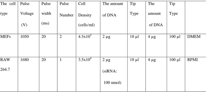
Table 3. 4 The Transfection Methods Depending On Cell Types ... 54
Table 3. 5 The Conditions For Electroporation Depending On Cell Types ... 55
Table 3. 6 Treatment Conditions For Cell Lines. ... 57
-- 1 --
1.
Introduction
1.1 Endoplasmic Reticulum Function and Biogenesis
The endoplasmic reticulum (ER), an extended membrane network, is the main organelle for protein folding and secretion, lipid synthesis and lipid droplet formation and a major store for calcium (Ca++). The ER controls a wide range of cellular process such as organogenesis, transcriptional activity, stress responses and apoptosis due to its many homeostatic functions9.
Secreted and transmembranes proteins are folded and reach maturation in the ER, which is the major gateway for the secretory pathway. The translation of proteins occurs in the ribosomes located on the cytosolic surface of the rough ER1. The proteins in the ER lumen or the newly synthesized, unfolded proteins being translocated into ER by Sec61 complex118 are covered with chaperones and associate with folding enzymes in the oxidizing and Ca++ rich environment of the ER. These nascent chains become N-glycosylated and assume their secondary and tertiary structures with the assistance of stabilizing protein disulfide isomerase (PDI). Endoplasmic reticulum oxidoreductin 1α and β (Ero1α and Ero1β) are responsible for the oxidation of PDI19, 113
. In the first step, a disulfide is introduced into the substrate protein with the concomitant reduction of a disulfide within the active site in PDI79. Then Ero1α or Ero1β catalyzes the reoxidation of the PDI active site in a reaction involving the reduction of oxygen to liberate hydrogen peroxide132, 43. In addition to these, the amino acid cis-trans isomerases, the chaperones such as the glucose regulated protein (GRP) 94 and immunoglobulin (Ig) heavy chain binding protein (BiP), N-glycosylation enzymes and the lectins, calnexin
- 2 -
and calreticulin; all reside in the ER and function in protein maturation. Mature proteins that pass quality control are transferred to Golgi, while the unqualified proteins become targets ER-associated degradation (ERAD). ER-associated degradation (ERAD) includes a translocation into cytosol by a translocon channel for proteosomal degradation. Recent studies show that the mammalian translocon associated proteins, with the help of Sec 61, can bind to improperly folded proteins and accelerate their degradation103. The other proteins as a translocon channel are derlins 86, 87, 168, and 155. Taken together, ER is an important organelle for cell homeostasis and protein metabolism.
ER is also a major store for Ca++; the concentration of Ca++ in rested ER can reach to three to four times higher than that in the cytosol. This difference is generated by the sarco (endo) plasmic reticulum Ca++ ATPase (SERCA) proteins that pump the Ca++ into ER lumen. The latter release of Ca++ is controlled by the signaling molecule, Ins (1, 4, 5) P3, and the ryanodine receptors9.
Furthermore, it was recently suggested that lipid droplet formation from ER membrane is mediated by ER resident proteins such as calnexin and the immunoglobulin heavy chain binding protein BiP117. Lipid droplets are the main intracellular storage sites for esterified fatty acids and neutral lipids such as triglycerides (TG) and cholesterol esters (CE). While the outer layer involves amphipathic lipids such as phospholipids (PL) and cholesterol (CL), the core is made of only neutral lipids93. The lipid droplets provide building blocks for biological membranes through hormone-dependent and independent-pathways33. The biogenesis mechanism of lipid droplets is not clear, but data shows it is mainly derived from the ER. The ER may assume a central role in lipid
- 3 -
droplet biogenesis through its enzymes like the diacylglycerol acyltransferase 1(DGAT) or 1-acylglycerol-3-phosphate acyltransferase (AGAT), which synthesize neutral lipids residing on the ER membrane. Additionally, Robenek and colleagues observed that a large portion of lipid droplets are covered by ER membranes 127. When fatty acids accumulate in the cell, TG is formed as a form of storing these fatty acids for future energy and neutralization. For this purpose, three fatty acids and one glycerol molecule are joined by enzymes resident in the ER membrane such as acyltransferases. The newly formed TG molecules are stored in lipid droplets that are surrounded by a single PL membrane layer and associated proteins. One hypothesis is that the lipid droplet originates from between two membranes of the ER and moves toward the outer cytoplasmic layer of the ER membrane via bud formation. This hypothesis could explain why the lipid droplet is surrounded by a PL monolayer 15. A contrasting second theory explains that the lipid droplet occurs outside of the ER but with the ER facilitating synthesis of the surrounding PL monolayer127. Regardless of the model, ER membranes and associated proteins are found to be integral components of lipid droplets.
Another crucial function of the ER is in lipid biosynthesis; ER is a major site for the production of phospholipids, cholesterol and ceramides96. For example, low intracellular cholesterol levels activate the ER resident sterol regulatory element-binding protein (SREBP 1 and 2), a transcriptional regulator of the fatty acid and cholesterol synthesis pathway, respectively, which works to upregulate the expression of enzymes responsible for cholesterol synthesis. Stearoyl coenzyme A (CoA) desaturase (SCD), which catalyzes the delta-9 desaturation of saturated free fatty acids (FFAs) is another enzyme located on the ER membranes. Furthermore, serine
- 4 -
palmitoyltransferase, which is rate –limiting enzyme of de novo ceramide production, is found on the ER membrane. Fatty acid elongation beyond the 16-Carbons (length of the palmitate) produced by the cytoplasmic Fatty acid synthase (FAS) is mainly catalyzed by enzymes such as DGATs [DAG (diacylglycerol) acyltransferases] for triacyl- glycerols) and/or ASATs (acyl-CoA: sterol acyltransferases) for SEs (steryl esters) associated with the ER48. These ER enzymes lengthen not only fatty acids produced by FAS but also the dietary polyunsaturated fatty acids. Formation of a double bond in a fatty acid involves the following endoplasmic reticulum membrane proteins such as desaturases, NADH-cyt b5 Reductase and Cytochrome b5.
The expansion of ER based on the increased demands on the exocytic pathway was documented in some specialized cells including the β cells of the pancreas and antibody-secreting plasma cells. However, the molecular mechanisms coordinating the protein and lipid components of the ER during this process remain unclear48. A study conducted by Sriburi and colleagues showed that spliced XBP-1 (X box binding protein-1) activated by UPR was enough to stimulate synthesis of phosphatidylcholine, the primary PL of the ER membrane138. Overexpression of the spliced XBP-1 in cells resulted in increased membrane PLs, surface area and volume of the rough ER, and enhanced activity of the cytidine diphosphocholine pathway of phosphatidylcholine biosynthesis138. This data showed that coordinating induction of phospholipid biosynthesis with up-regulated expression of ER resident proteins allows the mammalian UPR to both build and equip the ER, at least via XBP-1 activity. These findings demonstrate ER plays an important role in the maintenance of its own biogenesis.
- 5 -
The ER plays a role on insulin biosynthesis from pre-pro-insulin to pro-insulin90. The newly synthesized and signal tagged pre-pro-insulin in the cytoplasm is translocated into the lumen of the ER124, 125. After the signal peptide is cleaved in the ER by the endopeptidase called proprotein convertase 1, pro-insulin undergoes folding process in the ER lumen and the newly formed disulfide bonds lead to its stabilization and bioactivity124, 125. Then, this processed pro-insulin passes into secretory granules through Golgi apparatus. In final step, it gets native structure and then is secreted.
1.2 Endoplasmic Reticulum Stress
1.2.1 Endoplasmic Reticulum Stress : The Causes and Consequences
The ER participates in wide range of cellular activity from protein folding, lipid synthesis, calcium homeostasis to its own biogenesis. That’s why many factors that negatively impact cellular homeostasis leads to ER dysfunction and stress. Protein overload, accumulation of unfolded proteins, protein trafficking defects, chaperones deficiency, environmental toxins, viral infections, aging and chemicals such as tunicamycin, thapsigargin, and DTT destroy the balanced environment of the ER leading to a unique signaling cascade emanating from the ER and known as the accumulation of unfolded protein response (UPR) 30, 35. Physiological and pathological processes including aging105, glucose deprivation, aberrant calcium regulation, viral infection and hypoxia may alter the protein folding and cause ER stress70, 128. In the stressed ER, the misfolded or unfolded proteins are targeted for destruction through the ER-associated degradation (ERAD) pathway, which leads to their translocation to the cytosol for proteasomal degradation where they are degraded by ubiquitin-proteasome machinery70. Such proteins become subject to the ER quality system (ERQC), in which molecular chaperones are the main players aiding in proper folding and evaluation of
- 6 -
protein damage30. If the screened substrates fail to pass this quality test, they are marked as substrates for ERAD with the help of 76 amino acid peptide- specific E3 ubiquitin near or in ER membrane and then targeted to the proteasome, which is a multi-catalytic protease that resides in cytoplasm. If the ER stress can not be relieved, a specific stress response pathway, the UPR, is activated. A major function of the UPR is to combat with the accumulation of unfolded proteins through three complementary signaling pathways activated depending on the duration and the degree of the cellular insult. These functions of the UPR include reducing the protein load, increasing the capacity of the ER to handle the unfolded proteins and the decision to commit the cell to apoptotic death. This response is made of three signaling arms each governed by a proximal stress sensor: Inositol requiring enzyme 1 (IRE1), PKR resemble ER kinase (PERK) and activating transcription factor 6 (ATF6).
Recent studies showed that metabolic stress during obesity leads to ER stress and activation of the UPR especially in metabolically active sites such as liver, adipose tissues and pancreatic cells57. Nutrient or energy deprivation, in addition to its excess in obesity, can lead to ER stress. How obesity leads to ER stress remains a question of great interest. The maintenance of ER membrane function is supported by lipid metabolism, so one possibility is that obesity-driven perturbations in lipid metabolism can cause stress in the ER and lead to its metabolic dysfunction37. ER stress is a marker of early consequences of nutrient excess and a causally linked to the development of inflammation and insulin resistance in metabolic tissues including hepatocytes, cardiomyoblasts, pancreatic β cells and macrophages17, 18, 75, 158. Based on its duration, ER stress can be categorized as acute, periodic and chronic. The acute form of ER stress can be induced experimentally through treatments of chemicals such as tunicamycin,
- 7 -
DTT, calcium ionophores, thapsigargin and high levels of lipids such as saturated fatty acids or free cholesterol. An example of periodic ER stress is related to the rhythmic and transient physiological changes that occur during feeding and fasting cycles17, 116. After each cycle, the activation of the UPR is completely restored back to the basal levels. On the other hand, chronic ER stress indicates that the stress cannot be resolved and is continuous due to the presence of an ER stress inducer or as part of a vicious cycle, such as that created during obesity38.
1.2.2 The Unfolded Protein Response
Kozutsumi and colleagues suggested for the first time that unfolded proteins can activate a signal transduction pathway emanating from the ER78. This study observed that the expression of mutant influenza hemagglutinin lead to the induction of both unfolded proteins and the expression of ER resident proteins78. The delineation of this signaling pathway was first depicted in yeast, Saccharomyces cerevisiae, in which a 22-bp cis-acting element termed the UPR element was identified in the promoter of most genes up-regulated by UPR98, 99. The screening of the yeast mutants that induced this UPR element helped identify an ER trans-membrane protein, IRE1p. IRE1p turned out to be a bifunctional enzyme with both Ser/Thr kinase and endoribonuclease activities, which cleaves a transcriptional factor HAC1, in its C terminal domain26, 98. Originally, the IRE1p was identified as a gene required for inositol prototrophy in S. cerevisiae and hinting to its mammalian counterpart’s diverse functions in lipid metabolism107. In eukaryotic cells, ER homeostasis is controlled and communicated through the unfolded protein response (UPR) initiated by three ER membrane associated proteins, PERK (PKR-like eukaryotic initiation factor 2 α kinase), IRE1 (Inositol requiring enzyme 1), and ATF 6 (activating transcriptional factor 6) (Figure 1.1). To maintain ER
- 8 -
homeostasis, these proteins keep a dynamic control over the ER chaperones- BiP/GRP7810, 134. However, when ER homeostasis cannot be maintained, IRE1 and PERK are oligomerized and become fully activated to engage the downstream adaptive or destructive signaling pathways129. Furthermore, the ER resident transcription factor, ATF6, translocates to the Golgi where it is cleaved by a serine protease site -1 protease (S1P) and the metalloprotase site-2 protease (S2P) for full activation23 (Figure 1.1).
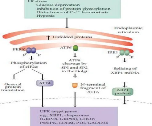
Figure 1. 1 ER stress and Unfolded Protein Response (reprinted with permission from Simone F., Gorman AM., Hori O., and Samali A. (2010). Cellular Stress Responses: Cell Survival and Cell Death. International Journal of Cell Biology, vol. 2010, Article ID 214074, 23 pages.)
ER stress caused by various disturbances leads to the activation of the three arms of unfolded protein response regulated by PERK (PKR resemble ER kinase), activating transcription factor 6 (ATF 6) and Inositol requiring enyzme 1 (IRE1). PERK phosphorylates eukaryotic initiation factor 2 alpha (eIF2) followed by global translation silencing and activation of activating transcription factor 4 (ATF 4) resulting in the expression of UPR target genes such as protein chaperones. ATF 6 goes to Golgi and is cleaved into the mature form, followed by transportation of nucleus where it regulates the expression of
- 9 -
UPR related genes. IRE1 phosphorylates itself and also catalyzes splicing of XBP-1 (X-box binding protein 1) mRNA through its RNAase activity.
Historically, the first discovered and conserved branch of the UPR is regulated by IRE1, which regulates the production of key ER chaperons through processing and activating the XBP1 mRNA20, 15. IRE1 has two homologs: IRE1α is expressed in a variety of tissues including placenta, liver, skeletal muscle, lung and especially pancreas145, but IRE1β is expressed only in the intestinal epithelia145, 156. IRE1 is activated by homo-oligomerization followed by auto-phosphorylation of the C-terminal kinase domain at serine 724114, 133, 159,161. The kinase activity of IRE1 has only one known substrate, itself. Although poorly understood, IRE1 has a unique effector function which leads to the endonucleolytic cleavage of its main substrate: mRNA encoding a transcription factor called HAC1 in yeast and XBP1 (X-box binding protein 1) in metazoans. IRE1’s endonucleolytic activity cleaves 26 nucleotides from the intron of XBP1-mRNA81, 135, 171
, resulting in a 41 kDA frameshift variant (sXBP1) that is an active transcription factor. XBP-1 regulates the expression of ER chaperones and the components of ERAD machinery92, 123. Furthemore, XBP1 upregulates the phospholipid synthesis enzymes leading to ER expansion, a hallmark of UPR. IRE1 also has a nonspesific RNAse activity that degrades mRNAs localized on ER membrane, reducing the synthesis and import of the corresponding proteins into the ER lumen53. Additionally, XBP-1 can upregulate the expression of P58IPK; a member of the Hsp40 (heat shock protein 40 kD) known as DNAJ in short name that is responsible of proper protein folding, acts as a co-chaperones and also negatively regulates PERK activity forming one of the many intersections between the three UPR braches167. The IRE1 dimers also interact with adaptor proteins such as TNF receptor-associated factor 2 (TRAF2) to induce the
- 10 -
apoptosis signal regulating kinase (ASK1) and subsequently activate the pro-apoptotic cJUN NH2-terminal Kinase (JNK) and p38 mitogen activated kinase (p38MAPK) 149 and caspase 12 Pro-caspase 12 forms a complex with TRAF2, but with activation of ER stress these two proteins dissociate leading to the activation of caspase 12 and resulting in cell death169. IRE1 can also modulate the activation of extracellular signal regulated kinases (ERKs) and nuclear factor қB (NF-қB) pathways62, 106 via JNK-AP1 pathways (Figure 1.2).
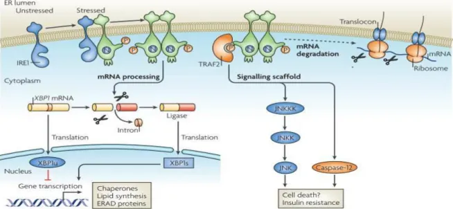
Figure 1. 2 IRE1 signaling (reprinted with permission from Ron, D., and Walter, P. (2007). Signal integration in the endoplasmic reticulum unfolded protein response. Nat. Rev. Mol. Cell Biol. 8, 519–529. © Nature publishing Group, 2007)
While inositol requiring enzyme 1 (IRE1) is in nonoligomerization state in unstressed conditions, IRE1 oligomerizes and autophosphorylates itself in stressed conditions. Upon stress induced activation, the RNAse activity of IRE1 cleaves X-box binding protein (XBP-1) in higher eukaryotes (HAC1 (homologous to ATF/CREB1) in yeast) into small RNA fragment encoding the active form of XBP-1 transcription factor, while unspliced XBP-1 encodes XBP1u, an inhibitor of UPR171. This is followed by the transcriptional upregulation of unfolded protein response (UPR) related genes. XBP-1 is also controls ER biogenesis. Alternatively, phosphorylated IRE1 recruits TRAF2 (tumour necrosis factor receptor (TNFR)-associated factor-2 resulting in activation of Jun N-terminal kinase (JNK) 149 and change
- 11 -
intracellular signaling such as insulin resistance109. Additionally, the IRE1-TRAF2 complex acts on caspase 12 activation and cell death171. ER stress can also lead to an ER-associated degradation (ERAD) of misfolded proteins.
Another arm of the UPR is initiated by the stress sensor known as the activating transcription factor 6 (ATF6), which is a 90kDA bZIP protein that becomes active after specific post-translational modifications. The disulfide and glycosylation bound luminal domain of ATF6 participates in sensing ER stress54, 102. Activation of ATF6 leads to its translocation to the Golgi, where it gets processed by site-1 and site-2 protease (S1P and S2P), which sequentially remove the luminal domain and the trans-membrane anchor of ATF6, respectively. After cleavage, now a 50 kDA protein, ATF6 translocates into the nucleus, where it binds to ER stress response element (ERSE; CCAAT(N)9CCACG)171 found in genes especially involved in the ERAD pathway, as well as some involved in lipid biosynthesis, ER expansion and protein folding45. A regulatory step for ATF6 activity involves the Wolfram Syndrome 1 (WSF1) protein, which marks ATF6 as a target for ubiquitination and proteasomal degradation by E3 ubiquitin ligase, HMG-CoA reductase degradation protein 1 (HRD1)36. Recent studies identified some homologs of ATF6 such as OASIS, CREBH, LUMAN, CREB4 and BBF2H7 can undergo similar processing at the Golgi and may have a role at tissue-specific cellular stress responses129 (Figure 1.3).
- 12 -
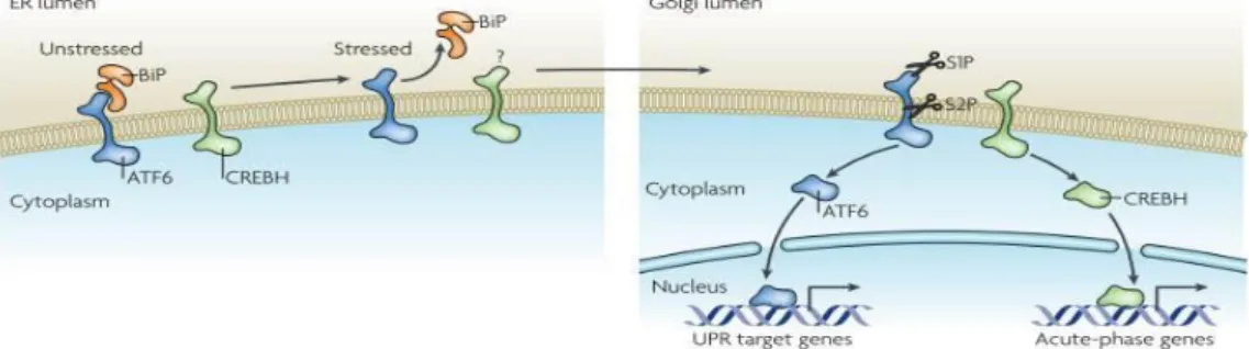
Figure 1. 3 ATF6 signaling (reprinted with permission from Ron, D., and Walter, P. (2007). Signal integration in the endoplasmic reticulum unfolded protein response. Nat. Rev. Mol. Cell Biol. 8, 519–529. © Nature publishing Group, 2007).
In unstressed conditions, activating transcription factor 6 (ATF6) and cylic AMP response element binding protein hepatocyte (CREBH) are located in the ER. Also, ATF6 is tethered to the ER by an ER chaperone immunoglobulin –binding protein (BiP) found in the lumenal domain of ER membrane. In stressed conditions, both ATF6 and CREBH translocate to the Golgi apparatus with the help of unknown vesicular transport system and are cleaved by first the lumenal site-1 protease and then the intra-membrane site 2 protease. After this activating cleavage, both proteins move to the nucleus, where ATF 6 activates UPR target genes and CREBH activates acute phase genes.
The third UPR arm is governed by the PKR-like eukaryotic initiation factor 2 α kinase (PERK), also known as pancreatic eIF2-alpha kinase (PEK). This is a type-1 trans membrane protein with ER luminal stress sensor and a cytosolic protein kinase domain. Under stress conditions, BiP dissociates from the N terminal of PERK, leading to its dimerization and autophosphorylation at the Threonine 981 on its kinase domain73. The C terminal kinase domain PERK shares similarities with other eukaryotic initiation factor 2 α a kinase such as double stranded RNA-dependent protein kinase R (PKR), activated by double stranded RNAs derived from viral infections, the general control non-depressible kinase 2 (GCN2), activated by uncharged tRNAs during amino acid deprivation, and the heme regulated inhibitor kinase (HRI), activated by heme limitation51, 136, 137. Under stress conditions, PERK phosphorylates α subunit of eIF2 at
- 13 -
serine 5152. The eIF2α is a heterodimer protein that is required for the transport of the initiating protein methoinyl-transfer RNA (met-tRNA) to the ribosome. Under stress conditions, PERK phosphorylates eIF2α, which inhibits translation initiation70, 52. PERK activity also stimulates some stress induced genes including the activating transcription factor-4 (ATF-4), the nuclear erythroid 2 p45-related factor 2 (Nrf-2)27 and nuclear factor kappa β (NF-қB) 28. ATF-4 regulates the expression of genes involved in amino acid import, glutathione biosynthesis, resistance to oxidative stress50 as well as the pro-apoptotic genes such as the CCAAT/enhancer binding protein (C/EBP) homologous protein (CHOP) and activating transcription factor-3 (ATF 3) 90, 65, 119. PERK dependent phosphorylation of the Nuclear factor eryhthroid-2-related factor-2) Nrf2/Keap1 (Kelch-like ECH-associated Protein 1) complexes leads to dissociation of this complex followed by Nrf2’s import into the nucleus27. The dephosphorylation of eIF2α is mediated by the growth arrest and DNA damage inducible gene (GADD34) interacting with catalytic subunit of protein phosphatase (PP) 1108 (Figure 1.4). Additionally, P58IPK that is induced by the activation of ATF6 binds to the cytosolic kinase domain of PERK and represses its activity151, 167. Interestingly, P58PIK expression occurs several hours after PERK activation and eIF2α phosphorylation. Thus, P58IPK
induction may be thought as a marking the end of UPR and the beginning of the alarm/apoptosis phase of response140 since at this point, if the ER stress can be handled, ER returns to normal function and the cell survives; but if the stress keeps going on, the blocking of the translational suppression by P58IPK might lead to apoptosis
- 14 -
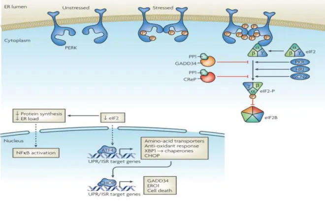
Figure 1. 4 PERK signaling pathway (reprinted with permission from Ron, D., and Walter, P. (2007). Signal integration in the endoplasmic reticulum unfolded protein response. Nat. Rev. Mol. Cell Biol. 8, 519–529. © Nature publishing Group, 2007)
Under stress conditions, protein kinase RNA (PKR) - like ER kinase (PERK) is activated, resulting in the phosphorylation of eukaryotic initiation factor 2 alpha (eIF2alpha) followed by general translation inhibition but selective translational activation of several UPR related genes. eIF2alpha can also be phosphorylated by other kinases including protein kine RNA (PKR), heme regulated inhibitor kinase (HRI) and general control non-derepressible-2 (GCN2) in an independent manner from ER stress. Hence, the larger stress response to a multitude of cellular insults and that includes the UPR is known as the integrated stress response (ISR).
1.2.3 The Adaptive and Destructive Outcomes of the Unfolded Protein Response UPR response involves three phases: adaptation, alarm and apoptosis (Figure 1.5). In the adaptation phase, UPR tries to re-establish homeostasis in cells by upregulating the expression of chaperone proteins that aid in protein folding. While the unfolded proteins degradation increases, global translation is attenuated in order to reduce the build up of further misfolded proteins in the ER. If this stage fails, UPR stimulates an alarm phase,
- 15 -
followed by the execution of the apoptosis program122. The alarm phase includes several signal transduction events, leading to the suppression of the expression and activity of pro-survival factors such as the B-cell lymphoma (Bcl2) protein. Beyond this stage, the stressed cell will undergo apoptosis. In addition ER stress can induce autophagy8, 39, 61, 69, 77, 111, and 170
. This fate decision from survival to death can be due to unresolved stress and incomplete recovery from the accumulation of misfolded proteins in the ER, ER Ca++ depletion, hampered disulfide formation and perturbed in redox state. All these stressors result in ER dysfunction and when prolonged, in apoptosis through the up regulation of the pro-apoptototic CCAAT/enhancer binding protein (CHOP), activation of JNK kinase and caspase 12 and via the interaction of IRE1 with other ER-localized pro-apoptotic B-cell lymphoma 2 (Bcl-2) family proteins141. However, the decision from survival to death in UPR may change from cell to cell since UPR is cell autonomous and context-dependent47. As an example, salubrinal, an inhibitor of eIF2α de-phosphorylation, protects pheochromocytoma cells from ER stress whereas it induces apoptosis in pancreatic β cells and renders them sensitive to free fatty acids25, 80.
Figure 1. 5 The switch from survival to apoptosis during ER stress (reprinted with permission from Jӓger R., Mathieu J.M., Bertran, Gorman A.M., Vandenabeele P., and Samali A. (2012). The unfolded Protein Response at the crossroads of cellular life and death during endoplasmic reticulum stress. Biol. Cell; 104, 259–270. © John Wiley and Sons, 2012
In early steps of ER stress, Both IRE1 and PERK favour cell survival, but in prolonged ER stress, adaptive signaling of IRE1 attenuates whereas PERK begins to engage pro-apoptotic pathways.
- 16 -
1.3 The Interface Between ER Stress And Inflammation
Recent studies have shown that the UPR may be linked to inflammatory signaling cascades and stress signaling pathways through multiple scenarios including activation of JNK and NF-қB pathways28, 62, the production of ROS and the generation of nitric oxide resulting in both ER stress and inflammation27, 42. These pathways also have a central role in obesity-induced inflammation and metabolic abnormalities, especially in abnormal insulin action56.
IRE1 is required for the ER stress induced activation of JNK, which regulates many inflammatory genes149. Earlier studies showed that pro-inflammatory cytokines such as TNF-alpha, IL6 and MCP-1 are suppressed in the absence of JNKincells and tissues, and that chronic JNK activation contributes to insulin resistance and type 2 diabetes55, 146 and 150
.
Other mechanism that leads to inflammation involves NF-қB-IKK activation by both IRE1α, through the interaction of TRAF2 (TNF receptor-associated factor 2) and apoptosis signal-regulating kinase 1 (ASK1), which, in turn, causes the phosphorylation and activation of JNK. This active JNK phosphorylates and induces the activity of transcription factor activator protein 1 (AP-1), which leads to the expression of inflammatory genes. Additionally, AP-1 recruits IқB kinase (IKK) causing release of NFқB and so promotes NFқB mediated inflammation. Moreover, PERK via PERK eIF2α mediated translation suppression of IқB can directly stimulate NFқB (negatively regulating nuclear factor-қB) activation. This activation results in increase in the ratio of NFқB to IқB, thereby allowing the excess NFқB to enter the nucleus to trigger
- 17 -
expression of inflammatory cytokines28, 62, and 138. Furthermore, recent studies also link the ATF6-regulated branch of UPR to NF-қB-IKK signaling, suggesting specific inflammatory signals may be induced through all three branches of the UPR166. Some studies in cellular systems report that experimental induction of UPR via free cholesterol or high amounts of free fatty acids68 may cause increased expression of pro-inflammatory molecules such as IL-8, IL-6, MCP-1 and TNFalpha82.
Another mechanism that ER stress may be linked to inflammation involves the transcription factor cyclic-AMP-responsive-element-binding protein H (CREBH), which also resides on the ER membrane and stimulates the production of acute phase response genes in the liver such as C reactive protein (CRP) and serum amyloid P-component (SAP) 174. CREBH and CHOP also may play an important role in the regulation of peptide hormone hepcidin, which may present a link between iron metabolism and inflammatory output via ER152. However, these interactions between ER stress and inflammation are not unidirectional, because recent studies in the brain provided proof that both ER stress and inflammation can activate each other, while inhibiting normal cellular metabolism174. In this particular study, activation of IKK-β led to ER stress and the activation of ER stress induced IKK-β.
More recently it was discovered that double stranded RNA-dependent protein kinase (PKR), which is another eIF2α kinase that is homologous to PERK, plays a unique role in linking over-nutrition to ER stress and inflammation in metabolic diseases104. Here, PKR was activated by lipids and was shown to have a central role in the activation of JNK and inflammatory responses during obesity. Moreover, PKR directly blocked insulin action via phosphorylating insulin receptor substrate-1 (IRS1). It is speculated
- 18 -
that PKR could assemble a putative metabolically-activated inflammatory complex called metabolic inflammasome or “metaflammasome” that integrates insulin action, pathogen responses and nutrient sensing. This scaffold may explain the functional overlap and between multiple signaling pathways, metabolic, stress and inflammatory, in modulating metabolism. Moreover, Baltzis et al. observed that PERK deficient mouse embryonic fibroblasts (MEF), which display much higher levels of vesicular stomatitis virus replication than that in the wild type MEFs, also exhibited partial PKR activation, suggesting a cross talk may occur between PKR and PERK5.
The interactions between ER stress and inflammation also involves oxidative stress caused by a toxic accumulation of ROS in the cells27. This can be due to protein chaperones uplegulated by UPR involving in disulfide bond formation in ER. Ero1p an essential for respiration and vegetative growth 2 (Erv2p), two enzymes responsible for disulfide bond formation, utilize oxidation/reduction reactions for reducing molecular oxygen. This reduced molecular oxygen also accumulates during increased improper protein folding in ER stress and is toxic to cells49. This increase in ROS levels due to UPR occurs through PERK mediated activation of antioxidant program with the help of transcriptional factor Nrf2 to neutralize the toxic effects of ROS. Hence, PERK to Nrf2 signaling may be another potential link between inflammation and ER stress.
Moreover, nitric oxide (NO) generation may be related to ER stress such that NO-induced S-nitrosylation of PDI, which catalyzes the thiol-disulphide exchange by facilitating disulphide bond formation and rearrangement reactions, inhibits its enzymatic activity and leads to the accumulation of polyubiquitinated proteins and activation of the UPR147. ER stress also can induce iNOS via activation of
pro-- 19 pro--
inflammatory responses through NKқβ activation165. However, the ER molecules that have direct effect on iNOS activation still remain unknown.
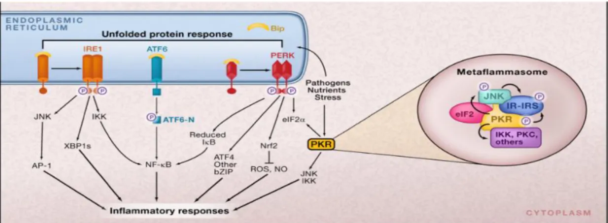
Figure 1. 6 ER stress and Inflammation (reprinted with permission from Hotamisligil GS. (2008b) Endoplasmic Reticulum stress and inflammatory basis of metabolic disease. Cell 140, 900–917. © Elsevier, 2010.
There are several potential pathways by which ER stress may be linked to inflammatory responses. Protein attenuation mediated by PERK activity can cause the release of NF-қB from its inhibitor IқB. This results in the transport of NF-қB to nucleus followed by changes in some genes involved in inflammatory pathways such as cytokines IL-1 and TNFalpha. Also, IRE1 recruits tumor necrosis factor 2 (TRAF2) and this complex causes activation of IқB and JNK resulting in activation of some inflammatory genes in downstream. The eIF2α kinase protein kinase RNA (PKR), induced upon ER stress, can form the core of a putative metabolic inflammasome called metaflammasome and has a direct interaction with some inflammatory kinases such as JNK and IKK, insulin receptor signaling components such as IRS1 and the translational machinery via eIF2.
The UPR may play a role in the dysfunction and death of macrophages under lipotoxic stress95. Also, it has been reported that lipid chaperones such as the adipocyte and macrophage fatty acid binding protein 2 (aP2) that can bind long chain fatty acids could be a link between toxic lipids and ER stress32. In the study by Erbay and colleagues, aP2-/- macrophages were found to be resistant to ER stress induced by high amounts of
- 20 -
saturated fatty acids and the lipid-induced toxicity was mediated by the aP2 protein. Lipotoxicity of immune cells such as macrophages is one of the critical features of obesity and atherosclerosis suggesting that the action of the lipid chaperones in controlling lipid-induced ER stress could be therapeutically targeted to treat these diseases and their complications32, 91. To this date, the precise molecular links between ER stress and inflammation and how ER stress in immune cells alters the progress of chronic metabolic diseases is not fully understood (Figure 1.6).
1.4 ER Stress And Inflammation In Cardiometabolic Syndrome Myocardial infarction and stroke are two consequences of the atherosclerotic disease and the leading cause of mortality and morbidity worldwide. Even though there have been important advances to understanding the pathogenic mechanisms underlying these metabolic diseases, due to very limited preventive and therapeutic strategies they still account for one third of all deaths annually. The signaling pathways that lie at the interface of chronic inflammation and nutrient metabolism strongly influence these chronic diseases as well as obesity, insulin resistance and type 2 diabetes59.
Atherogenesis is triggered with the subendothelial retention of apolipoprotein (apo) B-containing lipoproteins in focal areas of the arterial subendothelium162, 142. A series of maladaptive inflammatory responses are triggered against oxidized or alternatively modified lipoproteins162, 142, 89, 84 and 40. Blood-borne monocytes are attracted to focal sites of activated endothelial cells (ECs) overlying areas of lipoprotein retention, followed by monocyte differentiation into macrophages. The recruited macrophages ingest these modified lipoproteins, convert to foam cells, and are of pro-inflammatory nature89, 40, 41 and 84.
- 21 -
In most lesions, the detrimental clinical results of atherosclerosis are prevented by several adaptive processes: The remodeling of the vessel wall maintains the patency of the arterial lumen. The phagocytic clearance (“efferocytosis”) of dead cells, mostly apoptotic macrophages, prevents plaque necrosis. The scar formation by collagen-producing intimal smooth muscle cells (myofibroblasts) helps to defend against matrix protease–mediated erosion or rupture of the intima into the lumen. Despite all these measures, a minority of these lesions progress due to increased lesional macrophage apoptosis, defective efferocytosis, and death of collagen producing intimal smooth muscle cells. These plaques erode or rupture, leading to the exposure of the blood to coagulation and thrombotic factors released from the necrotic lesions and acute lumenal thrombosis 7, 83, 143 and 154. Studies conducted in animal models of atherosclerosis and, most importantly, through the analysis of human atherosclerotic lesions, support ER stress occurs in atherosclerotic plaques, particularly in the advanced stages of the disease leading to acute thrombosis101. ER stress occurs in lipid-laden macrophages at all stages of the disease34, 101 and 175. In Miyoishi and colleagues’ study human coronary artery lesions from autopsy samples and fresh human carotid endarterectomy specimens were examined in terms of lesion stage, UPR markers, and apoptosis, revealing a striking relationship among advanced lesion stage, CHOP expression, and lesional apoptosis101.
The possible connection between endoplasmic reticulum stress and metabolic disorders such as obesity, diabetes and atherosclerosis may occur through several ways. First, there is a direct relation between lipid metabolism and ER stress, including the existence of lipogenic enzymes in ER domains. Furthermore, XBP1 plays a role in ER phosphatidylcholine synthesis and ER membrane expansion139. Multiple studies also
- 22 -
show that ER stress promotes lipogenesis and hepatic lipid accumulation, but how the individual UPR branches plays a role in this remains unclear59. Second, ER stress leads to abnormal insulin action and hyperglycemia by causing insulin resistance, stimulation of hepatic glucose production and suppression of glucose disposal. Studies show ER stress is a potential link between obesity, type 2 diabetes and atherosclerosis121. Moreover, ER stress may lead to the production of inflammatory mediators and reactive oxygen species, which are destructive for insulin action, lipid metabolism and glucose homeostasis60.
It is known that primary macrophages play an important role in the progress of atherosclerosis97. Hence, the studies on the role of ER stress in macrophages may provide clues to ER stress contribution to atherosclerosis. ER stress can be induced by excess accumulation of free cholesterol, resulting with cell death in macrophages. The overload of fatty acid leads to their accumulation and stiffening of ER membranes, which are known to be poor in cholesterol content. Consequently, the stiffening of ER membranes activates the UPR and the PERK-mediated CHOP activation leads to apoptosis34.
Recently, it has been reported that obesity and insulin resistance may stimulate ER stress in lesion macrophages due to elevations of saturated fatty acids (SFAs) 32, 131. SFAs cause the loss of fluidity of the ER membrane bilayer, which is a known inducer of the UPR. However, the exact mechanism of SFA-induced ER stress is not fully understood. Erbay and colleagues demonstrated that intracellular lipid chaperones (or macrophage/adipoctye fatty acid-binding protein-4, aP2) mediates SFA- induced ER stress and apoptosis in macrophages. In this study, ApoE-/- ( apolipoprotein E) mouse
- 23 -
model fed by Western diet, when genetically deficient for aP2 were found to be protected against lipid induced PERK activation, XBP-1 splicing and apoptosis in macrophage-rich regions of atherosclerotic lesions. These results implicated aP2 as a central mediator of lipotoxic ER stress in macrophages during atherogenesis. Furthermore, reduction of ER stress by chemical chaperons reduced atherosclerosis in mice32. Collectively; the described findings show the important contribution of lipid-induced ER stress to atherosclerosis progression and offer new insights into metabolic disease pathogenesis.
1.5 Restoring ER Stress
Restoring ER function may become a promising future therapeutic strategy against metabolic diseases including insulin resistance, diabetes, fatty liver disease, obesity and atherosclerosis. For example, Qi et al. (2004) 120 and Vilatabo et al. (2005) 153 observed that 4-phenylbutyric acid (PBA), which is known as a chemical chaperon, has protective effects against cerebral ischemia and liver ischemia reperfusion injury by inhibiting ER-stress- related apoptosis. Another reagent tauroursodeoxycholic acid (TUDCA), a hydrophilic endogenous bile acid, also prevented diet-induced ER stress and ER-stress associated apoptosis in human liver cells through unknown mechanisms31. Additional studies in obese and diabetic mouse models showed that the administration of TUDCA and PBA can reduce ER stress, normalize hyperglycemia, increase systemic insulin sensitivity, resolve the fatty liver disease and enhanced insulin action in liver, muscle and adipose tissues110. Furthermore, administration of PBA to Apolipoprotein E null (ApoE−/−) mice with atherosclerosis exhibit ER stress and UPR activation in the lesions175 resulted in a dose-dependent reduction ER stress in macrophages and atherosclerotic lesions32. In this study, the ER stress markers phosphorylated-eIF2α,
- 24 -
phosphorylated-PERK and ATF3 expression were significantly reduced in the atherosclerotic lesions of mice treated with PBA, suggesting restoring ER function with chemical chaperone and reducing ER stress and death of macrophages in lesions can protect against the deleterious effects of toxic lipids and prevent atherosclerosis32. Additionally, it was also observed that gastric bypass induced weight loss results in the reduction of the ER stress markers, GRP78, sXBP1, phosphorylated-eIF2α and JNK-1 in the liver samples of these patients43. These observations indicate reduction in nutrient intake and in adipose tissue size may reduce the ER stress levels in metabolically active tissues in humans. Furthermore, a study conducted by Diakogiannaki showed that the administration of a monounsaturated fatty acid palmitoleate for 18 hrs., reduced ER stress induced by both a saturated fatty acid palmitate and also tunicamycin (a chemical stressor of the ER by blocking N glycosylation) in β cells29. When the two species of fatty acid palmitate and palmitoleate were given in combination, the extent of cell damage was less than that when palmitate was applied alone. In fact, examination of multiple images showed that the overall ER membrane area was reduced in palmitate and palmitoleate administration compared with in only palmitate administration; suggesting that mono unsaturated fatty acids entering membrane lipids is less destructive to membrane architecture than when saturated molecules exist29.
The possibility of applying these chemical chaperones to restore ER function and reduce ER stress in human metabolic diseases still remains to be fully explored. However, new studies are appearing with promising results. For instance, a study conducted by Kars et al. in 2010 displays TUDCA-stimulated increase in the muscle and hepatic insulin sensitivity of obese and insulin resistant patients71.
- 25 -
1.6 Chemical Genetics And Its Applications
Advances in high throughput chemistry and genetics have created a new field dubbed “chemical genetics”, which generates tools that can help elucidate and validate novel drug targets. This approach can also be successfully applied to deciphering signal transduction cascades, particularly, in discovering novel substrates of kinases74.
The traditional genetics approach can examine the function of a single protein at the level of the organism, a major reason why knock out mice have become popular tools for target analyses. However, this approach has some disadvantages. For example, genetic approach knocks out a specific gene without discriminate against the other proteins that associated with the targeted protein or in other words, leading to the disassembly of associating proteins in a protein complex (for instance, the targeted gene may be an important scaffold in a complex) or abolish an enzymatic function of the targeted protein. This unwanted effect renders it difficult to determine the direct and indirect effects of the gene knock-out approach. Second, the genetic approach lacks temporal control over the targeted protein. While, inducible knock outs and RNA based approaches (such as antisense and RNAi) have been devised, these lead to a slow change in protein activity compared to quick signaling responses that normally take place in cells. In addition, Causton et al. in 2001 showed that a temporal shift in yeast (from 25 oC and 37oC) leads to significant differences in genome expression22. This study indicates that conditional or inducible systems such as temperature sensitive alleles may cause secondary perturbations with unwanted side effects. Finally, some genes, which play significant roles in embryonic development result in lethal phenotype making these genetic models inapplicable, particularly valid for knockout mice63.