Abstract
A male Long-legged buzzard with a gunshot wound on his left wing was presented for treatment. However, the bird died shortly after, and then the routine necropsy was performed. At necropsy, numerous white-to-yellow nodular lesions sizing several mm to 1 cm in diameter were noted in liver, spleen, gizzard and lung. Microscopic examination of the nodules in lung and gizzard revealed classical formation of tubercles characterized by a caseous core surrounded by epitheloid cells, multinucleated giant cells, heterophils, macrophages, and outer fibrous capsule. Fibrous capsule formation was vague in tubercles located in liver and spleen. Acid fast bacteria were shown by Ziehl-Neelsen staining. Based on the observations a diagnosis of avian mycobacteriosis was made. This report indicates that avian tuberculosis might be an important disease in free living animals in Turkey as in other places, and more attention might be needed to monitor the disease.
Keywords: Avian tuberculosis, Long-legged buzzard
Doğada Serbest Yaşayan Bir Uzun Bacaklı Şahinde (Buteo rufinus)
Tuberküloz Olgusu
Özet
Sol kanadında ateşli silah yarası bulunan bir erkek Uzun bacaklı şahin tedavi amaçlı getirilmiş, ancak kısa sürede ölmüş ve rutin nekropsisi yapılmıştır. Nekropside; karaciğer, dalak, ventrikulus ve akciğerlerde çok sayıda, yarıçapı birkaç mm ile 1 cm arasında değişen büyüklüklerde beyaz-sarı renkli nodüler lezyonlar dikkati çekti. Mikroskobik bakıda akciğer ve ventrikulusta kazeifiye merkez etrafında epiteloid hücreler, çok çekirdekli dev hücreleri, heterofil lökositler, makrofajlar ve en dışta fibröz bir kapsül ile karakterize klasik tüberkel yapısı gözlemlendi. Fibröz kapsül yapısı karaciğer ve dalaktaki tüberkellerde belirgin değildi. Ziehl-Neelsen boyama ile aside dirençli bakterilerin varlığı belirlendi. Yapılan gözlemler sonucunda kanatlı tüberkülozu tanısı konuldu. Bu olgu sunumu; diğer pek çok yerde olduğu gibi ülkemizde de kanatlı tüberkülozunun doğada serbest yaşayan hayvanların önemli bir sorunu olabileceğini göstermiş ve bu alanda daha fazla ilgiye ihtiyaç duyulduğunu ortaya koymuştur.
Anahtar sözcükler: Kanatlı tüberkülozu, Uzun bacaklı şahin
A Case of Tuberculosis in a Free-living Long-legged Buzzard
(Buteo rufinus)
Hasan ÖZEN
1
Musa KARAMAN
2Serpil DAĞ
1Emin KARAKURT
1Yalçın AKBULUT
31 2 3
Kafkas University, College of Veterinary Medicine, Department of Pathology, TR-36100 Kars - TURKEY Balıkesir University, College of Veterinary Medicine, Department of Pathology, TR-10145 Balıkesir - TURKEY Kafkas University, College of Veterinary Medicine, Department of Anatomy, TR-36100 Kars - TURKEY
INTRODUCTION
Tuberculosis is a commonly seen disease affecting both human and animals [1]. Avian tuberculosis, also called avian
mycobacteriosis, is occasionally reported both in various domestic and wild birds as well. The causative agents of the disease in birds are mostly Mycobacterium avium subsp. avium and less frequently M. genavense [2]. However, there
are studies reporting that M. genevanse is the predominant mycobacterial species in pet birds [3,4]. Several other species
of mycobacteria such as M. tuberculosis, M. bovis, M. terrae, M. fortuitum, and M. scrofulaceum was rarely recorded in the etiology of avian mycobacteriosis [2,5-7].
Susceptibility of different species of birds to the disease varies greatly. Although birds were broadly classified into four groups, namely highly susceptible, less susceptible, moderately resistant and highly resistant, based on their susceptibility to the disease [8], the reason why they differ
is exactly not known. Birds living under the same housing
İletişim (Correspondence)
+90 474 2426836, Fax: +90 474 2426853
[email protected]KafKas Universitesi veteriner faKUltesi Dergisi JoUrnal Home-Page: http://vetdergi.kafkas.edu.tr
online sUbmission: http://vetdergikafkas.org
Case Report
Kafkas Univ Vet Fak Derg 22 (3): 473-476, 2016
DOI: 10.9775/kvfd.2015.14888
474
A Case of Tuberculosis in ...
conditions were reported to differ in contracting the disease, and hence genetic and environmental conditions were suggested [9,10]. Even feather color in doves was
shown to be a factor probably in relation to chromosomal association of immune response [11]. Countless stressors
such as overcrowding, malnutrition and concurrent infections that impair immune system are known to be some important predisposing factors [6,12].
Reports of mycobacteriosis in wild and captive birds are very common worldwide where affective monitoring and protection studies performed regularly [3,6,7,13,14]. However,
there are only limited numbers of reports describing avian tuberculosis in Turkey. Besides, most of these cases are on pigeons [15-18]. There are also reports in a hen [19],
chickens [20] geese [21], a peafowl and pheasants [10]. Avian
mycobacteriosis in raptors was only described once in Turkey before, however the subject of that case, which was also a Long-legged buzzard, was a captive bird held in a park under a protection program [12]. Therefore, a case of
avian tuberculosis in a raptor living freely in Turkey was found to be worth of reporting.
CASE HISTORY
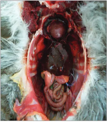
An adult male Long-legged buzzard (Buteo rufinus) was referred to Kafkas University Wildlife Safety, Rescue, Rehabilitation, and Research Center in Kars-Turkey for gunshot treatment. Clinically the bird was depressed and emaciated, however it died shortly after without the chance of treatment. Thereafter, routine necropsy was performed. In gross examination, body condition of the bird was poor with atrophied pectoral muscles, and there was an apparently old open wound at the level of left ulna, which was detected to be broken. Intestinal content revealed presence of diarrhea. No other gross lesions or abnormalities were observed in the head and the skin. Upon inspection of the internal organs, multiple white-to-yellow firm nodular lesions sizing several mm to 1 cm in diameter were seen in liver, spleen, gizzard and lung (Fig.
1), which caused the speculation of tuberculosis.
Tissue samples from all organs were collected and routinely processed for 10% formalin fixation and paraffin embedding. Tissue sections were then cut and stained with hematoxylin and eosin (HE) for evaluation of histopathological changes and with Ziehl-Neelsen for demonstration of acid-fast bacteria.
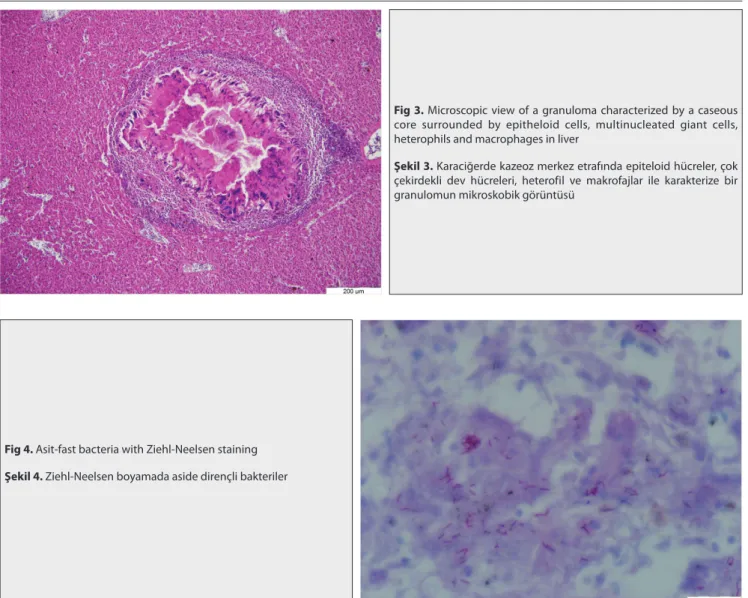
In microscopic examination of lungs (Fig. 2) and gizzard, typical tuberculoid granulomas characterized by non-mineralized or non-mineralized central necrosis, surrounded by heterophils, macrophages, lymphocytes, epitheloid cells, multinucleated giant cells and outer fibrous capsule were
Fig 1. White-to yellow nodules located on liver and lungs Şekil 1. Karaciğer ve akciğerlerde beyaz sarı renkli nodüller
Fig 2. Microscopic view of a granuloma characterized by a mineralized
caseous core surrounded by epitheloid cells, multinucleated giant cells, heterophils and macrophages with outer fibrous capsule formation in lung
Şekil 2. Akciğerde mineralize kazeoz merkez etrafında epiteloid
hücreler, çok çekirdekli dev hücreleri, heterofil ve makrofajlar ile karakterize çevresinde fibröz kapsül şekillenmiş bir granulomun mikroskobik görüntüsü
475 ÖZEN, KARAMAN, DAĞ KARAKURT, AKBULUT
observed. Fibrous capsule formation around the tubercles was vague in liver (Fig. 3) and spleen. Where tubercles not formed yet, sheets of inflammatory cells mostly composed of macrophages and lymphocytes were noted. Ziehl-Neelsen staining revealed the presence of acid-fast bacteria located both intracellular within the giant cells and as free (Fig. 4).
Based on the typical tuberculoid lesions and view of acid-fast bacteria, a diagnosis of avian mycobacteriosis was made. Microscopy of kidney revealed severe glomerular and tubular degeneration with infiltration of inflammatory cells and congestion.
DISCUSSION
Avian tuberculosis is a ubiquitous disease worldwide. It is common in gallinaceous poultry and wild animals living in captivity [6]. Although occasional case reports or
investigations on poultry, captive and wild birds are present elsewhere, there is only few in Turkey [10,15-18,20]. Avian
tuberculosis in a raptor was only reported once before in Turkey, and it was a captive Long-legged buzzard [12].
In the current case report, avian mycobacteriosis was
described also in a Long-legged buzzard, which was differently from the previous case, living free in nature.
Mycobacterial agents are highly resistant to environ-mental conditions and can survive several years in soil. Contaminated soil, water and infected prey in wild and housing equipment in captivity can help spreading of the infection [13]. Continuous shedding of microorganisms with
droppings and aerosol are the main sources for transmission to susceptible birds. Proper sanitation measures taken after if the disease is diagnosed in a flock of poultry animals or captive birds may easily help preventing the spread or the reoccurrence [22]. However, since such measures cannot
be applied in nature it is almost impossible to control the disease in free-living animals.
Lesions in avian tuberculosis are mostly seen in liver, spleen, and gastrointestinal system organs [11,23]. Lung
and facial lesions can also be observed occasionally [10,14].
In the present case, white to yellow tubercles were also noted in liver, spleen, gizzard and lung. Microscopic view of the lesions revealed classical formation of tubercles with macrophages, epitheloid cells, giant cells, lymphocytes
Fig 4. Asit-fast bacteria with Ziehl-Neelsen staining Şekil 4. Ziehl-Neelsen boyamada aside dirençli bakteriler
Fig 3. Microscopic view of a granuloma characterized by a caseous
core surrounded by epitheloid cells, multinucleated giant cells, heterophils and macrophages in liver
Şekil 3. Karaciğerde kazeoz merkez etrafında epiteloid hücreler, çok
çekirdekli dev hücreleri, heterofil ve makrofajlar ile karakterize bir granulomun mikroskobik görüntüsü
476
A Case of Tuberculosis in ...
and an outer fibrous capsule with a central caseous core as reported previously [24]. However, fibrous capsule
formation was not clear in liver and spleen. The reason of vague fibrous capsule formation in liver and spleen in opposite to lung and gizzard might be explained by extensive connective tissue presence in the later organs. In addition, we did not detect any lesions in intestines, where tuberculoid lesions are often reported. In birds, alimentary tract is the main route of transmission of the infection, since most lesions are generally localized in this system. However, presence of lung lesions in addition to liver, spleen and gizzard, might suggest that aerosol transmission were involved in the current case. On the other hand possibility of alimentary transmission cannot be omitted based on the size of tubercle in gizzard. In the previously reported case in a Long-legged buzzard in Turkey, lesions were detected in wing, spleen, intestines and lungs but no tubercles were reported in liver. In that case report, because of the close contact with other birds such as sparrows, starlings and crows, the disease was speculated to be contracted from such animals [12]. In the
present case, it is not even possible to speculate the source of infection since the animal was a free living animal and, besides no reports was before present from the region where the animal was located.
Avian tuberculosis is a chronic disease, and hence is seen mostly in adult animals. However, considerable amount of young companion birds with ages less than a year were previously reported to have tuberculosis [13].
In the current case the bird was also a mature animal. In wild birds with tuberculosis, mostly subclinical disease state is observed. Emaciation, stagnation, drowsiness, and diarrhea were often the recorded clinical signs in birds [2,12].
However, probable affects of gunshot and following unknown state of the bird for a while did not let the observation of any signs except pectoral muscle atrophy.
In conclusion, the aim of this case presentation was to provide awareness to the wild-life diseases besides describing the pathological changes in a tuberculoid bird detected coincidentally. Insufficient wild life facilities as well as absence of monitoring programs in Turkey probably limit the observation of diseases occurring in wild animals in general. Therefore, it is clear from the lack of enough disease reports in wild animals that more attention is undoubtedly needed for wild-life animals and the diseases. Control measures can only then be applied. In addition, it must be mentioned that since the infected wild birds can be a serious source for captive birds as well as poultry in avian tuberculosis, regular monitoring of the disease in wild can help estimating the risk for poultry.
REFERENCES
1. Ghodbane R, Drancourt M: Non-human sources of Mycobacterium
tuberculosis. Tuberculosis, 93, 589-595, 2013. DOI: 10.1016/j.tube.2013.09.005
2. Dhama K, Mahendran M, Tiwari R, Singh SD, Kumar D, Singh S,
Sawant PM: Tuberculosis in birds: Insights into the Mycobacterium infections. Vet Med Int, Article ID: 712369, 2011. DOI: 10.4061/2011/712369 3. Hoop RK, Böttger EC, Pfyffer GE: Etiological agents of mycobacterioses in pet birds between 1986 and 1995. J Clin Microbiol, 34 (4): 991-992, 1996.
4. Holsboer Buogo C, Bacciarini L, Robert N, Bodmer T, Nocolet J:
Vorkommen von Mycobacterium genavense bei Vögeln. Schweizer Arch
Tierheilkd, 139 (9): 397-402, 1997.
5. Smit T, Eger A, Haagsma J, Bakhuizen T: Avian tuberculosis in wild birds in the Netherlands. J Wildlife Dis, 23, 485-487, 1987. DOI: 10.7589/0090-3558-23.3.485
6. Tell LA, Woods L, Cromie RL: Mycobacteriosis in birds. Res Sci Tech Off
Int Epiz, 20 (1): 180-203, 2001.
7. Schmidt V, Schneider S, Schlömer J, Krautwald-Junghanns ME, Richter E: Transmission of tuberculosis between men and pet birds: a case report. Avian Pathol, 37, 589-592, 2008. DOI: 10.1080/03079450802428901 8. Hejlicek K, Treml F: Comparison of the pathogenesis and epizootiologic importance of avian mycobacteriosis in various types of domestic and free-living syntropic birds. Vet Med-Czech, 40, 187-194, 1995.
9. Cromie RL, Brown MJ, Price DJ, Stanford JL: Susceptibility of captive wildfowl to avian tuberculosis: the importance of genetic and environmental factors. Tubercle, 72, 105-109, 1991. DOI: 10.1016/0041-3879(91)90036-R
10. Kul O, Tunca R, Hazıroğlu R, Diker KS, Karahan S: An outbreak of avian
tuberculosis in peafowl (Pavo cristatus) and pheasants (Phasianus colchicus) in a zoological aviary in Turkey. Vet Med-Czech, 50, 446-450, 2005.
11. Saggese MD, Tizard I, Phalen DN: Mycobacteriosis in naturally infected ring-neck doves (Streptopelia risoria): Investigation of the associaition between feather colour and susceptibility to infection, disease and lesion type. Avian Pathol, 37, 443-450, 2008. DOI: 10.1080/ 03079450802210655
12. Atasever A, Beyaz L, Kibar M, Gümüssoy KS: A case of tuberculosis and aspergillosis in a Long-Legged Buzzard (Buteo rufinus). Revue Med
Vet, 157 (1): 26-29, 2006.
13. Manarolla G, Liandris E, Pisoni G, Sassera D, Grilli G, Gallazzi D, Sironi G, Moroni P, Piccinini R, Rampin T: Avian mycobacteriosis in companion birds: 20-year survey. Vet Microbiol, 133, 323-327, 2009. DOI: 10.1016/j.vetmic.2008.07.017
14. Mayahi M, Mosavari N, Esmaeilzadeh S, Parvandar Asadollahi K: Avian tuberculosis in naturally infected lofts of domestic pigeons, isolation, molecular identification and study of necropsy findings. Intern
J Appl Res Vet Med, 11 (3): 194-201, 2013.
15. Sezen İY, Erer H, Erganis O: Bir güvercinde tüberküloz olgusu. Selçuk Üniv
Vet Fak Derg, 2 (1): 163-166, 1986.
16. Kutsal O, Sağlam M: Güvercinlerde tüberkuloz olgularının
değerlendirilmesi. Ankara Üniv Vet Fak Derg, 35 (2.3): 545-552, 1988.
17. Gürel A, Arun SS, Yesildere T: Üç farklı evcil güvercin sürüsünde spontan tüberkülozis olguları. Istanbul Univ Vet Fak Derg, 23 (1): 131-139, 1997. 18. Terim Kapakin KA, Alçığır G: Bir güvercinde tüberküloz olgusu. Kakfas
Unıv Vet Fak Derg, 15, 477-479, 2009. DOI: 10.9775/kvfd.2009.061-G
19. Beytut E, Atabay Hİ, Akça A: Tuberculosis and Sarcosporidiosis in the periorbital location in a hen. Kafkas Univ Vet Fak Derg, 7 (2): 213-217, 2001. 20. Terim Kapakin KA, Sağlam YS, Altun S: Histopathological examinations of tuberculosis cases detected in chickens grown by a family enterprice.
Atatürk Univ Vet Bil Derg, 5 (3): 141-146, 2010.
21. Özcan K, Beytut E, Aydın F, Tuzcu M: Tuberculosis in geese (Anser
anser) in Turkey. Avian Dis, 45, 755-759, 2001. DOI: 10.2307/1592924
22. Gill IJ, Blandy ML: Control of avian tuberculosis in a commercial poultry flock. Aust Vet J, 63, 422-423, 1986. DOI: 10.1111/j.1751-0813.1986. tb15889.x
23. Millán J, Negre N, Castellanos E, de Juan L, Mateos A, Parpal L, Aranaz A: Avian mycobacteriosis in free-living raptors in Majorca Island, Spain. Avian Pathol, 39, 1-6, 2010. DOI: 10.1080/03079450903389945 24. Kriz P, Makovcova J, Skoric M, Huml O, Pokorny J: Avian myco-bacteriosis in an individual of the endangered Mauritian Pink pigeon
(Nesoenas mayeri) species: A case report. Vet Med-Czech, 60, 101-104, 2015.