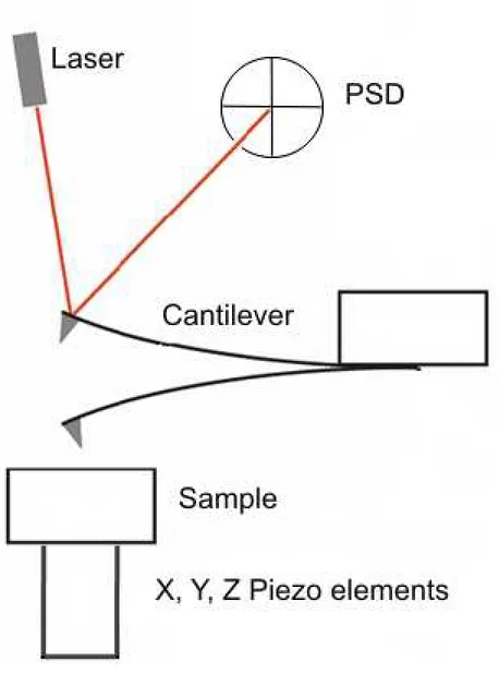
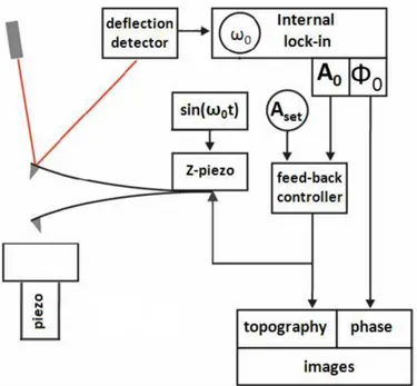
Novel techniques in multi-frequency atomic force microscopy and spectroscopy
Tam metin
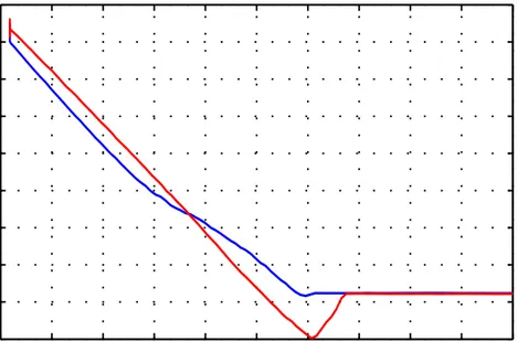
Şekil

Benzer Belgeler
Ovidius hem kendi sanat~~ için, hem de ~iirlerinin konusu (kendi a~k~) için uygun dü~tü~ü biçimde Cupido, Venus, Bacchus ve Apollo'nun kendine kar~~~ il- gisi oldu~unu ileri
As mentioned in the previous chapter, the solution concepts applied in multiple criteria decision making literature rely mainly on three ideas, namely: aggregating the
To this end, based on a sample of 409 Turkish employees and their 72 leaders, the current study investigates the effects of three dimensions of paternalistic leadership
The rest of this thesis is organized as following; chapter 2 is literature review of manufacturing system, it is focus on definition of manufacturing system
In this section, apart from the problem under the scope of this study, we have introduced additional hub network design problems with realistic features, namely, the multimodal
Given the 2-anonymized table, an attacker can at best link Luke into GPAs “3.72” and “2.34.” k-Anonymity does not enforce diversity on the sensitive information of equivalence
Apart from these we conducted 25 additional empirical tests used in statistical evaluation of random number packages and observed that Feigenbaum constants successfully pass all
If, instead, a fixed interval method had been used, the number of rescheduling points would then depend on the makespan of the schedule, which in turn would
