Res Cardiovasc Med. 2014 November; 3(4): e25715. DOI: 10.5812/cardiovascmed.25715
Published online 2014 November 26. Case Report
Acute Myocarditis or the Kounis Syndrome: Role of Cardiac MRI and
Speckle-Tracking Echocardiography in Diagnosis
Oguz Karaca
1,*; Beytullah Cakal
1; Sinem Deniz Cakal
1; Muhsin Turkmen
11Department of Cardiology, Faculty of Medicine, University of Medipol, Istanbul, Turkey
*Corresponding author: Oguz Karaca, Department of Cardiology, Faculty of Medicine, University of Medipol, Istanbul, Turkey. Tel: +90-5053557600, Fax: +90-2124607070, E-mail:
Received: November 29, 2014; Revised: December 2, 2014
Introduction: The Kounis syndrome and acute myocarditis are two distinct clinical entities, which could share nearly the same
symptomatology as well as ECG (electrocardiography) and laboratory findings.
Case Presentation: First case was a 39-year-old male presented with acute chest pain and inferolateral ST elevation on ECG. The second case
was a 29-year-old male presented with chest pain and diffuses ST elevation. Diagnosis of acute myocarditis was achieved by demonstrating subepicardial contrast enhancement as well as atypical involvement in both of our clinical cases.
Conclusions: We reported two cases in which specific imaging modalities (cardiac magnetic resonance imaging and speckle-tracking
echocardiography) were used when the signs and symptoms were indistinguishable.
Keywords:Kounis Syndrome; Myocarditis; Magnetic Resonance Imaging, Speckle-Tracking Echocardiography
Copyright © 2014, Rajaie Cardiovascular Medical and Research Center, Iran University of Medical Sciences. This is an open-access article distributed under the terms of the Creative Commons Attribution-NonCommercial 4.0 International License (http://creativecommons.org/licenses/by-nc/4.0/) which permits copy and redis-tribute the material just in noncommercial usages, provided the original work is properly cited.
1. Introduction
The Kounis syndrome and acute myocarditis are two distinct clinical entities, which could share nearly the same symptomatology as well as ECG (electrocardiogra-phy) and laboratory findings. After exclusion of signifi-cant coronary artery disease by coronary angiography, diagnostic dilemmas have usually been encountered in clinical practice. While history of atopy and allergenic substance exposure supports the Kounis syndrome known as "allergic myocardial infarction", preceding symptoms compatible with a viral infection are consid-ered to be related to acute myocarditis. We reported two cases in which specific imaging modalities were used when the signs and symptoms were indistinguishable.
2. Case Presentation
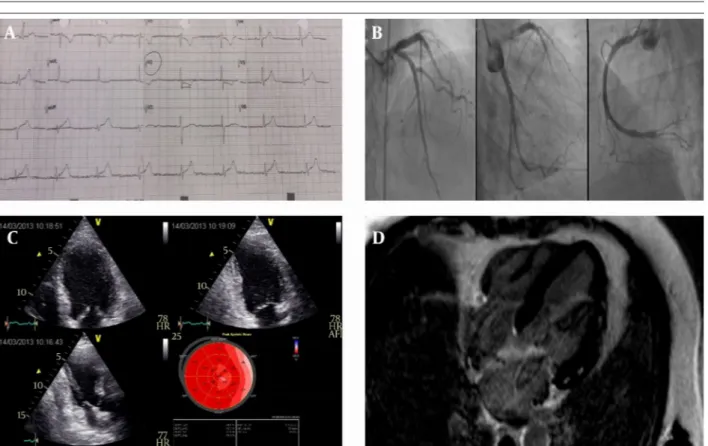
First case was a 39-year-old male presented with acute chest pain and inferolateral ST elevation on ECG (Figure 1 A). He was receiving amoxicillin because of pharyngi-tis. Angiography revealed non-critical plaques (Figure 1 B). TTE showed posterolateral hypokinesis with an EF of 45%. Acute myocarditis and the Kounis syndrome (al-lergic myocardial infarction due to amoxicillin) were considered as differential diagnoses. LV (left ventricular) strain analysis showed reduced global (GLS: global lon-gitudinal strain) and regional values incompatible with any coronary artery area (Figure 1 C). MRI showed
antero-lateral and posteroantero-lateral hypokinesia and edema with late gadolinium enhancement (LGE) at subepicardial regions (Figure 1 D). The second case was a 29-year-old male presented with chest pain and diffuses ST elevation (Figure 2 A) on ECG and elevated serum troponin level. He was receiving cefuroxime because of tonsillitis on admission. Angiography revealed normal coronary ar-teries (Figure 2 B). On transthoracic echocardiography, EF was 40% with hypokinesia of the anterior and lateral walls. 2D-speckle tracking echocardiography showed re-duced left ventricular global strain (LV-GLS) values and reduced strain values at the apical, anterior and infero-septal walls (Figure 2 C). MRI showed anterolateral and inferior hypokinesia and edema with LGE at the sub-epicardial regions (Figure 2 D). Diagnosis of acute myo-carditis was achieved by demonstrating subepicardial contrast enhancement as well as atypical involvement in both of our clinical cases.
3. Conclusions
Myocarditis and allergic myocardial infarction known as the Kounis syndrome should be always kept in mind in patients with acute chest pain, ECG changes and in-creased cardiac enzymes in case of normal coronary ar-teries (1). In the Kounis syndrome, subendocardial layer of the myocardium is affected compatible with the territory
Karaca O et al.
Res Cardiovasc Med. 2014;3(4):e25715 2
Figure 1. A, ECG of the patient showing marked ST segment elevations; B, coronary angiographic snapshots showing that the patient does not have signifi-cant coronary disease; C, ‘bull’s eye’ report of the longitudinal strain analysis showing heterogeneous impairment by speckle tracking echocardiography; D, magnetic resonance imaging demonstrating subepicardial contrast enhancement.
Figure 2. A, Diffuse ST elevations on the ECG; B, coronary angiography revealed normal coronary arteries; C, impairment in longitudinal strain is not compatible with a specific coronary artery territory; D, subepicardial LGE in cardiac MRI suggestive of acute myocarditis.
Karaca O et al.
3 Res Cardiovasc Med. 2014;3(4):e25715
of a particular coronary artery, while heterogeneous and subepicardial involvement is detected in acute myocarditis (1, 2). Combination of cardiac MRI and speckle-tracking echocardiography successfully differentiates these two clinical entities when ECG and laboratory findings are indistinguishable.
Authors’ Contributions
Oguz Karaca: Analysis and interpretation of data and drafting of the manuscript; Beytullah Cakal and Sinem
Deniz Cakal: acquisition of data. Muhsin Turkmen: ad-ministrative, technical and material support.
References
1. Almpanis GC, Mazarakis A, Dimopoulos DA, Tragotsalou NA, Kou-nis GN, KouKou-nis NG, et al. The conundrum of hypersensitivity car-diac disease: hypersensitivity myocarditis, acute hypersensitiv-ity coronary syndrome (Kounis syndrome) or both? Int J Cardiol. 2011;148(2):237–40.
2. Akoz A, Bayramoglu A, Uzkeser M, Kantarci M, Aksakal E, Emet M. Two questions for Kounis syndrome: can we use magnetic reso-nance imaging in the diagnosis and does ST elevation correlates with troponin levels? Am J Emerg Med. 2012;30(9):2086 e5–7.