REACTIVATION OF
TELOMERASE REVERSE TRANSCRIPTASE GENE
IN LIVER CANCER
A THESIS
SUBMITTED TO THE DEPARTMENT OF MOLECULAR BIOLOGY AND GENETICS
AND THE GRADUATE SCHOOL OF ENGINEERING AND SCIENCE OF BILKENT UNIVERSITY
IN PARTIAL FULFILLMENT OF THE REQUIREMENTS FOR THE DEGREE OF
DOCTOR OF PHILOSOPHY
BY
DİLEK ÇEVİK
SEPTEMBER, 2014
i
I certify that I have read this thesis and that in my opinion it is fully adequate, in scope and in quality, as a thesis for the degree of Doctor of Philosophy.
Assoc. Prof. Dr. Rengül Çetin-Atalay Prof. Dr. Mehmet Öztürk (Advisor) (Co-Advisor)
I certify that I have read this thesis and that in my opinion it is fully adequate, in scope and in quality, as a thesis for the degree of Doctor of Philosophy.
Prof. Dr. Tamer Yağcı
I certify that I have read this thesis and that in my opinion it is fully adequate, in scope and in quality, as a thesis for the degree of Doctor of Philosophy.
Assoc. Prof. Dr. Işık Yuluğ
I certify that I have read this thesis and that in my opinion it is fully adequate, in scope and in quality, as a thesis for the degree of Doctor of Philosophy.
Assoc. Prof. Dr. A.Elif Erson-Bensan
I certify that I have read this thesis and that in my opinion it is fully adequate, in scope and in quality, as a thesis for the degree of Doctor of Philosophy.
Assist. Prof. Dr. Ali Osmay Güre
Approved for the Graduate School of Engineering and Science:
Prof. Dr. Levent Onural Director of the Graduate School
ii
ABSTRACT
REACTIVATION OF
TELOMERASE REVERSE TRANSCRIPTASE GENE IN LIVER CANCER
Dilek Çevik
Ph.D. in Molecular Biology and Genetics Advisor: Assoc. Dr. Rengül Çetin-Atalay Co-Advisor: Prof. Dr. Mehmet Öztürk
September, 2014
Hepatocellular Carcinoma (HCC) is one of the major causes of cancer related deaths worldwide and its incidence has been increasing drastically, especially in western countries. HCC has a heterogeneous molecular and pathological background with various underlying risk factors and survival rate of HCC patients is very low due to late diagnosis and limited curative therapies. The mechanisms involved in hepatocellular immortality gains critical importance in order to develop preventive and therapeutic options against HCC. Telomerase reactivation is a keystone for HCC cells during transformation process. TERT promoter mutations activating its promoter by creating a novel activating motif were recently identified in different cancer types. In this study; we determined TERT promoter mutation frequency in HCC cell lines and tumors which are 67% (10/15) and 34% (15/44) respectively. High frequency of TERT promoter mutations in HCC indicated a possible functional role during hepatocarcinogenesis. We performed transcriptional factor search to find a candidate TF that could bind to mutant TERT promoter and STAT1 came out of that search. To study the role of STAT1 during reactivation of TERT expression, we activated STAT1 signaling by Interferon alpha (IFN-α) treatment and down regulated
iii
STAT1 with RNA interference in several HCC cell lines. We have found that IFN-α was able to upregulate TERT expression in the HCC cell lines carrying a TERT promoter mutation and STAT1 knockdown was enough to eradicate this upregulation. In case of wild type cell lines, IFN-α treatment and STAT1 knock down had no effect on TERT expression. Our data delineates the contributions of
TERT promoter mutations to hepatocellular immortality and gives insights into the
potential use of TERT as a target for chemoprevention of hepatocarcinogenesis.
Keywords: Hepatocellular Carcinoma, TERT, promoter mutations, STAT1,
iv
ÖZET
KARACİĞER KANSERİNDE TELOMERAZ REVERS TRANSKRİPTAZ GENİNİN REAKTİVASYONU
Dilek Çevik
Moleküler Biyoloji ve Genetik Doktora Tezi Danışman: Doç. Dr. Rengül Çetin-Atalay
Eş Danışman: Prof. Dr. Mehmet Öztürk
Eylül, 2014
Hepatoselüler Karcinoma (HSK) dünyada görülen kanser kaynaklı ölüm vakalarının ana sebeplerinden biridir. HSK farklı risk faktörlerine sahiptir ve heterojen bir moleküler ve patolojik temel üzerine oluşmaktadır. HSK’ ya karşı daha etkili tanı ve tedavi seçenekleri geliştirmek için, hepatoselüler immortaliteye sebep olan mekanizmaların açıklanması önem kazanmaktadır. Telomeraz geninin yeniden aktive olması HSK hücreleri için kritik bir aşamadır. Yakın zamanda yapılan çalışmalarda, TERT geninin promotör bölgesinde mutasyonlara rastlanmıştır ve bu mutasyonların normalde var olmayan bir promotör motifi oluşturarak TERT geninin ifadesini arttırdığı düşünülmektedir. Bu çalışmada, HSK hücre hatlarındaki TERT promotör mutasyon oranı %67 (10/15), tümör örneklerindeki mutasyon oranı ise % 34 (15/44) olarak belirlenmiştir. TERT mutasyonlarının yüksek oranları, HSK oluşumunda önem taşıdıklarına işaret etmektedir. Yaptığımız analizlerde STAT1 transkripsiyon faktörü, TERT mutasyonlarının oluşturduğu yeni motife bağlanabilecek bir aday transkripsiyon faktörü olarak ön plana çıkmıştır. STAT1’in TERT gen ifadesi üzerine olan etkisini araştırmak için HSK hücreleri interferon-α ile muamele edildi ve bunun TERT promotör mutasyonu taşıyan HSK hücre hatlarında TERT gen ifadesini arttırdığı belirlendi. STAT1 gen ifadesi RNA interferaz yöntemi ile düşürülerek bunun TERT gen ifadesi üzerine yaptığı etki araştırıldı. TERT promotör mutasyonu
v
taşıyan HSK hücre hatlarında STAT1 gen ifadesi düşürüldüğünde, TERT gen ifadesinin azaldığı ve IFN-α muamelesinin TERT gen ifadesi üzerine olan etkisinin de yok olduğu gözlendi. Mutasyon taşımayan hücre hatlarında ise, IFN-α muamelesinin ve STAT1 gen ifadesinin düşürülmesinin TERT gen ifadesi üzerine etkilerinin bulunmadığı tespit edildi. Bu sonuçlar, TERT promotör mutasyonlarının hepatoselüler immortalite üzerinde önemli bir etkisinin olduğunu ve bu mutasyonların HSK oluşumunun önlenmesi amacıyla kullanılabileceğini göstermektedir.
Anahtar kelimeler: Hepatoselüler Karsinoma, TERT, STAT1, promotör mutasyonu,
vi
vii
Acknowledgements
First and foremost, I would like to express my deepest gratitude to my advisor, Prof. Dr. Mehmet Ozturk for giving me the opportunity to work in this group, for his supervision and guidance. I have learnt a lot from his experience in the HCC field and wisdom for life throughout the course of my PhD.
I would like to extend my appreciation to my co-advisor Assoc. Prof. Dr. Rengul Cetin-Atalay for her support and guidance during my PhD.
I also would like to thank Dr. Stefan Dimitrov for giving me the opportunity to work in his laboratory at the Institute Albert Bonniot.
I am grateful to Dr. Hani Alotaibi for his contributions to this study during the last years of my PhD; he was very supportive and motivating.
I also would like to thank past Ozturk group members; Gokhan, Ozge, Mustafa, Aysegul, Cigdem, Yusuf İsmail, Merve Deniz, Umur and Dimitrov group members for the great working environment and for their friendship.
Moreover, I would like to thank all past and present members of MBG department, especially Uygar Tazebay, Bilge Kılıc, Fusun Elvan and Abdullah Unnu.
Here, I would like to express my deepest gratitude to my friends; Ece, Svetlana, Sevgin, Pelin, Aysegül, Yusuf İsmail, Defne and Duygu for their endless support throughout my PhD. Without their love and caring, I would not have been able to survive graduate school.
I am grateful to my family, especially my sister Arzu and my mother; I always felt their love and support and they were always with me whenever I needed them.
Finally, I would like to thank The Scientific and Technological Research Council of Turkey (TÜBİTAK) for supporting me during my PhD study through BİDEB 2211 scholarship.
viii
Table of Contents
ABSTRACT ... ii
ÖZET... iv
Acknowledgements ... vii
Table of Contents ... viii
List of Figures ... xiii
List of Tables... xv
Abbreviations ... xvi
1. Introduction ... 2
1.1 Hepatocellular Carcinoma ... 2
1.1.1 Epidemiology of Hepatocellular Carcinoma ... 2
1.1.2 Etiologies and Risk Factors of Hepatocellular Carcinoma ... 4
1.1.2.1 Hepatitis B Virus ... 5
1.1.2.2 Hepatitis C Virus ... 6
1.1.2.3 Alcohol Abuse ... 7
1.1.2.4 Aflatoxin Exposure ... 8
1.1.2.5 Non Alcoholic Fatty Liver Disease (NAFLD) ... 8
1.1.2.6 Obesity and Diabetes Mellitus ... 8
1.1.3 Molecular Mechanisms of Hepatocarcinogenesis ... 9
1.1.4 Genetics and Epigenetics of Hepatocellular Carcinoma ... 10
1.1.5 Liver Cirrhosis ... 11
1.1.6 Diagnosis and Prognosis of Hepatocellular Carcinoma ... 12
1.1.7 Treatment of Hepatocellular Carcinoma ... 12
ix
1.2.1 Replicative Senescence ... 14
1.2.2 Cellular Immortality in HCC ... 16
1.2.3 TERT Promoter Structure and Regulation ... 16
1.2.4 TERT Promoter Mutations in Different Cancers ... 17
1.3 Signal Transducer and Activator of Transcription 1 (STAT1) and Cancer .... 19
1.3.1 STAT Transcription Factor Family ... 19
1.3.2 STAT1 and Interferon (IFN) Signaling ... 20
1.3.3 STAT1 Signaling and HCC ... 21
1.3.4 IFN-α Therapy during HCC Treatment ... 22
1.4 Aim and Strategy ... 23
2. Materials and Methods ... 25
2.1 Materials ... 25
2.1.1 General Laboratory Reagents ... 25
2.1.2 Tissue Culture Reagents and Materials ... 25
2.1.3 Genomic DNA Isolation ... 26
2.1.4 Polymerase Chain Reaction (PCR) ... 26
2.1.5 Primers ... 26
2.1.6 Agarose Gel Electrophoresis ... 26
2.1.7 Spectrophotometry ... 27
2.1.8 Determination of Gene Expression ... 27
2.1.8.1 Total RNA Isolation ... 27
2.1.8.2 First Strand cDNA Synthesis ... 27
2.1.8.3 Quantitative Real Time PCR ... 27
x
2.2 Solutions and Media ... 28
2.2.1 General Solutions ... 28
2.2.2 Bacterial solutions ... 28
2.2.3 Tissue culture solutions ... 29
2.2.4 Sodium Deodecyl Sulphate (SDS)-Polyacrylamide Gel Electrophoresis (PAGE) and immunoblotting Solutions ... 29
2.3 Methods ... 30
2.3.1 Tissue Culture Methods ... 30
2.3.1.1 Cell Lines and Growth Conditions of Cells ... 30
2.3.1.2 Passaging Cells ... 30
2.3.1.3 Cryopreservation and Thawing of Cells ... 30
2.3.1.4 Transfection of siRNA with Lipofectamine RNAi MAX ... 31
2.3.1.5 Transfection of Plasmid DNA with Lipofectamine 2000 ... 32
2.3.1.6 Treatment of Cells with Interferon alpha ... 32
2.3.2 Isolation of Genomic DNA from Cultured Cells ... 32
2.3.3 Amplification of Genomic DNA ... 33
2.3.4 Agarose Gel Electrophoresis ... 34
2.3.5 Total RNA Extraction from Cultured Cells ... 34
2.3.6 First Strand cDNA Synthesis ... 35
2.3.7 Quantitative Real Time PCR (qRT-PCR) ... 36
2.3.9 Total Protein Extraction from Cultured Cells ... 36
2.3.10 Western Blotting ... 37
2.3.11 Statistical Analysis and Bioinformatics Tools ... 40
2.3.11.1 DNA Dynamo Sequence Analysis Software ... 40
xi
3. Results ... 41
3.1 TERT Promoter Mutations in HCC ... 41
3.1.1 TERT Promoter Mutations in HCC Cell lines ... 41
3.1.2 TERT Promoter Mutations in HCC Tumors ... 46
3.1.3 Geographic Distribution of TERT Promoter Mutations in HCC patients . 48 3.1.4 Association of TERT promoter mutations with patient characteristics ... 49
3.2 TERT Promoter Polymorphism (rs2853669) Status in HCC ... 51
3.2.1 rs2853669 Polymorphism in HCC cell lines ... 51
3.2.2 rs2853669 Polymorphism in HCC Tumors ... 52
3.3 STAT1 is a Candidate Transcription Factor for Mutant TERT Promoter ... 55
3.3.1 Transcription Factor Search for Mutant TERT Promoter ... 55
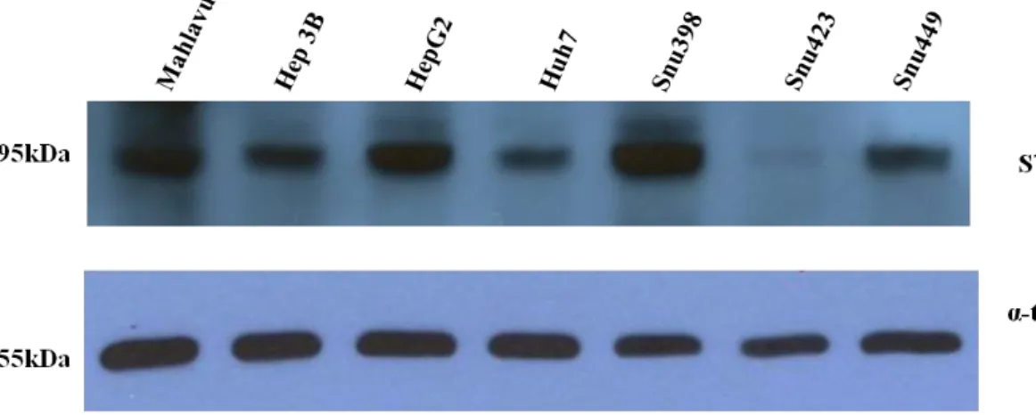
3.3.2 Detection of STAT1 protein level in HCC Cell Lines ... 58
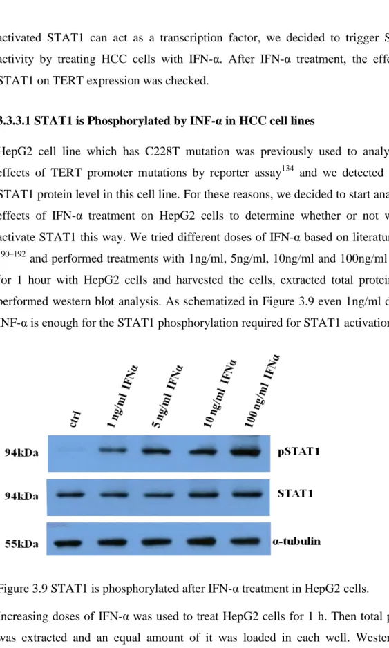
3.3.3 Activation of STAT1 by IFN-α in HCC Cell Lines ... 58
3.3.3.1 STAT1 is Phosphorylated by INF-α in HCC cell lines ... 59
3.3.3.2 IRF1 is upregulated by IFN-α in HCC cell lines ... 61
3.4 Regulation of TERT expression by IFN-α in HCC cell lines ... 64
3.4.1 TERT is upregulated by IFN-α in mutant HCC Cell Lines ... 65
3.4.2 TERT expression is not regulated by IFN-α in WT HCC cell lines ... 70
3.5 Regulation of TERT expression by STAT1 knockdown in HCC cell lines ... 73
3.5.1 TERT is down regulated by STAT1 knock down in HepG2 cell line ... 73
3.5.2 TERT is downregulated by STAT1 knock down in Mahlavu cell line .... 78
3.5.3 TERT is not regulated by STAT1 knock down in PLC cell line ... 81
4. Discussion ... 82
xii
References ... 89 Appendices ... 113 Curriculum Vitae and Publications ... 117
xiii
List of Figures
Figure1.1 Mortality and Incidence Rates of HCC across different geographical
regions. ... 3
Figure1.2 HCC is induced by multiple risk factors... 4
Figure1.3 Molecular Signaling Pathways Involved in Hepatocarcinogenesis ... 10
Figure1.4 Telomere induced senescence... 15
Figure1.5 Schematic representation of a part of the TERT promoter that contains hotspot mutations ... 18
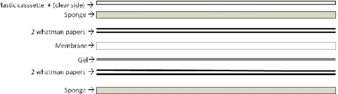
Figure 2.1 Placement of gel and membrane for blotting ... 39
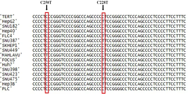
Figure 3.1 TERT sequencing alignment in HCC cell lines. ... 42
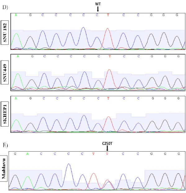
Figure 3.2 Sequence chromatograms of HCC cell lines comprising TERT promoter mutations C228T and C250T. ... 45
Figure 3.3 TERT sequencing alignment in HCC tumors. ... 47
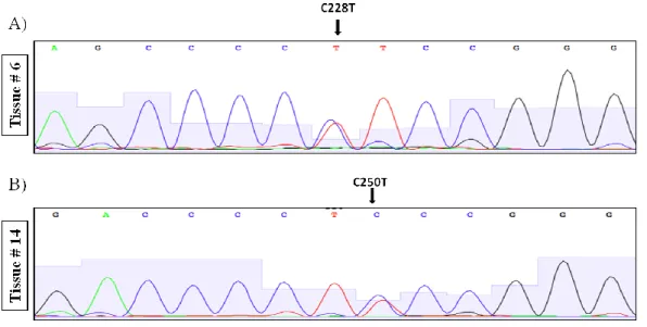
Figure 3.4 Representative sequence chromatograms of HCC tumor samples carrying TERT promoter mutations C228T and C250T ... 48
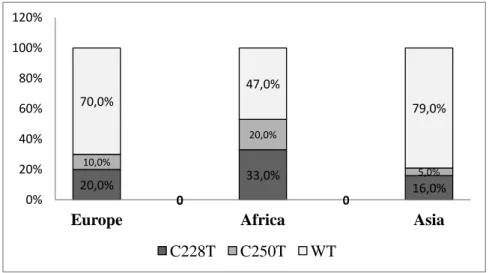
Figure 3.5 Geographic distibution of TERT promoter mutations across the world. .. 49
Figure 3.6 STAT Transcription Factor Family is a candidate TF family that might bind to common mutation motif. ... 56
Figure 3.7 STAT1 is among individual Transcription Factors that might bind to common mutation motif ... 57
Figure 3.8 STAT1 protein is differentially expressed in HCC cell lines. ... 58
Figure 3.9 STAT1 is phosphorylated after IFN-α treatment in HepG2 cells. ... 59
xiv
Figure 3.11 IRF1 expression is upregulated in response to IFN-α in HCC cells. ... 64
Figure 3.12 TERT expression is upregulated by IFN-α starting from 24h. ... 65
Figure 3.13 TERT expression is regulated by IFN-α in HCC cell lines. ... 66
Figure 3.14 TERT expression is regulated by IFN-α in HCC cell lines. ... 69
Figure 3.15 TERT expression is not regulated by IFN-α in wild type HCC cell lines. ... 71
Figure 3.16 TERT expression is not regulated by IFN-α in wild type HCC cell lines. ... 72
Figure 3.17 STAT1 is down regulated by STAT1 specific siRNA in HepG2 cells. . 74
Figure 3.18 TERT is downregulated after STAT1 knockdown in HepG2 cell line. . 75
Figure3.19 TERT expression is regulated by STAT1 knock down and IFN-α treatment in HepG2 cell line. ... 77
Figure 3.20 TERT is downregulated in response to STAT1 knockdown in Mahlavu cell line. ... 78
Figure3.21 TERT expression is regulated by STAT1 knock down and IFN-α treatment in Mahlavu cell line... 80
Figure3.22 TERT expression does not change in response to STAT1 knockdown in PLC cell line. ... 81
xv
List of Tables
Table2.1 Primers used in this study ... 26
Table2.2 Antibodies used in this study ... 27
Table 2.3 Preparations of Stacking and Resolving Tris Glycine Gels ... 37
Table 3.1 Association of TERT promoter mutations with patient characteristics ... 50
Table 3.2 TERT promoter mutations and rs2853669 polymorphism in HCC cell lines ... 52 Table 3.3 TERT promoter mutations and rs2853669 polymorphism in HCC tumors 53
xvi
Abbreviations
AH Alcoholic Hepatitis
ALD Alcoholic Liver Disease
E-Value Expected Value
HBV Hepatitis B Virus
HBx Hepatitis B virus X protein
HCC Hepatocellular Carcinoma
HCV Hepatitis C Virus
HSC Hepatic Satellite Cells
IFN-α Interferon alpha
NAFLD Non Alcohol Fatty Liver Disease
ROS Reactive Oxygen Species
STAT Signal Transducers and Activators of Transcription TERT Telomerase Reverse Transcriptase
2
Chapter 1
1.
Introduction
1.1 Hepatocellular Carcinoma
1.1.1 Epidemiology of Hepatocellular Carcinoma
Liver cancer is a global health problem since it is one of the most deadly cancers. Liver cancer has different hepatic neoplasms such as hepatocellular carcinoma (HCC), cholangiocarcinoma, hepatoblastoma, bile duct cystadenocarcinoma, haemangiosarcoma and epitheliod haemangioendothelioma1. Among these, HCC is the most commonly observed liver cancer type with 83% frequency and it is the sixth most common cancer worldwide. 2. HCC is two times more common in men compared to women. 782,000 new HCC cases occurred in 2012 worldwide and male patients comprised 554,000 while females comprised 228,000 of all the cases. HCC shows a non-uniform distribution across different continents. In men and women together, Eastern and South-Eastern Asia are the regions with the highest incidences of HCC; Northern and Western Africa have the intermediate rates while Northern Europe and South-Central Asia are the regions with the lowest mortality rates. Mortality rates are very similar to incidence frequency of HCC since the survival rate is very low due to limited treatment options (Figure 1.1). HCC is the second leading cause of cancer related deaths worldwide with an estimated number of nearly 746,000 deaths in 2012 which is 9.1% of the total cancer related deaths.
3
Figure1.1 Mortality and Incidence Rates of HCC across different geographical regions.
In all regions, incidence and mortality rates are quite similar. Eastern Asia has the highest incidence, Africa has the intermediate incidence and Europe and America has relatively lower incidences. Age standardized rate (W) per 100,000 cases. Adopted from GLOBOCAN.
Although HCC incidence is highest in China and other developing countries, the incidence of HCC has been increasing drastically in United States and Europe over the last decade 3. In European countries like France, England and Spain; HCC incidence shows an increasing trend while in Slovakia and Denmark, HCC incidence is more stable. In Asian countries such as Japan, China, Philippines and Singapore; HCC incidence has been decreasing while in India there is an increased tendency. In United States, Canada and Australia, HCC incidence has been increasing, on the other hand, Colombia and Costa Rica has unstable changes in HCC rates. The increase of HCC incidence in Western countries is mostly caused by cirrhosis associated with alcohol abuse, fatty liver disease caused by obesity and type-2 diabetes 4.
4
1.1.2 Etiologies and Risk Factors of Hepatocellular Carcinoma
The most common risk factors of hepatocellular carcinoma include Hepatitis B and C viruses, Aflatoxin B exposure and alcohol abuse. Non Alcoholic Fatty Liver Disease (NAFLD), diabetes and haemochromatosis can be classified among less critical risk factors5. Mechanisms induced by the presence of the most common risk factors are shown in Figure 1.3. Aflatoxin B1 and HBV act together to inactivate p53 which increases cell proliferation. Viral hepatitis and alcohol abuse induced inflammation causes necrosis and regeneration cycles. Viral and alcohol induced oxidative stress causes mutagenesis and alters signaling pathways in favor of tumorigenesis. Finally, all risk factors lead to HCC formation after accumulation of genetic alterations in hepatocytes 2.
Figure1.2 HCC is induced by multiple risk factors.
Hepatitis B and Hepatitis C viruses, Aflatoxin B exposure and alcohol induce hepatocarcinogenesis by acting on intersecting mechanisms such as inactivation of tumor suppressor p53, chronic liver inflammation, cirrhosis, oxidative stress and mutagenesis. Adopted from 2 .
5 1.1.2.1 Hepatitis B Virus
Hepatitis B virus (HBV) chronic infection is the primary cause of HCC worldwide since more than 50% of all HCC patients are infected with HBV 6. However, there is a high geographical heterogeneity in HBV incidence 7. In Asia and Africa, more than 70% of HCC cases are attributable to HBV due to high incidence of infection 8. HBV enhances the effects of aflatoxin exposure to trigger HCC formation, thus patients with HBV carry a greater risk of getting HCC if they are exposed to aflatoxin 9. In USA and Australia, HBV incidence is low in the general population, thus, 70–80% of the HCC cases that are linked to HBV are observed in immigrants from Asia 10. In Europe, HBV incidence is very low compared to Asia or Africa, therefore, it is a low risk factor of HCC in European population 11. Survival rates of HBV-related HCC is extremely low, thus HBV infection is a major public health problem 12. Mechanisms behind the involvement of HBV in hepatocarcinogenesis have been discussed since the first time HBV was claimed to be related to HCC formation at seventies 13. Most prominent effect of HBV during liver cancer progression starts with the integration of viral DNA into the host genome causing genetic instability and activation of oncogenes 14. HBV DNA integration occurs in the early acute phase of the infection since it is critical for the persistence of the infection in hepatocytes even though it is not necessary for viral replication 15. Apart from causing genetic instability, HBV also triggers hepatocarcinogenesis by synthesizing viral proteins such as HBx, PreS2/S, HBSP which will in turn cause genetic alterations, transactivation of oncogenic transcription factors, and deregulation of important cellular pathways such as cell proliferation, differentiation, and survival of hepatocytes 16. HBx acts as a pro-apoptotic protein by creating reactive oxygen species (ROS) or as an anti-apoptotic protein by inhibiting p53, Fas, TNF and TGF-β induced apoptosis17. C terminal truncated version of HBx protein also contributes to hepatocarcinogenesis through different pathways; creating oxidative stress and mitochondrial damage; increasing invasiveness and metastatic potential of HCC; and causing over expression of Centromere protein A in HCC tissue 18. HBx has another oncogenic effect by preventing repair of DNA damage by inhibiting base excision
6
repair system due to its structural similarity with human thymine DNA glycosylase , an important enzyme for the proper functioning of BER pathway 19.
1.1.2.2 Hepatitis C Virus
Hepatitis C Virus (HCV) is a positive RNA virus that belongs to the Flaviviridae family 20. HCV causes a chronic infection in most of the infected individuals; it stays dormant for decades and then causes liver fibrosis and cirrhosis at the final stages of hepatitis 21. There are 170 million people infected with HCV worldwide, thus, HCV is the second main risk factor for HCC and HCV related HCC cases comprise 10– 20% of HCC cases worldwide 22. Unlike HBV associated HCC, most HCV-related HCC cases develop after liver fibrosis and cirrhosis reach critical levels 23. Chronic inflammation caused by chronic HCV infection creates a great risk of developing HCC, a risk higher than the one caused by any other non-viral risk factors 24. HCV infection causes inflammation in the liver (hepatitis) and leads to infiltration of lymphocytes and Natural Killer cells 25. Recognition of viral RNA by RIG-I (retinoic acid-inducible gene 1) and TLR-3 (Toll like receptor 3) triggers NF-κB pathway and cells start to secrete interferon and other pro-inflammatory cytokines. Moreover, HCV polymerase is found to activate inflammatory signaling pathways causing cytokine release 26. Generation of ROS by HCV core protein leads to oxidative stress and DNA mutagenesis and acts as another mean of contribution to hepatocarcinogenesis 27. Other than chronic inflammation, there are more direct effects of HCV on HCC. Both tumor suppressor genes and proto-oncogenes are subject to deregulation by viral proteins. HCV polymerase NS5B interacts with one of the most critical tumor suppressors, the retinoblastoma protein (Rb), causes its degradation favoring entry of cells into S phase 28. HCV proteins interact with another critical tumor suppressor protein, p53, to inhibit apoptosis 29. Furthermore, HCV targets the WNT/β-catenin pathway by causing stabilization of β-catenin and preventing it from functioning 30. Transforming growth factor beta (TGF-β) is also a target of HCV proteins; HCV NS5A interacts with TGF-β receptor and prevents its signaling, leading to improper regulation of hepatocyte proliferation and contributes to further liver damage, fibrosis and transformation 31.
7 1.1.2.3 Alcohol Abuse
Severe alcohol consumption increases HCC risk up to 5 fold by a multistep process called Alcoholic Liver Disease (ALD) that leads to chronic liver disease, liver fibrosis and cirrhosis, and hepatocellular carcinoma 32,33. Alcohol induced fatty liver disease, also called steatosis is the first pathology caused by chronic alcohol intake. As the name implies, it is caused by the accumulation of fat in liver cells. Alcohol increases the storage of triglycerides, phospholipids, and cholesterol by preventing their oxidation in mitochondria 32. Another effect of alcohol on liver metabolism is to increase lipid uptake by the liver. Transcription factors regulating lipid metabolism are also subject to regulation by alcohol intake through induction of lipogenesis and inhibition of lipid oxidation. SREBP-1c (Sterol Regulatory Element-Binding Protein 1c) is a transcription factor favoring lipogenesis and it is upregulated by Ethanol in hepatocytes during severe alcohol intake 34. Alcoholic Hepatitis (AH) is another pathology of alcohol abuse; it is marked with liver inflammation and hepatic injury caused by infiltration of inflammatory cells into the liver 35. AH occurs after steatosis and is mostly accompanied by liver fibrosis in 10-35% of alcohol abusers. EtOH is metabolized in hepatocytes into acetaldehyde and acetate forms after acetaldehyde degradation 36. Reactive oxygen species and acetaldehyde generated during ethanol metabolism in hepatocytes cause hepatocyte injury 37. Acetate is not hepatoxic by itself but it upregulates pro inflammatory cytokine secretion triggering chronic inflammation in liver 38. AH causes hepatocyte apoptosis, activation of innate immunity, infiltration by neutrophils, activation of adaptive immunity and inhibition of liver regeneration 39. Liver fibrosis is the next step of alcoholic liver disease. It is a physiological healing process in response to chronic liver damage and it forms by accumulation of extracellular matrix proteins such as collagen, however excess amount of fibrotic liver tissue prevents liver from functioning 40. Chronic liver damage stimulates Hepatic Satellite Cells (HSCs), fibroblasts and myofibroblasts to synthesize collagen and other extracellular matrix proteins 41. Alcoholic cirrhosis leads to development of HCC as any other cirrhosis and it also increases the effects of HCV chronic infection 33.
8 1.1.2.4 Aflatoxin Exposure
Aflatoxins are main dietary risk factors triggering hepatocellular carcinogenesis; indeed 5 to 25% of HCC cases are associated with aflatoxin exposure 42. Aflatoxins are produced by Aspergillus flavus and Aspergillus parasiticus and mainly found in contaminated food such as maize, rice, soy bean and nuts 43. Aflatoxin is linked to hepatocellular carcinoma especially in sub-Saharan Africa, Southeast Asia and China where high exposure of this contaminant is observed in the general population 44. Aflatoxin B1, the major hepatotoxic agent, metabolizes into mutagenic products in the liver. It is the main cause of the transversion mutation (G:C to T:A) found at codon 249 of the tumor suppressor TP53 gene 45. Since it is a well defined carcinogen, it was used as a biomarker in a study based in China and a very significant correlation was found between the urinary concentrations of aflatoxin metabolic by products and HCC occurrence 9. To decrease HCC cases associated with aflatoxin exposure, aflatoxin content in food should be strictly controlled and any chemopreventive strategy should be applied in case of detection of the contaminant 44.
1.1.2.5 Non Alcoholic Fatty Liver Disease (NAFLD)
Non Alcoholic Fatty Liver Disease (NAFLD) is a disease marked with the accumulation of lipids in the liver of individuals who are not heavy alcohol drinkers
46
. NAFLD causes cirrhosis eventually, thus it is as dangerous as other triggers of cirrhosis that lead to HCC 47. Unlike Asia where most HCC cases are related to HBV or HCV infections, in USA and Europe, it is NAFLD which establishes a huge risk of developing HCC 48. As the percentage of people with obesity and metabolic syndrome increases, the incidence of NAFLD also increases accordingly 49.
1.1.2.6 Obesity and Diabetes Mellitus
Epidemiologic data suggests that both obesity and type 2 Diabetes Mellitus (T2DM) are associated with increased risk of developing HCC 50. Metabolic disorders and increased fat tissue in liver are common observations in obesity and DM patients.
9
NAFLD is also quite frequent in these patients and there is a good correlation between Body Mass Index (BMI) and cancer risk 51. Lipid peroxidation which occurs excessively as a result of NAFLD causes formation of mutagens from ROS and leads to both mutation accumulation and liver damage 49. Moreover, obesity and diabetes are both associated with insulin resistance and increased IGF (Insulin-like growth factor) amount triggering cell proliferation and cancer progression 52.
1.1.3 Molecular Mechanisms of Hepatocarcinogenesis
Pathogenesis and molecular mechanisms of hepatocarcinogenesis are quite complex due to involvement of various factors during the development of HCC 2. One of the first events through hepatic transformation is acquirement of mutations, genetic and epigenetic changes that lead to malignant transformation 53. Other than hepatocytes, there are other cells that occupy the liver such as cholangiocytes, Kupffer cells, sinusoidal endothelial cells, and hepatic stellate cells (HSCs). Exposure to hepatoxic substances such as aflatoxins and infection of the liver cells leads to inflammation in Kupffer cells and HSCs 54. In case of severe chronic inflammation; necrosis, fibrosis and cirrhosis occur. The underlying reason for cirrhosis can be viral infections such as HBV and HCV, alcohol abuse or NAFLD. Once cirrhosis occurs, there is no going back in most cases and cirrhosis turns into HCC55. Hepatic cells enter into damage-repair-regeneration cycle and this increases chromosomal instability and sensitivity of the cells against carcinogens that cause formation of dysplastic nodules and HCC eventually 56. Main pathways involved in hepatic transformation are as follows: tumor suppressor TP53, Retina Blastoma protein (Rb), Wnt-CDKN2A (β-catenin), IGF2R (Insulin-like growth factor receptor-2), PTEN (Phosphatase and tensin homolog), Axin1, PI3K (Phospatidiyl Inositol Kinase), JASTAT pathway, and K-ras/MAPK (K Rat Sarcoma) and TGF-β (Transforming Growth Factor Beta) 55. A summary of these pathways is depicted in Figure 1.5. Each pathway is given together with the underlying risk factor that leads to HCC.
10
Figure1.3 Molecular Signaling Pathways Involved in Hepatocarcinogenesis
These pathways act together in a synergistic or additive manner during HCC development. Depending on the etiology, different combinations of these pathways can be observed in a patient specific manner. Adopted from 55.
1.1.4 Genetics and Epigenetics of Hepatocellular Carcinoma
Chromosomal aberrations in the form of deletions and copy number variations are commonly observed in HCC 57. Most frequently amplified regions are the chromosomes 1q, 8q, 6p, 7, 8q, 17q and 20 while most frequently lost regions are 1p, 4q, 6q, 8p, 13q, 16, 17p and 21 chromosomal loci 58. 13q and 4q are also lost, but only in poorly differentiated HCC tumors 59. Chromosomal changes are not random but rather comprise the locations of critical genes such as p53 (17p) or Rb (13q) 60. Moreover, there are genetic variations caused by integration of HBV DNA into the host genome in HCC patients with underlying HBV infection 61. Integration of HBV DNA within or upstream of the TERT (telomerase reverse transcriptase) gene causes upregulation of TERT and provides cellular immortality in HCC patients 62. TP53 was the first gene discovered to be mutated in HCC 63. The two most frequently mutated genes are TP53 (coding for p53) with 35% of mutation frequency and CTNNB1 (coding for β-catenin protein) with close to 20% of mutation frequency
58,62,64
11
frequently mutated in HCC patients 65. There are also a group of less frequently mutated genes that are involved in pathways critical for hepatocarcinogenesis such as Wnt/b-catenin, p53, PI3K/Ras signalling, oxidative and endoplasmic reticulum stress pathways 55.
In addition to genetic regulation of gene expression, epigenetic pathways regulating gene expression are also deregulated in HCC 66. Like many other cancers, global hypomethylation and promoter hypermethylation are main epigenetic changes observed in HCC 67. Mutations at the genes involved in epigenetic regulation are also reported in HCC and most frequent mutations were found in ARID1A, ARID1B, ARID2 genes 58,68.
1.1.5 Liver Cirrhosis
Liver cirrhosis is the term used for the last stage of chronic liver disease associated with regenerative nodules, fibrotic and necrotic liver tissue 69. Regardless of the etiology behind it (HBV, HCV, alcohol abuse, toxic exposure etc.) liver cirrhosis is the major risk factor for hepatocarcinogenesis. The clinical complications of cirrhosis include impaired hepatocyte function, increased vascular pressure (portal hypertension), ascites formation 70. Regenerative nodules are composed of disorganized hepatocytes, fibrotic tissue and bile duct cells and have occasional dysplastic nodules. Small and large cell dysplasias are two forms of dysplasia observed in liver. Small cell dysplasia (SCD) is characterized by hepatocytes with an increased nucleocytoplasmic ratio whereas Large Cell Dysplasia (LCD) is characterized by a decrease in nucleocytoplasmic ratio 71. SCD is composed of cells with a high proliferation potential, and have common chromosomal alterations with neighboring HCC tissue thus it is considered to be the early precursor of HCC 72. Unlike SCD, LCD is not considered to be the precursor of HCC since hepatocytes forming LCD have normal nucleocytoplasmic ratio and low proliferation capacity together with relatively high apoptosis. Another finding supporting this hypothesis is the fact that LCD forms as a result of hepatocyte senescence triggered by necrosis, inflammation and regeneration 73.
12
1.1.6 Diagnosis and Prognosis of Hepatocellular Carcinoma
Early diagnosis of HCC is not very common due to asymptomatic disease progression 74. Late diagnosis is the reason of limited therapy options and low disease survival rate 75. Ultrasonograpy is the most common surveillance method since it is non invasive and available 76. Serum α-fetaprotein (AFP) is the most common serological test used for diagnosis of HCC; however, it has a low sensitivity
77
. Combination of ultrasound with AFP does not have a positive impact on diagnosis but increase false positive rates 78. Based on available data, ultrasound screening of the patient twice a year is recommended 79. When a nodule size exceeds 1cm in ultrasound, further diagnosis should be performed by biopsy or more accurate imaging methods such as CT (computer tomography) or MRI (magnetic resonance imaging) 80. These methods are only applicable for patients with cirrhosis or viral infections (HBV or HCV) without cirrhosis; liver biopsy is necessary for other patients 75.
Once the patient is diagnosed with HCC, determination of prognosis becomes critical. Early diagnosis and proper treatment provides a 5 year of survival rate for HCC patients. During the assessment of prognosis, several factors such as tumor stage, liver function, and cancer-related symptoms should be considered for proper assessment 81.
1.1.7 Treatment of Hepatocellular Carcinoma
After the diagnosis and classification of HCC, treatment options should be determined by qualified experts. Early diagnosed cases have treatment options such as surgical resection of the tumor, liver transplantation and ablation with high curative potential 81. Other therapeutic options are chemoembolisation and sorafenib; however, these treatments only increase survival, they are not curative for HCC 82. Systemic chemotherapy or drugs such as tamoxifen, octreotide, or antiandrogens have no effect on HCC 83. Surgical resection of the tumor is applicable to patients without cirrhosis and this group comprises a very low percentage of patients that is 5% in the USA and Europe and 40% in Asia 84. Liver transplantation is the best option to treat patients with one HCC tumor of 5 cm or smaller and for patients
13
having up to 3 nodules of 3 cm or smaller without metastasis 85. However, transplantation is limited with the number of donations 86. Tumor ablation, injection of chemicals such as ethanol or acetic acid to induce tumor necrosis, is also an effective option for early stage HCC. Survival rates after tumor ablation and resection is 5 years in majority of the patients thus they are considered as interchangeable therapeutic options 87. Chemoembolization (delivering a relatively large dose of chemotherapy directly to the liver tumor and cutting arterial blood supply) is only an option for patients with large tumors that are not suitable for resection or tumor ablation 88. The only FDA approved drug for HCC is Sorafenib which is a multikinase inhibitor and it increases patient survival for only three months compared to placebo group 89. In conclusion, there is an urgent need for novel biomarkers and therapeutic agents that can be used for HCC to increase the survival rate of both early and late diagnosed patients.
1.2 Telomerase and Cancer
Telomeres are repetitive DNA sequences (5′-TTAGGG-3′ in human) and a collection of proteins capping this DNA that are found at the end of chromosomes to protect chromosome ends from degradation and end-to-end chromosome fusions 90. Human telomere length is between 5 and 15 kb and it is mostly double stranded except a 30– 200 nucleotide GT-rich 3′ overhang 91 that forms a (T)-loop by annealing with a portion of double stranded region of the telomeric DNA. DNA polymerases cannot replicate the end of linear chromosomes due to lack of a proper primer sequence 92. This problem is known as end of replication problem 93. At the final stages of DNA replication from the lagging strand, a gap is formed after degradation of the very distal RNA primer. Moreover single stranded 3′ region of overhang of telomere has an intrinsic exonuclease activity and removes the 5′ end of the complementary strand
94
. At the end of each DNA replication, telomere DNA gets shorter due to these two mechanisms 94. Telomerase is a ribonucleoprotein containing Telomerase Reverse Transcriptase (TERT) enzyme, the catalytic part of the complex and RNA component (TERC), the template used during synthesis of new telomeric DNA 95. There are other proteins necessary to form the whole telomerase such as DKC
14
(dyskerin), (NOP10NOP10 ribonucleoprotein), GAR1 (GAR1 ribonucleoprotein homolog, NHP2 (NHP2 ribonucleoprotein), reptin and pontin 96. Theoretically, telomerase can solve the end of replication problem by adding lost repeats to the chromosome ends, however, most normal somatic cells, except germ cells lose TERT expression 95. Most tumor cells recover telomerase activity to overcome end of replication problem which will be discussed in detail in the next sections 97.
1.2.1 Replicative Senescence
The term of cellular senescence is first used by Hayflick and Moorhead in 1961 when they discovered that human fibroblasts cannot divide infinitely when they are continuously cultured 98. However, what they had called cellular senescence is now called replicative senescence and described as a type of cellular senescence which is associated with telomere shortening 99. Senescence phenotype is associated with growth arrest, apoptosis resistance, limited and altered gene expression, change in cell metabolism 100. Senescent cells also carry one or more of senescence markers such as senescence associated β-galactosidase, p16 and senescence associated DNA damage foci (SDF) and senescence associated heterochromatin foci (SAHF) 101. Oncogene induced senescence, ROS induced senescence, DNA damage induced senescence and replicative senescence are the main types of cellular senescence and they involve p53 and Rb pathways together with Cyclin dependent kinase inhibitors p16, p15, p21 and p27 102.
Telomeric DNA gets shorter after each cell division due to end of replication problem, when shortening of telomeres reach a critical level, cells can no longer divide and enter replicative senescence 103. Replicative senescence starts as critically short telomeres act like DNA double-stranded breaks that are sensed by 911 (RAD9/HUS1/RAD1) and MRN complex (MRE11–RAD50– NBS1), then DNA damage signal is sent to ATM-ATR and they activate CHK1 and CHK2 kinases which in turn activate p53 to make the cells enter a permanent cell cycle arrest 104. Most tumor cells gain telomerase activity to bypass senescence; however, TERT expression can only overcome telomere induced senescence but not oncogene induced senescence or other stress induced senescence types 105. The relationship
15
between telomere length, cell division and senescence is shown in Figure 1.6. In normal cells, there is no or very little TERT expression, that’s why they enter into senescence when telomere length reaches a critically short level. In contrast to normal cells, germ cells and tumor cells have telomerase activity that keeps telomeric DNA length in the normal level, thus these cells continue dividing 100.
Figure1.4 Telomere induced senescence.
Normal somatic cells are telomerase negative, thus they enter into senescence as a response to shortened telomere length. Germ cells and cancer cells express telomerase and they can divide infinitely without entering into telomere induced senescence. Adopted from 100 .
Replicative senescence was studied in normal liver, cirrhotic liver and HCC and it was found that replicative senescence was a rare event in normal liver and common in cirrhotic liver. In HCC, medium levels of senescent cells were detected 106. Since telomere length is an obstruction that leads to replicative senescence, it is expected to observe shorter telomere length in cirrhotic tissue that is already defined to have a high percentage of senescent cells. Indeed, telomere length was found to be significantly shorter in cirrhosis samples than that of the normal liver’s regardless of the etiology behind the disease and degree of shortening of telomeres and senescence significantly correlated with increase in fibrosis and cirrhosis 107.
16 1.2.2 Cellular Immortality in HCC
Replicative senescence induced by the shortening of telomeres should be bypassed by HCC cells for cellular transformation. HCC cells also need to recover TERT expression in order to maintain telomere length and gain immortality. They achieve bypassing senescence by acquiring mutations in p53 gene and Rb pathway genes (p16 INK4a, p15INK4b or RB1 genes) 108. p53 mutations are detected frequently in HCC 109.Rb pathway genes are targets of mutations and are mainly silenced through epigenetic mechanisms such as promoter metylation65.Acquiring telomerase activity is necessary and present in all kinds of cancers including HCC 110. Indeed, 90% of HCC cases have increased telomerase activity 111,112. One known mechanism to re-establish TERT expression in HCC is through integration of HBV viral DNA into TERT gene, however this is restricted to a very low number of HCC cases 113. Viral proteins HBx and PreS2 also reported to de-repress TERT expression in HCC 114,115. Oncogenes such as c-MYC, NF-κB and β- Catenin are transcription factors that induce TERT expression in several cancers 116. In addition to re-gaining TERT expression, HCC cells also up-regulate expression of other telomeric proteins in order to preserve functional telomeres 117. As an alternative to re-establishing TERT expression, tumor cells restore telomere length by a mechanism called Alternative Lengthening of Telomeres (ALT) that is similar to homologous recombination 118. 1.2.3 TERT Promoter Structure and Regulation
Telomerase reverse transcriptase (TERT) gene is a large gene (40 kb) located on chromosome 5p15.33 and it has 15 introns and 16 exons 119. TERT expression is regulated tightly due to its protein’s critical function. TERT core promoter is 260 base pairs and it does not contain TATA and CAAT boxes 120. TERT promoter has binding sites for great number regulatory factors. It has E-boxes that are subject to binding and activation by c-MYC. BRCA1 is also known to down-regulate TERT transcription together with (Nmi) N-Myc interacting protein and c-Myc 121. This inhibitory function of BRCA1 is compromised in some mutants 122. TERT transcription is repressed by p53 tumor suppressor 123. ETS transcription factor family members activate TERT transcription 124 and they are activated by several
17
oncogenes such as EGF, Her2/Nez, Ras and Raf 125. One other property of TERT promoter is the fact that it is highly GC rich which makes it a target for zinc finger transcription factor, Sp1 126 and this GC rich promoter is suitable for epigenetic regulations such as promoter methylation and chromatin remodeling 127. Finally, TERT transcription is also controlled by hormones such as Estrogen and cytokines such as TGF-β 128. During cancer formation, oncogenes are activated whereas tumor suppressors are repressed; these two events contribute to immortalization by regulating TERT expression in favor of tumor cells 129. Moreover, recent reports discovered a critical relationship between obesity related hormone leptin and telomerase activity in breast cancer patients 130. Leptin hormone is found to increase TERT expression in HepG2 cells (HCC cell line) and MCF-7 cells (breast cancer cell line) 131,132. These results indicate that obesity may cause cellular immortality by increasing telomerase activity 127.
1.2.4 TERT Promoter Mutations in Different Cancers
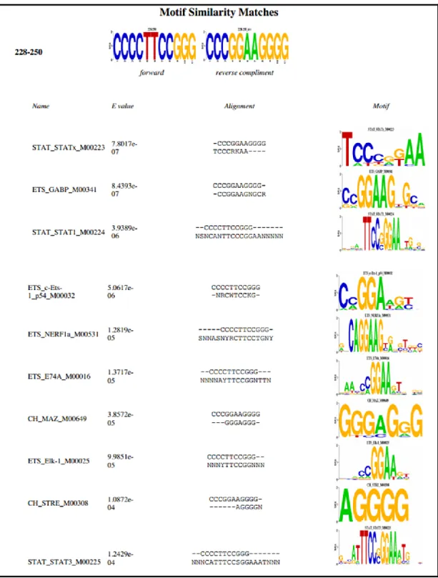
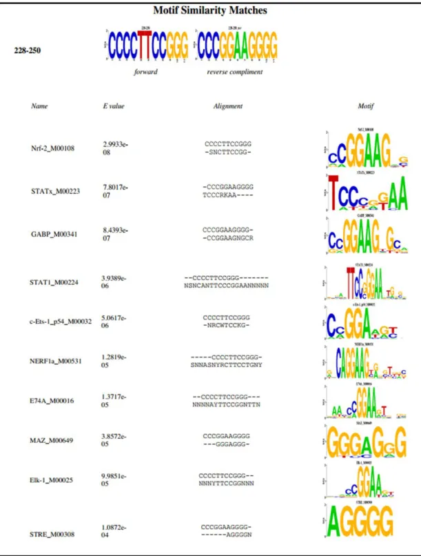
Recently two groups detected highly recurrent germ line and somatic mutations in the promoter region of TERT gene in melanoma patients and cell lines 133,134. Horn et al. first detected a single nucleotide mutation, A > C (T > G) at the -57 bp from transcription start site (Chromosome 5: 1,295,161) in a familial melanoma case. They detected this mutation to be in allelic linkage with the common polymorphism (rs2853669) found at the -245 bp from ATG start site in the TERT promoter 135. Then they analyzed melanoma cell lines and other patients unrelated to the first family and detected two more mutations located at -124 bp (1,295,228; depicted as C228T) and -146 bp (1,295,250; depicted as C250T) of TERT promoter 133. In an independent study by Huang et al. they have detected the same somatic promoter mutations at the TERT promoter as a result of whole genome sequencing analysis This group also discovered additional CC > TT mutations at the -124 and -146 bp 134. These frequent promoter mutations are suggested to create a common binding motif (CCGGAA/T) for E-twenty six/ ternary complex factors (ETS/TCF) transcription factors thus increasing TERT expression which helps tumor cells during transformation proces133,134. Representative image of the TERT promoter including hotspot mutations is given in Figure 1.7.
18
Figure1.5 Schematic representation of a part of the TERT promoter that contains hotspot mutations
Mutations are marked with red and new binding motif is marked with bold characters.
Discovery of frequent promoter mutations in TERT gene in melanoma led other scientists to search for the same mutations in different cancers, indeed they have found the same mutations 136. Mutation frequency has been different in different types of cancers. Melanoma, pleomorphic dermal sarcoma, myxoid liposarcoma, glioma, urothelial cell carcinoma of bladder, basal and squamous cell carcinoma of skin and liver cancer are among the cancers with high frequency of TERT promoter mutations 136–146 In other cancers, TERT promoter mutations exist in a very low rate
136,139
. Two reports suggested that TERT promoter mutations are found at higher frequencies in tumors originated from tissues with low self renewal capacity 136,147. TERT promoter mutations are high in frequency, they are suggested to be gain of function mutations and they are observed in many different cancers; thus, they are considered to be mutations driving cancerogenesis but not random mutations 129. There is some evidence showing that these mutations are increasing promoter activity when tested in reporter assays together with controls which do not have mutated base 133,134,148. In glioma and thyroid cancer, TERT promoter mutations have been associated with advanced disease and poor prognosis 136,143,149.
Other than promoter mutations, a T>C single-nucleotide polymorphism (SNP) also attracted attention of the scientists. This SNP is detected at position−245 bp from transcription start site of TERT gene (genomic loci 1295349) and it is represented by rs2853669 or T349C 133,134. Hsu et al. reported that carriers of the TERT rs2853669 CC genotype had a significantly lower telomerase activity compared to ones with TT genotype in lung cancer since it disrupted an existing Ets2 transcription factor
19
binding site at TERT promoter 135. In another study, TERT rs2853669 CC allele was found to be associated with a significantly increased risk of lung cancer 150. In breast cancer, there is one study showing that there is no significant association between
TERT rs2853669 SNP and breast cancer risk 151. However, there is a more recent study demonstrating that the TERT rs2853669 CC genotype was correlated with a low risk of breast cancer 152. There is also promising data about the possible use of
TERT promoter mutations as biomarkers together with rs2853669 polymorphism in
bladder cancer. It is found that a variant allele of rs2853669 polymorphism counteracts with the effect of TERT promoter mutations C228T and C250T in bladder cancer 148. In the presence of the variant allele of rs2853669 polymorphism together with TERT promoter mutations; the patients have a higher survival rate compared to the patients with TERT promoter mutations but without the variant allele of rs2853669 polymorphism 148. This data is supported with the observation that the variant allele of rs2853669 polymorphism prevents the binding of Ets2 to its non-canonical target site found in the vicinity of the polymorphism which is located near an E-box 135,153.
TERT promoter mutations provide an advantage to the bearing cells during
transformation process provided that necessary transcription factors are available and functional to up-regulate TERT protein expression154. Increased expression of ETS transcription factors in many cancers 155,156 can be considered as a supporting event for the hypothesis that ETS transcription factor binding site is created by C228T and C228T mutations, however, there is no experimental data supporting this event yet.
1.3 Signal Transducer and Activator of Transcription 1 (STAT1)
and Cancer
1.3.1 STAT Transcription Factor Family
The signal transducer and activator of transcription (STAT) transcription factors are composed of several members such as STAT1, STAT2, STAT3, STAT4, STAT5A, STAT5B and STAT6 157. STAT family member proteins have 5 functional domains composed of one amino-terminal domain, one coiled-coil domain, one DNA-binding
20
domain, one SH2 domain and one carboxy-terminal transactivation domain 158. Phosphorylation of two amino acids inside the carboxy-terminal transactivation domain activates STAT transcription factors. One of these sites (tyrosine) is critical for the dimerization function while the other site (serine) is for activation of the transcriptional regulatory function of STATs 159. STAT transcription factors regulate transcription with the signal coming from cytokines and growth factors. STAT transcription factor activation starts with the interaction of a ligand with its receptor. The next step is phosphorylation of the receptor and recruitment of STAT proteins to this site. Then, specific tyrosine kinases (growth factor receptors, Janus kinases (JAKs) or SRC family kinases) phosphorylate STATs on the tyrosine residue. Dimerization of two phosphorylated STAT proteins takes place and STAT dimers localize to the nucleus to bind to specific promoters and regulate gene transcription. STAT induced transcription activation is antagonized by SOCS (suppressors of cytokine signalling) and PTPs (protein tyrosine phosphatases) 160. STAT proteins have major roles during normal cellular functioning such as cell growth and differentiation, development, apoptosis, immune responses and inflammation 161. 1.3.2 STAT1 and Interferon (IFN) Signaling
STAT1, the first member of STAT transcription factor family is activated by both types of Interferon (IFN). IFNs are important cytokines with critical functions such as antiviral signaling, prevention of cell proliferation, anti-tumoral activity and immunomodulation 162. IFNs are classified into two groups; Type I and Type II IFNs. Type I IFNs are composed of a large group of molecules; IFN-alpha (which has 13 subtypes: IFNα1, α2, α4, α5, α6, α7, α8, α10, α13, α14, α16, α17 and -α21), IFN-beta, IFN-omega, IFN-tau, IFN-kappa, IFN-lambda, and IFN-zeta. Type II interferon group however, only has one member which is IFN-gamma 163. IFN signaling is the leading arm of STAT1 signaling pathway since STAT1 activation starts with binding of IFN to its receptor 162,164. The fact that STAT1 knockout mice cannot respond to IFN 165 is critical for the establishment of the proof of concept that STAT1 is mediating antibacterial and viral functions of the cells through IFN signaling 166. IFN mediated STAT1 signaling is schematized in Figure 1.8. 163 The first step of both type I and type II IFN signaling is the activation of JAKs through
21
autophosphorylation once dimerization of Interferon alpha receptor subunits 1 and 2 (IFNAR1 and IFNAR2) occurs. JAK1 and TYK2 then phosphorylate STAT1 and in low amounts STAT2, STAT3 and STAT5. STAT1 forms homodimers or heterodimers (with STAT2), translocates into the nucleus and starts transcription of target genes on promoters containing GAS (Gama activated sequence) or ISRF (Interferon stimulated response element). STAT1 and STAT2 dimers interact with another protein p48 or IRF9 to form a heterotrimeric transcription factor called ISGF3 (Interferon-stimulated gene factor 3) once they are in the nucleus 158.
1.3.3 STAT1 Signaling and HCC
Immune cell infiltration is a critical event in the continuance of hepatocyte injury during chronic liver disease induced by viral infections, alcohol or hepatoxins 167. Activated T cells, both CD4+ and CD8+ are involved in liver injury during chronic hepatitis B or C 168,169 and their amount correlates with regenerating nodules, inflammation, and fibrosis in ALD 170. STAT1 signaling is the key pathway for immune cell infiltration and induction of liver injury. Injured hepatocytes activate CD4+ and Natural killer T cells and stimulate secretion of interferon which in turn induces STAT1 phosphorylation through JAKs. pSTAT1 contributes to further liver injury by activating more CD4+ and NK T cells which will re-start the circle by secreting more interferon and inducing more damage 167. Consistent with this phenomenon, STAT1 expression is found to be significantly higher in HCC tumors compared to its neighboring normal liver tissue. In parallel with the expression data, phosphorylated STAT1 level is higher in HCC samples with poor prognosis compared to the ones with good prognosis 171. SOCS1 (suppressor of cytokine signaling) is the negative regulator of STAT1 signaling. Up-regulation of STAT1 activity and down regulation of STAT3 activity are the two reasons behind increased fibrosis and severe liver damage that are observed in SOCS1 knockout mice. This data provides another evidence for the contribution of STAT1 TF to hepatocancerogenesis 172.
22 1.3.4 IFN-α Therapy during HCC Treatment
IFN-α which is an antiviral cytokine, is used to treat Hepatitis infections which are main causes of HCC. Early diagnosed HCC cases are mostly treated by surgical resection, ablation or liver transplantation 173, however, nearly 20% of HCC cases reoccur in the first year after therapy and this value increases to 80–90% in a five year window 174. Moreover, HCC recurrence is observed in a higher rate in the patients infected with Hepatitis B and C compared to those without a viral induced hepatitis. Thus, IFN-α is used as therapy in post operative HCC patients to decrease the risk of tumor recurrence175. IFN-α has also been shown to decrease proliferation and prevent angiogenesis hence IFN-α is suggested as an anticancer agent that will be able to decrease tumor recurrence rate in HCC patients 176. There is controversial data about the efficacy of the interferon-α treatment for the patients with viral induced HCC. Some studies reported that IFN-α decreased tumor recurrence rate thus contributed to disease free survival 177,178. On the other hand, others could not recapitulate the same kind of data after two randomized control trials (RCTs) and suggested that IFN-α does not have a significant effect on disease free survival and overall survival of the patients viral induced HCC after curative therapy 179,180. In a recent report, Meta analysis of interferon therapy case studies is performed to determine the contributions of interferon-α on survival rate of HCC patients with underlying HBV or HCV induced hepatitis 181. The results indicated that IFN-α therapy has improved survival of HCC patients with HCV induced hepatitis compared to the control group, however, there was no significant improvement in HCC patients with HBV induced hepatitis 181.
23
1.4 Aim and Strategy
Hepatocellular Carcinoma is one of the top reasons of cancer related deaths worldwide. HCC incidence has been increasing in USA and Europe, however, the highest incidence is observed in China, Middle Africa, and Japan 3. HCC develops on a heterogeneous background with varying underlying risk factors in different geographical regions. Hepatitis B virus and Hepatitis C virus are considered as the main risk factors in Asia and Africa. In Europe and USA, alcohol abuse and obesity are the leading causes of HCC2. HCC patients are mostly diagnosed at later phases of cancer since HCC is mostly asymptomatic until the very end of tumorigenesis 74. There are limited therapeutic options such as surgical resection, ablation and liver transplantation, but they are all suitable for early diagnosed cases and disease recurrence is very common after these therapies75. Due to late diagnosis and limited curative therapies, survival rate of HCC patients is very low 75. Genetic mechanisms of hepatocarcinogenesis are very complicated yet critical to investigate in order to be able to find therapeutic targets against HCC. Unfortunately, most of the mutations discovered in HCC patients are “loss-of-function” mutations and they not appropriate therapeutic targets 57. Sorafenib is the only FDA approved drug against HCC, however it only provides a three month of disease free survival compared to control group 89. Telomere shortening problem is an obstacle for a cell during transformation process 103. Tumor cells solve this problem by two ways; the first and most common way is re-gaining telomerase activity by up-regulating TERT expression 97 and the other is through ALT (alternative lengthening of telomeres), a mechanism similar to homologous recombination118. TERT up-regulation is very common and observed in 90% of HCC cases 111,112; however molecular mechanisms behind TERT up regulation in HCC is still a mystery except for some cases with the involvement of Hepatitis B virus and loss of a region of chromosome 10p. HCC cells are reported to increase telomerase activity with allelic loss of chromosome 10p which codes for a telomerase repressor protein 113. HBV involvement is more common than allelic loss; HBV DNA is integrated into TERT gene and increases its expression 113. Yet, there is significant number of HCC cases which occur without an underlying HBV infection and mechanism of TERT activation is needed to be solved in those patients
24
for a better understanding of the telomerase activation in HCC. Recently, two frequent mutations in TERT promoter region have been reported in many tumors together with HCC 133,134,136–141,143,144,182,183. And these mutations are suggested to have a functional role rather than being random genetic events. It is proposed that, the presence of TERT promoter mutations create a novel ETS transcription factor binding site and increase TERT expression through this mechanism 133,134. Previously reported TERT promoter mutations in HCC were restricted to samples from USA136 and France146 with 44 % and 59% of frequencies. However, there was no data about the occurrence of TERT promoter mutations in different geographical regions such as China, Japan and Africa where HCC incidence is higher compared to USA and Europe. For this reason, we wanted to explore the frequency of TERT promoter mutations across the world, especially in Asia and Africa to have a deeper knowledge about this frequent genetic event.
The first objective of this study is to analyze our HCC cell line panel composed of 15 HCC cell lines together with 44 HCC tumors collected from different geographical regions to search for two common mutations in the promoter region of TERT. Next, we wanted to know if the presence of mutations in TERT promoter has a functional role on TERT expression or not. For this purpose, we have performed a bioinformatics analysis to search for possible transcription factors that can bind to mutant promoter sequence and we discovered that STAT1 is one of the candidate transcriptional factors. Considering the critical roles of STAT1 during liver carcinogenesis, we decided to continue analyzing this finding by designing experiments to see the effects of STAT1 activation or knockdown on the expression of TERT expression. Interferon alpha is the cytokine that triggers STAT1 signaling; therefore, we hypothesized that treatment of HCC cell lines with IFN-α would activate STAT1 signaling which will provide binding of activated STAT1 dimers onto mutant TERT promoter to increase expression of TERT enzyme. In conclusion, we analyzed TERT promoter mutations in HCC in a functional manner to unravel the molecular mechanism behind the possible upregulation of TERT expression by these mutations. This data will provide insides about the contributions of TERT promoter mutations into cellular immortality in hepatocellular carcinoma.
25
Chapter 2
2.
Materials and Methods
2.1 Materials
2.1.1 General Laboratory Reagents
General laboratory reagents such as methanol, ethanol, and Bradford reagent were all analytical grade and were mainly purchased from Sigma-Aldrich (St. Louis, MO, USA) or Merck (Darmstadt, Germany). Bradford reagent, methanol and ethanol were from Sigma-Aldrich (St. Louis, MO, USA). ECL Prime western blotting detection kit and Hybond nitrocellulose western blot membranes were from Amersham Pharmacia Biotech Company. DMSO and Ponceau S were purchased from Applied Biochemia (Darmstadt, Germany). Interferon alpha 2a human with catalog number SRP4594-100UG was purchased from Sigma-Aldrich (St. Louis, MO, USA).
2.1.2 Tissue Culture Reagents and Materials
All cell culture media such as Dulbecco’s modified Eagle’s medium (DMEM) and Roswell Park Memorial Institute (RPMI) 1640 medium and OptiMEM were purchased from GIBCO (Invitrogen, Carlsbad, CA, USA). Medium supplements like L-glutamine, Penicillin/streptomycin antibiotics, Non essential amino acids (NEAA), fetal calf serum (FCS) and Trypsin-EDTA were also purchased from the same company, GIBCO. Plastic materials used in cell culture such as Petri dishes, flasks, multiple well-plates, cryotubes were purchased from Corning Life Sciences Inc. (USA). Sterile serological pipettes were purchased from Costar Corporation (Cambridge, UK). Transfection reagents Lipofectamine 2000 and Lipofectamine RNAi Max were purchased from Invitrogen (Carlsbad, CA, USA).
26 2.1.3 Genomic DNA Isolation
Purelink Genomic DNA isolation kit (K1820-02) was purchased from Invitrogen (Carlsbad, CA, USA).
2.1.4 Polymerase Chain Reaction (PCR)
Recombinant Taq DNA polymerase enzymes (EP0401) were purchased from Thermo Scientific (MA, USA). AccuPrime GC-Rich DNA polymerase was purchased from Invitrogen (Carlsbad, CA, USA).
2.1.5 Primers
Primers for amplification of hTERT genomic DNA were the same as used by Horn et al, 2013133.Apart from those, all primers were designed using Primer3 online tool (http://frodo.wi.mit.edu/primer3/input.htm) and shown in Table 2.1
Table2.1 Primers used in this study
Gene name Forward Primer Reverse Primer
TERT 133 ACGAACGTGGCCAGCGGCAG CTGGCGTCCCTGCACCCTGG
TERT CGGAAGAGTGTCTGGAGCAA GGATGAAGCGGAGTCTGGA
hIRF1 GAGGAGGTGAAAGACCAGAGCA TAGCATCTCGGCTGGACTTCGA
STAT1 CACGCACACAAAAGTGATGA AGAGGTCGTCTCGAGGTCAA
GAPDH GGCTGAGAACGGGAAGCTTGTCAT CAGCCTTCTCCATGGTGGTGAAGA
2.1.6 Agarose Gel Electrophoresis
Horizontal gel electrophoresis apparatus was Thermo EC Midicell Primo EC330 Electrophoretic Gel System and the power supply was EC250-90 both from Thermo Scientific (MA, USA). Other power supplies used were PAC300 and Power-PAC200 which were from Bio Rad Laboratories (CA, USA).
27 2.1.7 Spectrophotometry
Spectrophotometer was from Beckman. 2.1.8 Determination of Gene Expression
2.1.8.1 Total RNA Isolation
Nucleospin RNA II total RNA isolation kit (740955.250) was from Macherey-Nagel (Duren, Germany).
2.1.8.2 First Strand cDNA Synthesis
RevertAid First Strand cDNAs Synthesis Kit (# 1622) was obtained from Fermentas-Thermo Scientific (MA, USA).
2.1.8.3 Quantitative Real Time PCR
Dynamo HS SYBR Green qPCR Kit (F-410L) was purchased from Thermo Scientific (MA, USA). The instrument used for gene expression studies was Stratagene Mx3000P qPCR System from Agilent Technologies (CA, USA).
2.1.9 Antibodies
Primary and secondary antibodies used in this study were purchased from different companies. All information including company name, catalog number and working conditions are shown in Table 2.2.
Table2.2 Antibodies used in this study
Antibody Name Catalog number Western Blot Dilution
Calnexin Sigma-Aldrich, C4731 1:5000
α-tubulin Calbiochem, CP06 1:5000