i
IDENTIFICATION AND UTILIZATION OF
AUTOLOGOUS ANTI-TUMOR ANTIBODIES
FOR THE DIAGNOSIS AND PROGNOSIS OF CANCER
A THESIS SUBMITTED TO
THE GRADUATE SCHOOL OF ENGINEERING AND SCIENCE OF BILKENT UNIVERSITY
IN PARTIAL FULFILLMENT OF THE REQUIREMENTS FOR THE DEGREE OF
DOCTOR OF PHILOSOPHY IN
MOLECULAR BIOLOGY AND GENETICS
By
Şükrü Atakan
December 2015
ii
IDENTIFICATION AND UTILIZATION OF AUTOLOGOUS ANTI-TUMOR ANTIBODIES FOR THE DIAGNOSIS AND PROGNOSIS OF CANCER By Şükrü Atakan
December 2015
We certify that we have read this thesis and that in our opinion it is fully adequate, in scope and in quality, as a thesis for the degree of Doctor of Philosophy.
_________________________________ Ali Osmay Güre (Advisor)
_________________________________ Özlen Konu ________________________________ Serkan Göktuna ________________________________ Uğur Özbek _________________________________ Eda Çelik Akdur
Approved for the Graduate School of Engineering and Science:
___________________________ Levent Onural
iii
ABSTRACT
IDENTIFICATION AND UTILIZATION OF AUTOLOGOUS ANTI-TUMOR ANTIBODIES FOR THE DIAGNOSIS AND PROGNOSIS OF CANCER
Şükrü Atakan
Ph.D. in Molecular Biology and Genetics Advisor: Ali Osmay Güre
December 2015
Lung cancer is the leading cause of cancer related death worldwide. Current diagnostic methods have limited power and unable to extend patient life significantly. SCLC; the most aggressive subtype of lung cancer is an immunogenic cancer type and able to elicit an immune response of which autologous antibodies are a measurable component. These antibodies are elicited even when the tumor is microscobic and impossible to be diagnosed clinically by the current methods of diagnosis thus antibodies can be utilized for early diagnosis. We aimed to develop a method to identify novel autologous antibodies, identify these antibodies for SCLC, Colorectal, Gastric and Ovarian cancers and validate these antibodies for SCLC diagnosis and prognosis and investigate their utility for autoimmune disease.
We have developed and optimized PA screening for novel autologous antibody discovery. We have screened PA with serum pools of cancer patients (SCLC, Colorectal, Gastric and Ovarian), BD and healthy controls since PAs have many advantages compared to other discovery methods like SEREX. We have also performed sensitivity and specificity evaluations by screening custom PAs by individual sera. Image analysis softwares developed by collaboration utilized for evaluation of the screenings. The filtered valuable clones were ordered from the PA manufacturer and HisTagged protein expression and purification was performed with these clones. Pure proteins were screened with 3 independent SCLC and 2 Healthy control cohorts by an iterative ELISA approach for validation of these antibodies as valuable biomarkers. ELISA results were also confirmed by Western blotting. Monte Carlo, SVM and PC were utilized for cut-off determination, panel formation and ROC plotting. AUC was compared for evaluation of diagnostic power. Kaplan-Meier, UCR and MCR analysis was performed for prognostic analysis of the valuable antibodies. Seperately protein expression and autologous antibody presence correlation was evaluated by comparison of IHC and ELISA. The same autologous antibody identification strategy was utilized as a collaborative support for an independent study for identification of NBD specific biomarkers.
iv
We have identified 23 distinct autologous antibody biomarkers for SCLC after evaluation of PA and custom PA screenings. For 8 of these antibodies we have completed ELISA screening for all 3 SCLC and 2 healthy control cohorts. 6 of these autologous antibodies were shown to be valuable as a panel for SCLC diagnosis both by MC and SVM. Utilization of 4 of these antibodies; SOX2, p53, POLB and C11orf20, as a panel resulted in superior AUC thus high sensitivity and specificity values (55% sensitivity, 90% specificity). PC method resulted in higher AUC even only by combination of SOX2 and p53 (82% sensitivity, 90% specificity). Although individual correlations were identified, we were unable to show a significant correlation of seropositivity with survival for any of the antibodies which is common to all cohorts. We have identified a significant correlation between SOX2 antigen expression intensity and autologous antibody presence. Mtch1 was identified as a NBD specific autologous antibody by the utilization of our autologous antibody discovery and validation methodology.
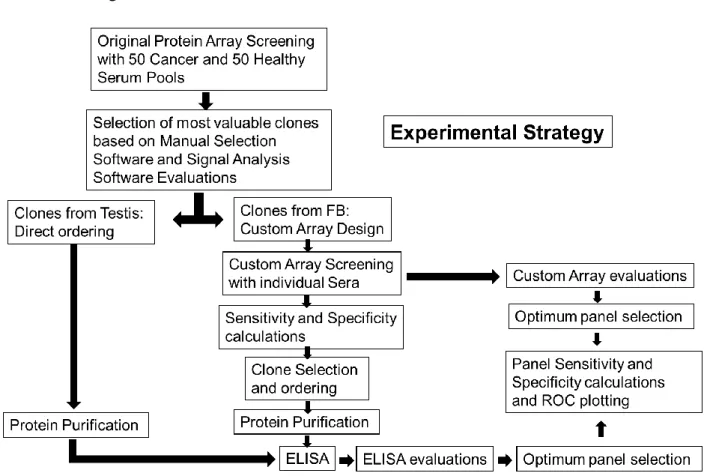
We were able to identify a panel of 4 antibodies; SOX2, p53, POLB and C11orf20, which resulted in 55% sensitivity at 90% specificity for SCLC. 2 of these antibodies were identified by this study as novel biomarkers; POLB and C11orf20. The panel is capable of exceeding the diagnostic power of the only commercially available diagnostic kit; EarlyCDT-Lung. PC method is very promising since a sensitivity value of 82% was reached at 90% specificity which is a diagnostic power comparable that of low-dose CT. As a future perspective we are planning to apply PC method to all the PA data and develop a kit based on this method to be utilized for SCLC diagnosis.
Keywords: SCLC, protein array, diagnosis, prognosis, autologous antibodies, biomarker, ELISA, sensitivity, specificity.
v
ÖZET
KANSERİN TEŞHİS VE PROGNOZUNDA KULLANILMAK ÜZERE OTOLOG ANTİ-TÜMÖR ANTİKORLARIN BELİRLENMESİ VE KULLANIMI
Şükrü Atakan
Moleküler Biyoloji ve Genetik, Doktora Tez Danışmanı: Ali Osmay Güre
Aralık 2015
Akciğer kanseri dünya çapında en çok ölüme yol açan kanserdir. Mevcut teşhis yöntemleri kısıtlı güce sahiptir ve hasta ömrünü anlamlı olarak uzatamamaktadır. Küçük hücreli akciğer kanseri (KHAK); en agresif akciğer kanseri alt türü olarak bağışıklık yanıtına yol açmaktadır ve bu yanıtın ölçülebilir ajanlarından biri otolog anti-tümör antikorlarıdır. Bu otolog antikorlar tümör henüz mikroskobik aşamadayken dahi ortaya çıkmaktadır ve bu sayede diğer teşhis yöntemlerinin tümörü tespit edemeyeceği durumlarda dahi tümör varlığını erken aşamada teşhis etmeye yarayabilir. Çalışmamızda otolog anti-tümör antikorları keşfetmek için yeni bir yöntem geliştirmeyi, KHAK, Kolorektal, Mide ve Over kanserlere özgü otolog antikorları keşfetmeyi, KHAK teşhisi ve prognozunda yararlı olabilecek otolog anti-tümör antikorlarını belirlemeyi ve doğrulamayı ve otolog antikorların otoimmün hastalık için yararlılığını araştırmayı hedefledik.
Çalışmamızda bilinmeyen otolog anti-tümör antikorları keşfetmek için PA taraması temelli bir yöntem geliştirdik ve optimize ettik. Kanser (KHAK, kolorektal, mide ve over) hastaları, Behçet hastalığı ve sağlıklı kontrollerden oluşturulan serum havuzları ile PA taraması gerçekleştirdik. PA taramasının SEREX gibi diğer otolog antikor teşhis yöntemlerine göre üstün yönleri vardır. Havuz taramalarından seçilmiş klonlardan oluşan arraylerin tek tek serumlar ile tekrar taranması ile hassasiyet ve özgüllük değerlendirmeleri gerçekleştirdik. Kolaborasyon ile oluşturulmuş görüntü işleme yazılımları tarama çalışmalarının sonuçlarının değerlendirilmesinde kullanılmıştır. Değerlendirmeler sonucu seçilen klonlar PA üreticisinden temin edilmiştir ve bu klonlar ile His ile işaretlenmiş protein üretimi ve saflaştırması gerçekleştirilmiştir. Saf proteinler ile 3 farklı KHAK ve 2 farklı kontrol grubu için sonuçların her defasında tekrar değerlendirildiği ELISA taramaları yapılmıştır. Bu taramalardaki amaç belirlenen otolog antikorların değerli birer biyobelirteç olduğundan emin olmakdır. ELISA sonuçlarımız Western blot yötemiyle doğrulanmıştır. MC, SVM ve PC yöntemleri eşik değer belirlemek, panel oluşturmak ve ROC eğrisi çizimi için uygulanmıştır. Teşhis gücünün kıyaslanması için AUC değerleri analiz edilmiştir. Kaplan-Meier, UCR ve MCR yöntemleri antikorlar varlığı ile yaşam süresi arasındaki ilişkinin değerlendirilmesi için uygulanmıştır. Ayrıca protein ifadesi ve otolog antikor varlığı arasındaki ilişki IHC ve ELISA değerlerinin
vi
analizi ile araştırılmıştır. Otolog antikor tespiti için geliştirdiğimiz yöntem ayrıca kolaboratif bir destek olarak NBD’ye özgü biyobelirteçlerin bulunmasında uygulanmıştır.
KHAK için 23 farklı otolog antikor biyobelirteç PA taramaları sonucunda anlamlı olarak seçilmiştir. Bu antikorların 8’i için 3 KHAK ve 2 sağlıklı grubu için tüm ELISA taramaları tamamlanmıştır. Bunların altısından oluşturulacak bir panelin değerli olacağı MC ve SVM ile gösterilmiştir. Bu 6 antikorun dördünden oluşturulan panelin; SOX2, p53, POLB ve C11orf20, AUC değeri tüm 6 antikor ile oluşturulan AUC değerinden çok farklı değildir ve yüksek hassasiyet ve özgüllük elde edilebilmektedir (55% hassasiyet, 90% özgüllük). PC yöntemi ile yalnızca SOX2 ve p53 içeren panel ile oluşturulan AUC değeri diğer tüm AUC değerlerinden daha yüksektir (82% hassasiyet, 90% özgüllük). Tek başına değerlendirildiğinde genel yaşam süresi ile istatistiksel olarak anlamlı şekilde ilişkili bulunmasına rağmen hiçbir antikor varlığı tüm gruplar için genel yaşam süresi ile ilişkili bulunmamıştır. Çalışmamızda SOX2 protein ifade yoğunluğu ile otolog antikor varlığı arasında istatistiksel anlamlı ilişki bulduk. Mtch1’in NBD’ye özgü bir otolog antikor olarak keşfi ve doğrulanması ile yöntemimizin otoimmün hastalıklarda da yararlılığı anlaşılmıştır.
Belirlediğimiz 4 otolog antikor ile oluşturduğumuz panel; SOX2, p53, POLB ve C11orf20, KHAK teşhisi için 55% hassasiyette 90% özgüllüğe sahiptir. Bu otolog antikorların ikisinin ilk defa bu çalışma ile SCLC teşhisinde kullanılabilirliği gösterilmiştir; POLB ve C11orf20. Dört antikorluk panel akciğer kanseri için piyasada mevcut tek otolog antikor temelli kit olan EarlyCDT-Lung’dan daha yüksek teşhis gücüne sahiptir. PC yöntemi ile 90% özgüllük için 82% hassasiyet değerine ulaşılabilmektedir. Bu teşhis gücü düşük doz bilgisayatlı tomografinin teşhis gücüne eşdeğerdir. Gelecek planı olarak PC yöntemini şimdiye kadar oluşturduğumuz tüm PA sonuçlarına uygulamayı ve bu yöntem temelli çalışan bir KHAK teşhis kiti geliştirmeyi hedefliyoruz.
Anahtar sözcükler: KHAK, protein array, teşhis, prognoz, otolog antikorlar, biyobelirteç, ELISA, hassasiyet, özgüllük.
vii
ACKNOWLEDGEMENTS
I would like to express my gratitude to my advisor Assist. Prof. Dr. Ali Osmay Güre for his invaluable contributions to this work and to my personal development. I appreciate so much his sincere and honest attitude, his patience, always encouraging positive criticisms and his influential didactic style. He gave me the chance to learn by trial and error which is very valuable for me. He has also been very supportive for my extracurricular activities in context of enterpreneurship thus gave me an opportunity to discover novel possibilities for my career besides autologous antibodies.
I would like to thank Assist. Prof. Dr. Özlen Konu and Prof. Dr. Uğur Özbek for their constructive and positive criticisms about my Ph.D. project.
I would like to thank Alper Poyraz for his invaluable contributions to the project and his support, encouragements and executive personality. I also appreciate his partnership in business by his positive, energetic and influential mood.
I would like to thank Serhat Yıldırım for his tremendous efforts for the project. He helped the project by providing hard work and performing attention and precision requiring, labor intensive and long lasting experiments.
I would like to thank Prof. Dr. Burçak Vural for her contributions to this project, her encouragements, positive attitude and her friendship. I would like to thank Elif Uğurel, Filiz Çavuş and Elçin Şehitoğlu for their friendships and contributions to the project.
I would like to thank Prof. Dr. Burçak Vural, Dr. Funda Demirağ, Dr. Hülya Bayiz and Prof. Dr. Pınar Saip for providing serum samples and the clinical data.
I would like to thank Assoc. Prof. Dr. Çiğdem Gündüz Demir and Can Fahrettin Koyuncu for their collaboration in image processing software development.
I would like to thank former and present members of AOG Lab; Alper, Barış, Kerem, Sinem, Seçil, Waqas, Mehdi, Zeynep, Murat, Duygu, Aydan, Esen, Rasim and Derya for their kind contributions to the project and their friendship.
I would like to thank the senior students who have contributed to this project; Alper, Serhat, Deniz, İpek, Berrak, Dilce, Fatma Zehra, Onur, Deniz, Duygu, Anıl, Çağrı, Ferhat, Erdi, Ezgi and Gökhan.
viii
I would like to thank Alper and Barış specially for their efforts for contributing my extracirricular R&D activities during their undergraduate study.
I would like to thank all my instructors in Bilkent MBG for their contributions to my scientific development. It is a honor for me to be a graduate of Bilkent MBG and doing my PhD as well and Bilkent’s contributions to my career as well as my philosophy about life.
I would like to thank to my friends; Burak, Duygu, Ender, Ceyhan, Kerem, Sinem, Gurbet, Nilüfer, Zeynep, Gizem and Aydan for our wonderful memories.
I would like to thank all the members of the Bilkent MBG family as a community for their contributions to my knowledge, my personal development and the warm family environment.
I would like to thank Rengül Hoca and Volkan Hoca for accepting me as a member of their family during my PhD study and giving me the opportunity to meet my wife.
I would like to thank my wife, Işıl for her love, her mental and emotional support and her patience during my PhD.
I would like to thank my family for all their support and encouragement about my education and my decisions.
I would like to thank to TÜBİTAK for the BİDEB 2241 scholarchip during my PhD.
I would like to thank to PRZ BioTech and Sentegen for employing me and encouraging and supporting my PhD studies in parallel to their employment.
ix
I dedicate my thesis firstly to my advisor Assoc. Prof. Dr. Ali Osmay Güre and all my instructors whose lectures I have attended during my undergraduate and PhD studies at Bilkent University. They have tremendously contributed to my scientific and philosophical development.
x
Contents
1
INTRODUCTION ... 1
1.1 LUNG CANCER AND IT’S DIAGNOSIS ... 1
1.1.1 Lung Cancer Risk factors, Incidence and Mortality ... 1
1.1.2 Current methods for dignosis of lung cancer ... 1
1.1.3 Potential of biomarkers for lung cancer diagnosis / early diagnosis ... 2
1.2 IMMUNE SYSTEM AND CANCER ... 2
1.2.1 Immune response against cancer ... 2
1.2.2 Serum reactivity against cell surface antigens ... 3
1.2.3 Serum reactivity against intracellular antigens ... 3
1.2.4 Methylcholanthrene induction causes tumor cells to be rejected by the host ... 3
1.2.5 Immunoediting is a driving force for less imunogenic tumor development ... 4
1.2.6 Autologous CTLs lyse antigen presenting tumor cells ... 4
1.2.7 Autologous antibodies as part of the immune response to cancer ... 4
1.2.8 Autologous antibody elucidation mechanisms ... 5
1.2.9 Antigen expression and autologous antibody elucidation ... 6
1.2.10 Autologous antibodies and PNS ... 6
1.2.11 Autologous antibodies in SCLC with/without PND... 7
1.3 DIAGNOSIS AND EARLY DIAGNOSIS OF CANCER ... 8
1.3.1 Autologous antibodies precedes PND and clinical cancer diagnosis ... 8
1.3.2 p53 antibody level stability / fluctuations can predict cancer development ... 9
1.3.3 Sensitivity and specificity of autologous antibodies ... 10
1.3.4 Autologous antibodies already in clinical use for diagnosis of cancer ... 10
1.4 AUTOLOGOUS ANTIBODY DISCOVERY METHODS ... 10
1.4.1 Methods for autologous antibody discovery ... 10
1.4.2 Limitations of SEREX ... 13
1.4.3 Theoretical comparison of SEREX with PAs ... 15
1.5 AUTOLOGOUS ANTIBODIES AND PROGNOSIS ... 16
1.5.1 Autologous antibodies can be utilized for prognosis ... 16
1.5.2 Autologous antibody positive sera have cytotoxic effect on cancer cell line ... 16
1.5.3 Autologous antibody positivity correlates with in-vivo cytotoxicity ... 16
1.5.4 Autologous antibodies and survival ... 17
xi
1.5.6 Anti-SOX1 autologous antibodies are specific to cancer not PND ... 17
1.5.7 SOX2 antibodies and prognosis of multiple myeloma (MM) ... 17
1.5.8 SOX2 and ZIC2 antibodies and survival in SCLC... 18
1.6 SENSITIVITY AND SPECIFICITY OF PANELS ... 19
1.6.1 Diagnosis by evaluating a panel of antibodies ... 19
1.6.2 Sensitivity and Specificity of already identified autologous antibodies ... 20
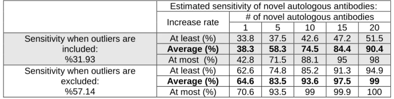
1.6.3 Predicted Sensitivity and Specificity of novel autologous antibodies ... 22
1.6.4 Commercially available diagnostic technologies ... 23
2
AIM, RELEVANCE AND RATIONALE ... 24
2.1 AIMS ... 24
2.2 RELEVANCE ... 25
2.3 RATIONALE ... 25
3
STUDY DESIGN ... 27
4
MATERIALS AND METHODS ... 28
4.1 METHOD DEVELOPMENT... 28
4.1.1 Optimization Experiments for PA screening protocol determination ... 28
4.1.2 Serum Selection for Pooling... 28
4.1.3 Serum Dilution ... 28
4.1.4 Pool Dilution ... 29
4.1.5 2o Antibody dilution ... 29
4.1.6 Q-Dot dilution ... 29
4.1.7 Strategy for autologous antibody discovery ... 29
4.1.8 Protein Arrays ... 31
4.2 NOVEL AUTOLOGOUS ANTIBODY IDENTIFICATION ... 32
4.2.1 Screening Strategy and Cohorts ... 32
4.2.2 Patient and Healthy Control Cohorts ... 32
4.2.3 PA Screening ... 33
4.2.4 Clone Selection Strategy based on PA Screening Results ... 34
4.2.5 Manual Selection Software (MSS) Development for selection of signals from PA screening results ……… ... 34
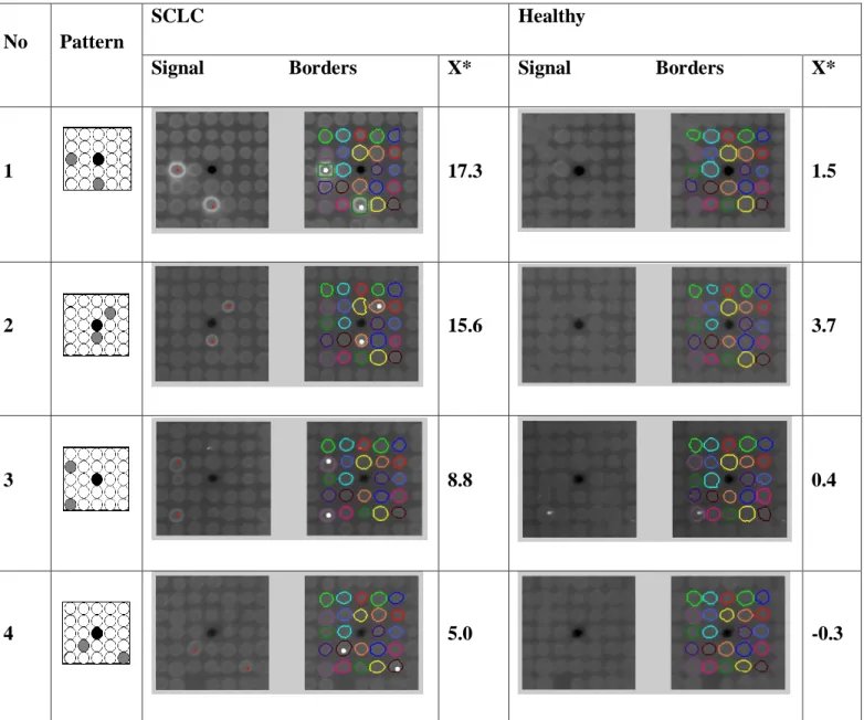
4.2.6 Singnal Analysis Software (SAS) and Pixel Count (PC) Method for Numerical Analysis... 35
4.2.7 Custom PA Screening ... 37
xii
4.3 INTER-ASSAY VALIDATION ... 38
4.3.1 Insert analysis of pQE plasmids ... 38
4.3.2 Clone protein expression analysis by IPTG induction and SDS-PAGE ... 38
4.3.3 HisTagged Protein Expression and Purification... 39
4.3.4 ELISA ... 39
4.3.5 ELISA Data Normalization ... 40
4.3.6 Sensitivity and Specificity calculation ... 40
4.3.7 ELISA Evaluations ... 41
4.3.8 Western blotting ... 41
4.3.9 Optimum cut-off determination for ELISA results by MC Method... 42
4.3.10 Optimum antigen panel determination from Custom Array Screening Evaluations by MC Method …………... 42
4.4 PROGNOSTIC ANALYSIS OF AUTOLOGOUS ANTIBODIES ... 43
4.4.1 R packages utilized for diagnostic and prognostic analysis ... 43
4.5 ANTIGEN EXPRESSION AND ANTIBODY CORRELATION ... 43
4.5.1 Patient and control population ... 43
4.5.2 Enzyme-linked immunosorbent assay ... 44
4.5.3 Western analysis of SOX2 seroreactivity ... 44
4.5.4 Immunohistochemistry ... 44
4.5.5 Statistical analysis ... 45
4.6 AUTOLOGOUS ANTIBODIES IN AUTOIMMUNE DISEASE ... 45
4.6.1 Identification of autologous antibodies in NBD ... 46
4.6.2 ELISA and Western blot validation of autologous antibody levels ... 46
5
RESULTS ... 47
5.1 METHOD DEVELOPMENT... 47
5.1.1 Serum selection for pooling ... 47
5.1.2 Serum dilution ... 48
5.1.3 Pool dilution ... 49
5.1.4 2 Ab dilution ... 50
5.1.5 Q-Dot dilution ... 51
5.2 NOVEL AUTOLOGOUS ANTIBODY IDENTIFICATION ... 51
5.2.1 Quality Control of PA Screening... 51
5.2.2 Clone selection from pool screenings ... 53
xiii
5.2.4 Clones from FB pool screening ... 56
5.2.5 Sensitivity and Specificity of custom PA results ... 57
5.3 INTER-ASSAY VALIDATION ... 62
5.3.1 Insert Analysis of pQE plasmids ... 62
5.3.2 IPTG induction control of the selected clones ... 64
5.3.3 Iterative ELISA Approach ... 65
5.3.4 Stepwise clone selection by ELISA evaluations ... 66
5.3.5 Normalization for ELISA data ... 67
5.3.6 Coefficient of Variation (CV) Analysis ... 68
5.3.7 Quality control for serum integrity ... 69
5.3.8 ELISA Data ... 70
5.3.9 Manual cut-off determination ... 72
5.3.10 Cut-off determination by Monte Carlo ... 73
5.3.11 ELISA and Western blot comparison ... 74
5.3.12 ELISA and custom PA comparison ... 76
5.3.13 Custom PA and Western blot comparison ... 78
5.3.14 Sensitivity and Specificity analysis ... 79
5.3.15 ROC analysis ... 81
5.3.16 Accuracy of antibody combination ... 83
5.3.17 Comparison of evaluation methods ... 84
5.4 PROGNOSTIC ANALYSIS OF AUTOLOGOUS ANTIBODIES ... 88
5.5 ANTIGEN EXPRESSION AND ANTIBODY CORRELATION ... 100
5.6 AUTOLOGOUS ANTIBODIES IN AUTOIMMUNE DISEASE ... 104
5.6.1 Autologous antibody identification for NBD ... 104
6
SUMMARY ... 107
6.1 Significance of the study ... 107
6.2 Major findings ... 108
7
DISCUSSION AND CONCLUSION ... 112
7.1 Method Development and Protein Arrays ... 112
7.2 PA Screening Evaluations... 113
7.3 Contradictions between PA and custom PA Screening Evaluations ... 114
7.3.1 Clones not recognized by pool but recognized by individual sera ... 114
xiv
7.4 Novel autologous antibody identification and their diagnostic value ... 114
7.5 The literature survey for the most valuable 6 antigens ... 115
7.5.1 SOX2 ... 116 7.5.2 p53 ... 116 7.5.3 C11orf20 ... 116 7.5.4 POLB ... 117 7.5.5 ALMS1 ... 118 7.5.6 TUSC1 ... 119
7.6 Prognostic analysis of autologous antibodies ... 119
7.7 Correlates of antigen expression, presence of autologous antibodies and clinical parameters ... 120
7.8 Evaluation of autologous antibodies in autoimmune disease ... 122
8
FUTURE PERSPECTIVES ... 124
8.1 Application of PC to the PA data ... 124
8.2 Diagnostic kit development ... 124
8.3 Colorectal, Gastric and Ovarian cancers ... 125
8.4 Utilization of antibodies for early diagnosis of cancer ... 125
xv
List of Figures
Figure 1. Conventional techniques for novel autologous antibody discovery and validation ... 11
Figure 2. Anti-SOX2, HuD, CRMP5, p53, NY-ESO-1 and/or recoverin autologous antibodies in SCLC diagnosis ... 19
Figure 3. Anti-SOX2, HuD, CRMP5, p53, NY-ESO-1 and/or recoverin autologous antibodies in NSCLC diagnosis ... 20
Figure 4. Complete experimental strategy for discovery and validation of novel autologous antibodies ... 30
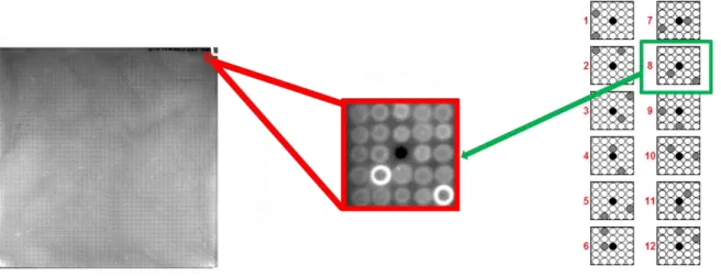
Figure 5. PA format and a representative positive signal... 31
Figure 6. PA Spotting pattern is composed of 12 proteins in duplicate and a reference InkDot. .. 32
Figure 7. Representative output of MSS ... 35
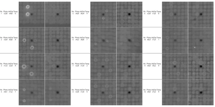
Figure 8. Evaluation method of PC ... 37
Figure 9. Non-specific background staining by some SCLC sera (left) and their corresponding ELISA values (right) ... 47
Figure 10. SCLC sera causing undesirable background staining ... 48
Figure 11. Final serum dilution ratio determination for ideal signal / background ratio ... 49
Figure 12. Final serum dilution ratio determination by pool dilution ... 50
Figure 13. 2o Ab dilution ratio comparison ... 51
Figure 14. Q-Dot dilution ratio comparison ... 51
Figure 15. Repeats of custom PA screening by the same serum for reproducibility assessment .. 52
Figure 16. Valuable clones selected from Testis pool screenings and the overlap for different cancers ... 53
Figure 17. Valuable clones selected from FB pool screenings and the overlap for different cancers ... 54
Figure 18. Our iterative ELISA strategy for identification of best antigens by performing the minimum number of screening ... 65
Figure 19. Stepwise antigen selection strategy by utilizing standart deviation cut-offs ... 66
Figure 20. ELISA data normalization for plate and serum ... 67
Figure 21. Quality control for the serum antibody integrity ... 69
Figure 22. Scatter dot plot of ELISA values of the most valuable 6 antigens ... 71
Figure 23. Manual cut-off determination for seropositivity for the ELISA results ... 72
xvi
Figure 25. Confirmation of SOX2 ELISA with Western blot for a representative group of
sera………..…..74
Figure 26. ELISA and Western blot correlation for a group of patients for the 6 antigens ... 75
Figure 27. ELISA and custom PA correlation ... 77
Figure 28. Correlation of PA with Western blotting for representative clones ... 78
Figure 29. SVM and MC comparison for ELISA values ... 82
Figure 30. Feature selection for the antigen panel accuracy evaluation ... 83
Figure 31. Comparison of SAS and ELISA for ROC by MC ... 85
Figure 32. ELISA, PC by MC and Chest X-ray and Low-dose CT comparison for ROC ... 87
Figure 33. Seropositivity and survival correlation for different cut-offs ... 94
Figure 34. Volcano plots; seropositivity and survival correlation for SCLC cohorts ... 97
Figure 35. Seropositivity and overall survival correlation by Kaplan-Meier graphs. ... 98
Figure 36. SOX2 autologous antibody and SOX2 expression correlation. ... 101
Figure 37. SOX2 protein expression and autologous antibody correlation analysis ... 103
xvii
List of Tables
Table 1: Autologous antibodies in cancer and PND ... 7
Table 2: p53 anbibody levels in case of cancer development and controls ... 9
Table 3: Correlation between SOX2 phage and ELISA results ... 12
Table 4: Technical limitations of SEREX (A) ... 13
Table 5: Technical limitations of SEREX (B)... 14
Table 6: mRNA representation limitations of SEREX ... 14
Table 7: Summary of limitations of SEREX ... 15
Table 8: Sensitivity and specificity of anti-tumor antibodies in lung cancer ... 21
Table 9: Sensitivity values of autologous antibodies useful in SCLC diagnosis at 100% specificity ... 21
Table 10: Estimated effect of novel autologous antibodies in sensitivity of SCLC diagnosis ... 22
Table 11: Sensitivity and specificity values of autologous anti-tumor antibodies for other solid tumors ... 22
Table 12: Representative output of SAS ... 36
Table 13: List of clones ordered from Testis pool screening ... 55
Table 14: List of clones ordered from FB pool screening ... 56
Table 15: Final list of clones screened with ELISA ... 61
Table 16: CV for ELISA data as quality control (A: Medium Signals) ... 68
Table 17: CV for ELISA data as quality control (B: High Signals) ... 68
Table 18: Individual sensitivity values of the most valuable 6 antigens in 100%-90% specificity range by MC ... 79
Table 19: Sensitivity and specificity values of the antigen panels for the best MC cut-offs ... 80
Table 20: Univariate Cox regression analysis for overall survival ... 88
Table 21: Antibody frequency and overall survival (cut-off: Mean + 3 * StDev) ... 90
Table 22: Cox Regression analysis and overall survival ... 92
Table 23: Final list of valuable clones after custom PA screening of BD ... 105
xviii
Abbreviations
AUC Area under the curve
BD Behçet’s Disease
CT Computerized Tomography
CV Coefficient of Variation
ELISA Enzyme Linked Immunosorbent Assay
FB Fetal Brain
IHC Immunohistochemistry
MC Monte Carlo
MCR Multivariate Cox Regression
MSS Manual selection software
NBD Neuro Behçet’s Disease
NSCLC Non-small cell lung cancer
PA Protein Array
PC Pixel Count
PNPP p-Nitrophenyl Phosphate
SAS Signal analysis software
SCLC Small cell lung cancer
SD Standard Deviation
SOX2 SRY (Sex Determining Region Y)-Box2
SVM Support Vector Machine
1
Chapter 1
1 INTRODUCTION
1.1 LUNG CANCER AND IT’S DIAGNOSIS
1.1.1 Lung Cancer Risk factors, Incidence and Mortality
Lung Cancer caused 1.1 million male and 491.200 female deaths in 2012 as the primary cause of cancer related death worldwide [1]. Tobacco smoking is the main reason of Lung cancer, responsible for 90% of the lung cancers [2, 3], and the other causes are radon exposure, asbestos exposure, passive smoking, arsenic and polycyclic hydrocarbons [3]. The main reason why Lung cancer is so deadly is that it is detected mostly when the disease is disseminated. SCLC constitutes 20% of all lung cancer cases [4, 5] and is the most aggressive subtype. Overall 5-year survival rate for SCLC is only 6% [3].
One of the highest number of yearly new cancer cases belongs to lung cancer among all cancers and the highest number of cancer related mortality belongs to lung cancer. The earlier stages of lung cancers correlate with good overall survival compared to the later stages. The biggest problem about lung cancer is that the disease doesn’t cause any symptoms at early stages and at later stages any type of therapy doesn’t cure the cancer. The survival rate of lung cancer drops from 37% for the first year to 8% for the 5. year [6]. So that early novel diagnosis methods for Lung Cancer are urgently required.
1.1.2 Current methods for dignosis of lung cancer
Currently the method used for lung cancer diagnosis is CT [4]. More than 60% of patients are diagnosed at advanced stages when a cure is not possible [7]. SCLC patients have 5-year relative survival rates of 31% for Stage 1, 19% for Stage 2, 8% for Stage 3 and only 2% for Stage 4 [1]. Since early diagnosis can increase the overall survival of SCLC patients [8-10] a novel method which can be practical, non-invasive and cheap is urgently required.
Currently, after presentation of symptoms, diagnosis of SCLC is confirmed by histological evaluation of bronchoscopy samples and study of fine-needle aspiration,
2
endoscopic ultrasound guided fine-needle aspiration or transbronchoscopic needle aspiration [11-14]. Utilization of these methods lack certain pathological discrimination of SCLC from other lung malignancies [12] and sampling errors are the most common cause of false negatives in FNA cytology [12, 15]. Since it is difficult to diagnose SCLC accurately, development of a method for SCLC diagnosis based on molecular biomarkers is crucial and this method can increase the efficacy of SCLC diagnosis.
1.1.3 Potential of biomarkers for lung cancer diagnosis / early diagnosis
Biomarkers are valuable tools to be utilized for diagnosis, however to be useful in the clinic biomarkers should fit some criteria: The biomarkers should (1) be quantifiable and reproducible, (2) have good PPV and NPV, (3) be measurable in suitable material, in small amounts and with little effort, (4) indicate a disease state, (5) have validated clinical use, (6) be accepted by the market, (7) be economical; and (8) be reimbursed by insurance companies [16]. There can be biomarkers for the risk detection for lung cancer as well as biomarkers for distinguishing malignant from benign tumors. Identifying individuals at high risk may prevent the false-positive rates and decrease the overall number of CT-scans to be performed both for the benefit of cost reduction and accurate diagnosis. Similarly a biomarker that can distinguish a malignant tumor from a benign tumor would be invaluable since it can reduce the number of unnecessary surgical operations which is applied to 24% of indeterminate pulmonary nodules and 30% of lung cancer patients.
1.2 IMMUNE SYSTEM AND CANCER
1.2.1 Immune response against cancer
Immune system and cancer interaction and interplay is valuable in determining the type of biomarkers to be utilized for cancer diagnosis. There is an interplay between cancer and immune system during cancer development which is called Immunosurveillance which both eliminates and manipulates the development of cancer. Immunosuppressed individuals have higher incidences of cancer subtypes compared to immunocompetent ones [17]. Immunosurveillance leads to the understanding of immunoselection; which constitutes the stages of elimination, equilibrium and escape by which cancer is manipulated to evolve by the immune system. Immunosubversion also contributes to a favorable growth of the cancer by suppressing the immune system [18]. So that immune system plays a major role during the
3
evolution of the tumor and there is a continuous immune response against cancer throughout its development. Both innate and humoral immunity takes role in this active response.
1.2.2 Serum reactivity against cell surface antigens
Some of the early studies about cancer immunity addresses the immune responses against the tumor cell surface antigens. These studies utilized hemadsorption and immune adherence assays to show serum reactivity profiles with surface tumor antigens. Absorption tests with cells from autologous, allogeneic and xenogeneic sources, determine the specificity of reactions between melanoma cells and autologous serum. By these studies it was shown that there is a heterogeneity of serum reactivity against surface antigens in melanoma. Patients have different serum reactivity profiles in terms of the tumor cell surface antigen identity [19].
1.2.3 Serum reactivity against intracellular antigens
Serum reactivity was not only observed against cell surface antigens but also against intracellular antigens. p53, one of the most famous tumor suppressor genes was originally identified as a transformation related antigen in induced sarcomas as a tumor associated antigen. p53 is a transformation related antigen consistently identified in methylcholanthrene induced sarcomas, MuSV and SV40 transformed cell lines, spontaneously transformed fibroblasts and leukemias, but not in nontransformed cells. Either induced or not, p53 autologous antibodies were consistently present in the sera of individuals that harbor a tumor transformation. Autologous serum from Meth A mice were shown to immunoprecipitate p53 from Meth A sarcoma extracts [20]. So that serum reactivity can also develop against intracellular antigens.
1.2.4 Methylcholanthrene induction causes tumor cells to be rejected by
the host
Mutagenic agents like methylcholanthrene were also shown to cause the tumor cells to be attacked by the immune system. Methylcholanthrene induction of a nonimmunogenic malignant mouse teratocarcinoma resulted in immunogenic variant cells that were incapable of forming tumors in syngenic mice. These variants undergo a process of immunerejection and confer a long lasting immunity. The inability of the induced cells to form tumors was specifically due to the immune response since they were able to survive in the gamma
4
irradiated syngenic mice. Methylcholanthrene induction makes the non-immunogenic tumor cells express strong transplantation antigens which leads tumor rejection by the host [21].
1.2.5 Immunoediting is a driving force for less imunogenic tumor
development
Immune system is known to be continuously shaping the tumor throughout its development. Immunoediting of tumor is a driving force for less imunogenic tumor development. Tumors developed in normal BALB/c mice are less immunogenic compared to tumors developed in T-cell deficient nude mice. The number of tumors that were not rejected by the normal mice were 2 fold of the ones that were originating from nude mice. This indicates that tumors are shaped by immunoediting toward a less immunogenic phenotype in time. Chemically induced sarcomas from nude mice are more immunogenic than similar sarcomas from congenic normal mice [22].
1.2.6 Autologous CTLs lyse antigen presenting tumor cells
Immune system is not only involved in less immunogenic tumor development, but also well known to actively kill tumor cells by a CTL response. MAGE-1 is a cancer-testis (CT) antigen against which a CTL response is developed through tumorigenesis. Autologous CTLs specifically recognize an immunodominant epitope of the protein which is a nonapeptide. The presentation of the nonapeptide on HLA molecules causes lysis of the tumor cells by the CTLs. The nonapeptide has superior effect on the lysis capacity of CTLs compared to the octapeptides lacking the N- or C- terminal amino acids thus the CTL response is specifically targeting the nonapeptide MHC complexes. MAGE-1 specific CTLs lyse tumor cells which present MAGE-1 nonapeptide on MHC Class I, but not the octapeptides lacking the N- or C- terminal amino acids [23].
1.2.7 Autologous antibodies as part of the immune response to cancer
Together with CTLs autologous anti-tumor antibodies are part of the humoral immune system that has the potential to be efficiently utilized in clinic as an indicator of cancer if profiled clearly. This is because autoantibodies are valuable since they are more stable and persistent in serum and so they can be measured by methods like ELISA compared to autoantigens causing their elucidation which are transient in duration due to short lived
5
changes in tumor site so requires a biopsy to monitor. However the antibody profiling is complex since tumor development is an evolutionary process under the influence of the immune system and autoantibody repertoire in the serum is highly probable to be patient specific and so it is hard to establish a general evaluation methodology to apply to all patients [18].
Autoantibody biomarkers have advantages over TAAs since they are stable in serum, can be present in high concentrations compared to a low level expression of the TAA which elicits the antibody, and a panel of the autoantibodies can have increased sensitivity and specificity values for cancer diagnosis [17]. Moreover these autoantibodies can also be utilized for the prognosis and immunotherapy of cancer as well as defining personalized therapy regimes [17]. Early development of autoantibodies implies their role in the elimination step of immunosurveillance. So that it is important to investigate the autoantibody elucidation mechanisms and reasons parallel to tumor progression for the sake of understanding the biology of tumorigenesis.
1.2.8 Autologous antibody elucidation mechanisms
Elucidation of a humoral immune response against tumor is not a well-established phenomena. However autoimmunity against tumor antigens is generated based on 4 known possible mechanisms: 1) a missense mutation in ORF of a gene causing a new epitope to be presented by the MHC class I for recognition as a foreing epitope by the T-cells, 2) a mutation in ORF of a gene causing a new epitope for recognition by TCR, 3) overexpression of a self protein, 4) ectopic expression of a self protein. Even intracellular proteins can become immunogenic by these mechanisms by usual immuno-monitoring mechanisms, as well as aberrant tumor cell death and consequent release of intracellular proteins into an immunogenic tumor environment and tumor cell microvesicle shedding [17].
Most of the autologous antibodies are elicited against intracellular proteins. Detailed mechanism of this phenomenon is not well understood. The current hypothesis is; the major component of the immune response against intracellular proteins is a CTL response and the B cell response is a side effect of this mechanism [24]. The tumor cells destructed in the tumor microenvironment either by mechanical force, phagocytosis, autophagy, apoptosis or necrosis result in recognition of the tumor specific antigens by the immune system. These antigens are primarily presented to CTLs and CD4+ T-cells and antibody elucidation developes as a secondary response.
6
1.2.9 Antigen expression and autologous antibody elucidation
Previously we have identified 2 proteins; SOX2 and ZIC2, against which high titer autologous antibodies are elicited in SCLC. SOX proteins are generally expressed within the developing nervous system of the vertebrate embryo, and their expression either ceases or is reduced at later stages of development. It was shown that in both chicken and mouse, Sox2 is expressed predominantly in the developing central nervous system and are down-regulated as neural differentiation progresses. Outside the nervous system, low-level mouse Sox group B gene expression has been reported in gut epithelium, developing limb buds, and genital ridges of the embryo. Mouse Zic genes, although unrelated to Sox group B genes, have similar temporal expression patterns, and Sox2 and Zic1 are subject to common developmentally regulatory elements. All Zic genes are expressed from the gastrula stage on and show different temporal expression patterns. Xenopus Zic2 expression commences earlier than XZic1 and 3. In the adult mouse, Zic2 expression is restricted to the granule cells of the cerebellum. Although SOX2 mRNA can be detected in some adult tissues, ZIC2 is expressed only in brain and testis of humans. Most probably various gene activation or derepression mechanisms are involved in the tumor expression of ZIC2 and maybe SOX genes. A likely explanation of why SOX2 and ZIC2 antibodies are elicited is that SCLC expresses a range of neuroectodermal antigens, including SOX group B and ZIC2, primarily restricted to the nervous system and that immune tolerance has not been established against these antigens. Growing outside the brain–blood barrier, SCLC would provide a potent immunogenic stimulus to the host. Of the SCLC cell lines tested in the study 80% were expressing ZIC2 mRNA wheras 50% were expressing SOX2 mRNA. SCLC patients tested in this study were 41% seropositive against SOX2 and 29% seropositive against ZIC2. This study showed that high-titered (1:106) IgG response to tumor antigens is not a rare event and such responses are not limited to patients with Paraneoplastic Neurological Disease (PND). None of the patients having SOX2 or ZIC2 antibodies have PND in the patient population. However the correlation between autologous antibody elucidation and antigen expression was not clarified with this study. So further study needs to be performed to evaluate the potential of these biomarkers as tumor biomarkers and the mechanisms triggering antibody elucidation [25].
1.2.10 Autologous antibodies and PNS
Autologous antibodies against tumor-associated antigens (TAA) is part of this immune response [26-32] and has the potential for early tumor diagnosis since it is well-known that
7
these antibodies are elicited even when the tumor is at its microscobic state [29, 33-35]. The autologous anti-tumor antibodies correlate with PNS in a tiny percent of cancer patients and these patients can be diagnosed with a cancer even after 10 years of PNS emergence [36]. In the case of SCLC-associated PNS the neurological disease is known to be symptomatic before the associated SCLC is symptomatic and thus diagnosed [37, 38].
There are a number of known autologous anti-tumor antibodies in SCLC. Most of these antibodies are indicators of presence of a PNS. About 6% of the SCLC patients are reported to have PNS (22); 3-4% of SCLC develop LEMS [39, 40] which is associated with VGCC autoantibodies. However SCLC patients without PNS are also shown to elicit autologous anti-tumor antibodies. For example SOX2, p53, NY-ESO-1, CAGE, GBU 4-5, Annexin 1 are shown to have sensitivity values of 11, 13, 13, 12, 9, 9 respectively, in SCLC patients without PNS [26].
1.2.11 Autologous antibodies in SCLC with/without PND
Autologous anti-tumor antibodies were observed in many cancers. Lung cancer is among these cancers and the autologous anti-tumor antibodies are not only elicited against tumor associated antigens (TAA) but also in the case of PND high titers of autologous antibodies are frequently observed. Table 1 summarizes the autologous antibody frequencies in cancer with and without PND.
Table 1: Autologous antibodies in cancer and PND
Antibody Cloned genes Tumor type Paraneoplatic disease Frequency in SCLC w/o PND Frequenc y in SCLC with PND 1 Anti-Hu (ANNA-1) HuD, HuC, Hel-N1 SCLC, Neuroblastoma, Prostate Encephalomyelits, sensory neuropathy, cerebellar degeneration, autonomic disfunction 18.5-25.5 %[41] 75 %[42] 2 Anti-ZIC ZIC4 (ZIC1,3?) SCLC Cerebellar degeneration 16 %[42] 29 %[42] 3 Anti-Recoverin Recoverin SCLC, melanoma, gynecological tumors Retinal degeneration 15 %[43] 66 % 4 Anti-CRMP5 CRMP5 (POP66) SCLC, thymoma Encephalomyelits, sensory neuropathy, cerebellar degeneration, corea 9.5 %[42] 27%[42] 5 Anti-VGCC P/Q type VGCC SCLC Lambert-Eaton myasthenic syndrome 5 %[41]-18 %[44] 91 %[44]
8 6 Anti-VGCC N type VGCC SCLC Lambert-Eaton myasthenic syndrome 12 %[45]- 22 %[44] 73 %[44] 7 Anti-Amphyphy sin
Amphiphysin SCLC, Breast Encephalomyelitis,
stiff-man syndrome 1.4 %[46] 2.9 %[46] 8 Anti-PCA-2 ? SCLC Cerebellar degeneration ? 9 Anti-VGKC Potassium channels SCLC, thymoma Neuromyotonia ? 10 Anti-Enolase Enolase SCLC, breast, pancreas Retinal degeneration ? 29 %[47]
11 Anti-p35 p35 SCLC Retinal degeneration ? 5 %[47]
12 Anti-Yo (PCA-1) CDR34, CDR62 Lung, ovarian, breast Cerebellar degeneration 3 %[45] 13 Anti-Ri (ANNA-2)
Nova1, 2 Lung, breast,
ovarian, bladder, gynecological
Ataxia with or witout
opsoclonus-myoclonus
3 %[45]
14 Anti-Ma1 Ma1 Lung Brain-stem
encephalitis, cerebellar degeneration
?
15 ANNA-3 ? Lung Sensory neuropathy, encephalomyelitis
? 16 Anti-Tr MAZ Hodgkin’s
lymphoma, brain, lung, testis
Cerebellar degeneration
16 %(non-SCLC tumors)[48]
17 Anti-Ma2 Ma2 Testicular Limbic brain-stem encephalitis
? 18 Anti-MAG MAG Waldenstrom’s
macroglobuline mia Peripheral neuropathy ? 19 Anti-mGluR1
Glu receptor Hodgkin’s
lymphoma
Cerebellar degeneration
The autologous antibodies associated with PND are commonly elicited in lung cancer; specifically in SCLC. So that SCLC is an immunogenic cancer. The frequency of autologous antibodies in SCLC with PND is always higher than the frequency of autologous antibodies in SCLC without PND. However SCLC patients without PND also elicit antibodies and this data suggests that there might be other autologous antibodies which are independent of PND. Thus the autologous antibodies that are specific to SCLC without PND can be exploited for diagnosis/early diagnosis of SCLC.
1.3 DIAGNOSIS AND EARLY DIAGNOSIS OF CANCER
1.3.1 Autologous antibodies precedes PND and clinical cancer diagnosis
The timing of elucidation of autologous antibodies is critical for their utilization for early diagnosis, thus their presence in the early stages of tumor development defines their potential. PND coexists with a tumor for a significant number of SCLC patients. Anti-Hu
9
antibodies are elicited in almost 70% of the SCLC patients having a coexisting PND. The symptoms of PND is observable in average of 6.5 months before cancer and PND diagnosis. Data strongly suggests that autologous anti-tumor antibodies are detectable at least 6.5 months before SCLC diagnosis [49]. Not only anti-Hu antibodies but several other antibodies are also detectable up to 5-10 years before cancer and PND diagnosis [49-51]
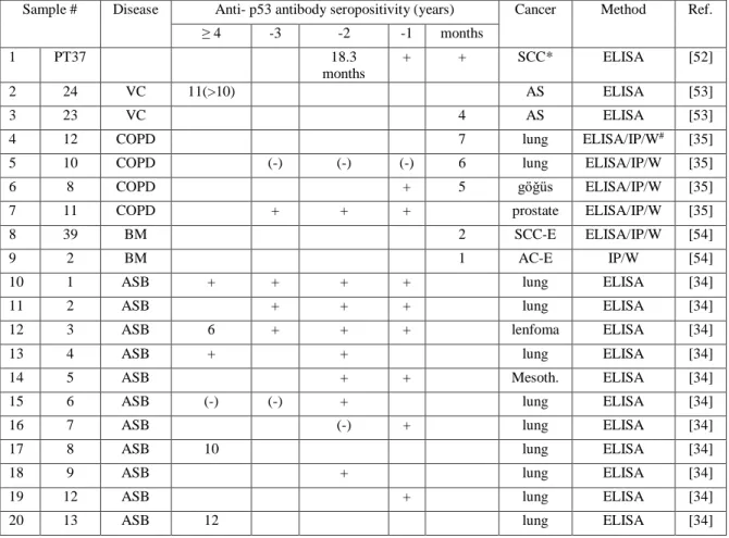
1.3.2 p53 antibody level stability / fluctuations can predict cancer
development
Not only antibodies associated with PND precede clinical cancer diagnosis, but also antibodies independent of PND; p53 antibodies were shown to precede cancer diagnosis. Table 2 shows the p53 antibody levels in a time course of up to 12 years in case of cancer development or controls.
Table 2: p53 anbibody levels in case of cancer development and controls
Cancer
Sample # Disease Anti- p53 antibody seropositivity (years) Cancer Method Ref. ≥ 4 -3 -2 -1 months 1 PT37 18.3 months + + SCC* ELISA [52] 2 24 VC 11(>10) AS ELISA [53] 3 23 VC 4 AS ELISA [53] 4 12 COPD 7 lung ELISA/IP/W# [35]
5 10 COPD (-) (-) (-) 6 lung ELISA/IP/W [35] 6 8 COPD + 5 göğüs ELISA/IP/W [35] 7 11 COPD + + + prostate ELISA/IP/W [35] 8 39 BM 2 SCC-E ELISA/IP/W [54] 9 2 BM 1 AC-E IP/W [54] 10 1 ASB + + + + lung ELISA [34] 11 2 ASB + + + lung ELISA [34] 12 3 ASB 6 + + + lenfoma ELISA [34] 13 4 ASB + + lung ELISA [34] 14 5 ASB + + Mesoth. ELISA [34] 15 6 ASB (-) (-) + lung ELISA [34] 16 7 ASB (-) + lung ELISA [34] 17 8 ASB 10 lung ELISA [34] 18 9 ASB + lung ELISA [34] 19 12 ASB + lung ELISA [34] 20 13 ASB 12 lung ELISA [34]
10
Healthy
Sample # Patient No. Disease Anti- p53 antibody seropositivity Method Ref. 1.year 2.year 3.year 4.year 5.year
1 14 ASB* + (-) (-) (-) (-) ELISA [34] 2 15 ASB (-) (-) + (-) ELISA [34] 3 16 ASB + (-) ELISA [34] 4 17 ASB (-) (-) + (-) ELISA [34]
These results indicate that anti-p53 autologous antibodies’ presence can precede cancer diagnosis even up to 12 years. Also p53 antibody levels are stable if there is an underlying cancer development. However p53 antibody response is sporadic in persons without cancer and the antibody levels fluctuate.
1.3.3 Sensitivity and specificity of autologous antibodies
Even though autologous anti-tumor antibodies can be utilized for early diagnosis, the sensitivity values of single antibodies cannot exceed 15% at 97% specificity. Published data strongly suggests that these autologous anti-tumor antibodies are not limited to a group of cancer patients so that their utilization in early cancer diagnosis has a high potential [55]. It is strongly suggested that the evaluation of autologous antibodies as a panel can increase the sensitivity at a certain specificity for early cancer diagnosis.
1.3.4 Autologous antibodies already in clinical use for diagnosis of cancer
There is already a diagnostic kit for lung cancer; Oncimmune’s EarlyCDT-Lung. The kit has a Sensitivity value of 40% at a Specificity value of 93% for lung cancer. SCLC has a relatively higher sensitivity compared to NSCLC, which also suggests that SCLC is a more immunogenic tumor type. There is no significant difference in terms of sensitivity of the kit between late stage and early stage of lung cancer or between limited and extended disease categories of SCLC or between NSCLC Stages I, II, III and IV [56] This is a strong indication that autologous anti-tumor antibodies can be utilized for diagnosis of any type of lung cancer independent from stage.
1.4 AUTOLOGOUS ANTIBODY DISCOVERY METHODS
1.4.1 Methods for autologous antibody discovery
Since autologous antibodies are valuable tumor markers new autologous antibodies need to be identified. There are a number of methods for novel autoantibody discovery. These
11
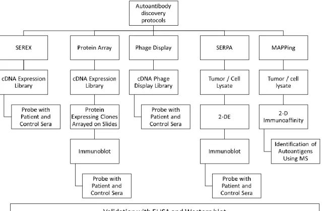
methods include serological analysis of expression cDNA libraries (SEREX), serological proteome analysis (SERPA), multiple affinity protein profiling (MAPPing), reverse-capture microarray, phage display, protein micro- and macro-array screening [17]. Each of these methods have their own specific limitations. By SEREX CT antigens (NY-ESO-1, SSX2, MAGE, etc), mutational antigens (p53, etc), differentiation antigens (tyrosinase, SOX2, ZIC2, etc) and embryonic antigens have been identified so far in cancers as immunogenic [25, 57, 58]. However the antigens identified by SEREX have low sensitivity values [17] and SEREX method; even though is one of the most powerful methods, has some limitations in identifying autoantigens [59]. PA (PA) screening has the potential for identification of many immunogenic clones in a specific cancer in a comperative analysis with the samples of benign form of the disease or healthy controls and in most cases futher validation and analysis of these clones is possible by subarraying [60].
Figure 1. Conventional techniques for novel autologous antibody discovery and validation. As common features all techniques include a cDNA expression library or
12
Figure 1 summarizes the procedures of different available methods for identification of new autologous anti-tumor antibodies. The methods start with a cDNA library or a tumor/cell lysate and investigate the affinity of autologous anti-tumor antibodies to the starting material. To identify tumor specific antibodies cancer and healthy sera are screened in parallel and compared afterwards. Even though all these methods have differences in the way of their application at the end validation of the identified autologous anti-tumor antibodies with ELISA or Western blot is common to all methods. Otherwise it is not possible for these newly diagnosed antibodies to be utilized in clinic.
Table 3: Correlation between SOX2 phage and ELISA results
Previously we have investigated the correlation of our SEREX and ELISA results (Table 3). There was a significant correlation and this indicates if more valuable autologous anti-tumor antibodies can be identified by SEREX these will most probably correlate with ELISA.
Previously many autologous anti-tumor antibodies have been identified by SEREX and they are stored in a database named; Cancer Immunome Database. Even though more than 1500 cancer specific autologous anti-tumor antibodies have been identified till now most of them have a limited sensitivity and specificity. Because of this reason new methodologies need to be investigated to identify more valuable autologous anti-tumor antibodies.
A number of methods have been developed to identify the antigens against which the autologous antibodies are elicited. SEREX is one of the most powerful methods to identify novel tumor antigens. SEREX was performed for many cancers like colon, lung, melanoma, renal and Hodgkin’s disease. The antigens identified by SEREX can be categorized into many different categories like mutational, differentiation, amplified or overexpressed, retroviral, splice variants and CT antigens. The CT antigens are mostly located on the X chromosome and almost all of them represent a family of genes [61].
SOX2 Phage SOX1 Phage
Number of XY Pairs 90 90
Pearson r 0.6855 0.7181
95% confidence interval 0.5575 to 0.7817 0.6003 to 0.8055
P value (two-tailed) < 0.0001 < 0.0001
P value summary *** ***
Is the correlation significant? (alpha=0.05) Yes Yes
13
One of the first autologous antibodies identified by our group is NY-ESO-1, which is a CT antigen. It was identified by utilization of SEREX in esophageal squamous cell carcinoma patients. NY-ESO-1 is a CT antigen, expressed mainly in testis, uterus and a diverse range of tumors. Northern blot results indicate NY-ESO-1 expression in Testis and melanoma cell lines. NY-ESO-1 is not expressed by the normal tissues. NY-ESO-1 has limited expression in Testis and cancer cell lines, but not in the other healthy tissues. [62] CT antigens behave very similar to NY-ESO-1 in terms of expression regulation. They were defined as ideal targets for immunotherapy because of their limited expression to tumors and the above mentioned tissues together with eliciting autologous anti-tumor antibodies in cancer patients.
1.4.2 Limitations of SEREX
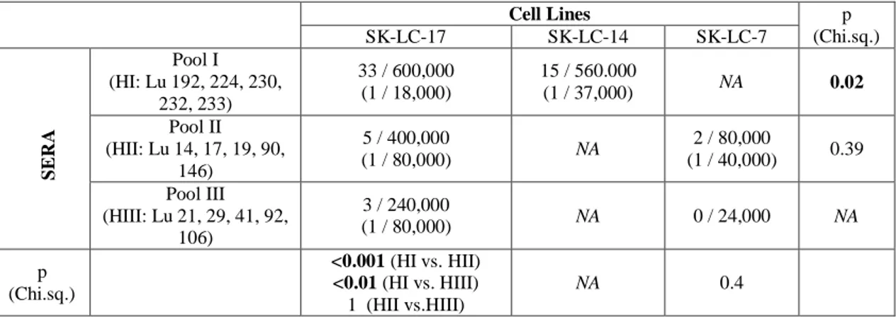
Even though SEREX was widely used for identification of new autologous anti-tumor antibodies it has dramatic limitations. For example cDNA libraries utilized in SEREX obtained from lung cancer cell lines represent different number of seropositive clones when these libraries were screened with the same serum (Table 4). Besides the same cell line represents different number of seropositive clones when screened with 3 different serum pools. This indicates that a single cell line has limited representation of mRNA diversity and likewise a serum pool has a limited representation of antibody diversity in SEREX.
Table 4: Technical limitations of SEREX (A)
Cell Lines p (Chi.sq.) SK-LC-17 SK-LC-14 SK-LC-7 SER A Pool I (HI: Lu 192, 224, 230, 232, 233) 33 / 600,000 (1 / 18,000) 15 / 560.000 (1 / 37,000) NA 0.02 Pool II (HII: Lu 14, 17, 19, 90, 146) 5 / 400,000 (1 / 80,000) NA 2 / 80,000 (1 / 40,000) 0.39 Pool III (HIII: Lu 21, 29, 41, 92, 106) 3 / 240,000 (1 / 80,000) NA 0 / 24,000 NA p (Chi.sq.) <0.001 (HI vs. HII) <0.01 (HI vs. HIII) 1 (HII vs.HIII) NA 0.4
14
Table 5: Technical limitations of SEREX (B)
SERA
Pool IV
Anti-NY-ESO-1 Ab (+)
Pool V
Anti SOX1, 2, 3 Ab(+)
C el l Li nes SK-LC-14 (NY-ESO-1+) (2.4 x 10⁴ pfu) NY-ESO-1 (-) NA NCI-H740 (SOX1,2,3+) (5.6x10⁵ pfu) NA SOX3 (-)
SK-LC-14 is a cell line known to express NY-ESO-1 mRNA by qRCR and Pool IV was shown to contain anti-NY-ESO-1 antibodies by ELISA, however no reactivity was observed with SEREX (Table 5). Similarly NCI-H740 is known to express SOX3 mRNA by qRCR and Pool V was shown to contain anti-SOX3 antibodies by ELISA, however no reactivity was observed with SEREX. Both NY-ESO-1 and SOX3 are previously shown to be expressed in mid-abundance (100-500 copies in 500.000 pfus) in these cell lines by qPCR, meaning that they are not even rare transcripts. So that SEREX has a limited mRNA representation capacity and the serum pools cannot be used more than once so that it is highly unlikely to detect many novel autologous antibodies by SEREX.
Table 6: mRNA representation limitations of SEREX
Screened pfu numbers Expected in 500,000:
Error rate: if 1:3 of all ORFs are correct:
Genes mRNA
number
mRNA percent (%)
Number of proteins having the correct amino acid sequence
Ef1α 4,399 0.88% 1,466 Cytochrom Cox 1 1,802 0.36% 601 Clone 190B1 1,670 0.33% 557 Tubulin β 1,361 0.27% 454 40S Riboprotein S6 1,203 0.24% 401 40S Riboprotein S4 658 0.13% 219 60S Riboprotein L4 559 0.11% 186 GAPDH 539 0.11% 180 Ef1β 441 0.09% 147 Calmodulin 210 0.04% 70 HSP KD71 184 0.04% 61 HSP KD90 171 0.03% 57 TNFR 79 0.02% 26 Clone 244D14 53 0.01% 18 Clone 241F 17 13 0.00% 4
Table 6 shows the number of mRNA numbers for different genes in a representative SEREX cDNA library on the left panel. The middle panel shows the percentage of these numbers in 500.000 pfus. And the right panel indicates the number of proteins having the correct amino acid sequence in an ideal case since only one third of the mRNA copies can
15
result in expression of the true ORFs. Based on these numbers one can expect the lower half of these genes on the table cannot be represented by the plaques in SEREX which indicates a significant limitation of the technique.
1.4.3 Theoretical comparison of SEREX with PAs
Common antibody signature for all cancers can be identified if a large number of signals can be analyzed simultaneously as in the PA screening format [60]. Antibody signatures can discriminate cancer from healthy as well as benign diseased individuals of the same organ. These antibody signatures can pave the way to identify antigens that take place in general metabolic pathways and targeted by the immune system. PA screening can result in identification of both in-frame an non in-frame clones to be immunogenic. However the newly identified antibodies are required to be validated by independent assays which is the bottleneck in utilization of an antibody biomarker in clinical diagnosis [60]. Previously many studies have been performed to identify accuracy, sensitivity and specificity values to discriminate cancer from healthy controls by many different methods [60].
Table 7: Summary of limitations of SEREX
Method SEREX PA
Technique Unnormalized mRNA library screening Screening of 15.744 different human proteins Ideal serum dilution 1:100 1:25.000 Serum amount
required 5 ml / run 0.2 ml / run
Sensitivity 1 50
As shown in Table 7, PA includes more than 15.000 different human proteins as opposed to an unnormalized cDNA library screening by SEREX. Serum can be diluted to 1:25.000 for PA screening compared to 1:100 diluted for SEREX which indicates a major advantage for PA screening in terms of required sample amount. 25 times less serum is enough for one PA screening compared to SEREX; 200 ul for PA screening and 5 ml for SEREX. Finally if the sensitivity of SEREX is assumed to be 1 while PA screening is at least 50 times more sensitive.
16
1.5 AUTOLOGOUS ANTIBODIES AND PROGNOSIS
1.5.1 Autologous antibodies can be utilized for prognosis
Autologous anti-tumor antibody response is a component of the anti-tumor immune response. One of the oldest studies addressing these antibodies shows a nice correlation, even though it is not statistically significant, between antibody response and good overall survival for the SCLC patients. In this study a number of antigens were shown to be recognized by the autologous antibodies in the patient serum according to Western blot data. After this study a major aim of the research became the identification of the individual antigens recognized by the autologous anti-tumor antibodies. This study suggested that autologous antibody response can be useful as a prognostic factor for cancer outcome prediction [63].
1.5.2 Autologous antibody positive sera have cytotoxic effect on cancer cell
line
Previously antibody positive sera were shown to have cytotoxic effect in vitro. Anti-Hu antibody positivity is not only associated with PND but also represents a strong anti-tumor immune response from the anti-cancer perspective. Anti-Hu antibody bearing sera are shown to have cytotoxic effect on NT2 cells. Whereas this effect is not observed with the tested normal serum. Anti-Hu negative SCLC sera were not as toxic as anti-Hu positive serum, and the difference is statistically significant. The cytotoxic effect was not caused by the anti-Hu antibodies themselves, since the serum was also toxic to Hu negative cell lines and IgG depleted serum was still cytotoxic to cell lines [64].
1.5.3 Autologous antibody positivity correlates with in-vivo cytotoxicity
Cytotoxic effect of the immune response coexisting with the autologous antibodies is also observable in vivo. A patient with anti-Yo positive paraneoplastic cerebellar degeneration (PCD) have lost all her Purkinje cells in the brain. However the Purkinje cells are clearly observable and morphologically normal in a cancer patient without neurologic involvement [65]. This indicates that the antibody seropositivity can be a measure of the cytotoxic immune response.
17
1.5.4 Autologous antibodies and survival
Different studies investigated the existence of a correlation between Hu, anti-VGCC and anti-p53 antibodies and survival. No correlation was identified between anti-Hu and anti-VGCC antibodies and survival for the SCLC patients. However a correlation was identified between anti-p53 antibodies and worse overall survival for NSCLC. This correlation was also observable for Squamous cell carcinoma whereas there was no correlation for Adenocarcinoma [41, 66]. So that a clear correlation between autologous antibody response and survival could not be formed yet.
1.5.5 Autologous anti-tumor antibodies identified by our previous studies
As a group we have also identified 2 antigens against which autologous anti-tumor antibodies are elicited; anti-SOX1,2,3 and anti-ZIC2. SOX proteins have more than 90% homology in their DNA binding HMG box domain. Thus autologous antibodies which recognize any of the SOX 1, 2 or 3 more than >90% of the cases recognize the other two antigens. SOX2 and ZIC2 autologous antibodies are high affinity and are present in high titer in serum; these antibodies are even detectable by SEREX at a million times diluted concentration [25].
1.5.6 Anti-SOX1 autologous antibodies are specific to cancer not PND
Autologous anti-SOX1 antibodies correlate with the existence of SCLC in Lambert-Eaton Myasthenic Syndrome. The seropositivity rate in the Paraneoplastic LEMS is 64% whereas none of the idiopathic patients are seropositive against SOX1. This data indicates that autologous antibody response against SOX proteins is highly specific to SCLC [67].
1.5.7 SOX2 antibodies and prognosis of multiple myeloma (MM)
Anti-SOX immune response is not limited to SCLC most probably since SOX2 is a stem cell antigen and is responsible for self renewal of the stem cells. Monoclonal gammopathy of undetermined significance (MGUS) represents a precursor lesion to MM. Detection of anti-SOX2 T-cells predicts a favorable outcome in patients with asymptomatic plasmaproliferative disorders. The patients with anti-SOX2 T-cells also had a significantly lower likelihood of disease progression, with a 2-year progression-free survival rate of 100% versus 30% compared with patients lacking anti-SOX2 T-cells with a statistically significant p
18
value. Therefore, immunity to SOX2 predicts the clinical outcome in patients with asymptomatic plasmaproliferative disorders [68].
1.5.8 SOX2 and ZIC2 antibodies and survival in SCLC
SOX2 and ZIC2 antibodies are among the most frequent antibodies elicited in SCLC patients. Previous study shows, although not statistically significant there is a trend for better overall and progression free survival for seropositive patients against SOX2 and/or ZIC2 [69]. According to this study most SOX1, 2 and 3 autoantibodies in serum remained consistent even after 6 months from SCLC diagnosis. All patients seropositive for SOX1 had SOX2 antibodies, and most were reactive with SOX3. None of the anti-SOX1 antibody-negative patients gained seroreactivity over the period of study. Over the periods tested, antibody titers remained within 10-fold in all seropositive patients. This indicates autologous SOX2 anti-tumor antibodies are reliable and stable biomarkers through the course of SCLC.
19
1.6 SENSITIVITY AND SPECIFICITY OF PANELS
1.6.1 Diagnosis by evaluating a panel of antibodies
Combined evaluation of 6 of the SEREX-identified autologous antibodies has 30% sensitivity at 100% specificity for SCLC diagnosis (Figure 2). If evaluated individually sensitivity of each antibody doesn’t exceed 15%. This indicates that combined evaluation of more autologous antibodies thus forming a panel can increase the sensitivity for diagnosis even more. To increase the sensitivity a panel should be formed and new valuable autologous antibodies needs to be identified to be incorporated in the panel.
Figure 2. Anti-SOX2, HuD, CRMP5, p53, NY-ESO-1 and/or recoverin autologous antibodies in SCLC diagnosis. Green line indicates x=y, blue line is the ROC line.
Sensitivity and specificity values were shown and AUC was indicated by confidence interval for p<0.001.
20
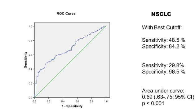
The situation is similar for NSCLC in which a sensitivity of 30% can be reached at 97% specificity with the same antigens evaluated for SCLC (Figure 3). So novel autologous anti-tumor antibodies needs to be identified to be utilized as a panel for cancer diagnosis
1.6.2 Sensitivity and Specificity of already identified autologous antibodies
The already identified autologous antibodies have significant sensitivity and specificity both for SCLC and NSCLC (Table 8). However these values values are not adequate for any of them or a panel of them to be utilized in clinical screenings for diagnosis.
Figure 3. Anti-SOX2, HuD, CRMP5, p53, NY-ESO-1 and/or recoverin autologous antibodies in NSCLC diagnosis. Green line indicates x=y, blue line is
the ROC line. Sensitivity and specificity values were shown and AUC was indicated by confidence interval for p<0.001.