A Study About Determining the Changes in the Structural
Characteristics of the Digital Cushion in Heifer and Multipar Dairy
Cows: A Preliminary Report
Celal İZCİ *
Muharrem EROL * Ebru GÖKŞAHİN *
* Selcuk Univercity, Faculty of Veterinary Medicine, Department of Surgery, TR-42075 Selcuklu, Konya - TURKEY
Makale Kodu (Article Code): KVFD-2010-3083
Summary
Claw horn lesions are crucial problems for dairy cows and causes serious financial losses. The changes in the structure of digital cushion are considered to be the trigger for such lesions. The purpose of this study is to determine these changes in heifer and multipar dairy cows. The structural changes in the digital cushion were determined by ultrasonography and gas chromatography. As a result, it can be mentioned that thickness of the digital cushion can be measured by ultrasonography and, also digital cushions of heifers have a lower percentage of fat than cows have and a higher rate of saturated fatty acid in their digital cushion. It can be asserted that these changes of digital cushion increase the traumatic effects inside the claw which may cause sole ulcer and white line disease.
Keywords: Dairy cow, Digital cushion, Lameness, Claw horn lesion
Düve ve Süt İneklerinde Ökçe Yastığının Yapısal Özelliklerindeki
Değişikliklerin Belirlenmesi: Ön Çalışma Raporu
Özet
Süt ineklerinde boynuz tırnak lezyonları önemli bir problemdir ve ciddi ekonomik kayıplara yol açar. Bunların oluşumunda ökçe yastığının yapısındaki değişikliklerin etkili olduğu düşünülür. Bu çalışmada; düve ve süt ineklerinde ökçe yastığının yapısal özelliklerindeki değişikliklerinin belirlenmesi amaçlanmıştır. Ökçe yastığının yapısındaki değişiklikler ultrasonografik ve gaz kromatografi yöntemi ile belirlendi. Ökçe yastığının kalınlığı ultrasonografik olarak ölçülebilir. Düvelerin ökçe yastığının yağ oranı sığırlardan daha düşük olup, doymuş yağ asitleri oranı daha yüksektir. Oluşan değişikliklerin tırnak içi travmatik etkiyi artırarak taban ülseri ve beyaz çizgi hastalığının oluşumunda etkili olabileceği söylenebilir.
Anahtar sözcükler: Süt ineği, Ökçe yastığı, Topallık, Boynuz tırnak lezyonu
INTRODUCTION
Claw horn lesions are crucial problems for dairy cows pressure acts on claws during the fi rst gestation period as and causes serious financial losses especially for highly a result of an upsurge of the weight of udder and fetus 11.
producti ve animals 1-3. As it is the case for sole ulcer and
Most of the body weight of a cow is carried by the white line disease, many claw lesions occur as a result of
supportive apparatus called “digital cushion” 12. Digital
traumas that affect the corium inside the horn capsule 4-7.
It is proven by epidemiological studies that heifers cushion is a complex structure that absorbs the pressure which give birth for the fi rst time have a higher rate of under the distal phalanx. It is constituted by loose claw lesions than cows which give birth twice or three connective tissue and different amounts of fatty tissue. times 8-10. It is emphasized, that unbalanced mechanic It consists of three segmental parts named axial, medial/
İletişim (Correspondence)
+90 332 2233579160
A Study About Determining ...
central and abaxial (Fig. 1) 12-15. Any corruption of the
absorbing characteristic of digital cushion predisposes the sensitive corium under the distal phalanx against injuries. Factors like standing and walking on hard flooring surfaces, diatery fatty acid composition and unwillingness of the animals to lie down raises predisposition. For this reason, digital cushion is considered as an important etiological factor to generate claw horn lesions and lameness 10,12,15,16. The purpose of this study is to
determine the periodical changes of digital cushion’s structure in heifers and dairy cows.
Fig 1. Appearance of the segmental parts (axial, medial/central and abaxial) of the digital cushion “(Sarel van Amstel& Jan Shearer 18)
Şekil 1. Ökçe yastığı’nın segmental parçalarının (aksiyal, medial / sentral ve abaksiyal) görünümü
MATERIAL and METHODS
This study was performed according to the ethical board approved by the Faculty of Veterinary Medicine University of Selcuk, Konya, Turkey (Approval No: 2010/043).
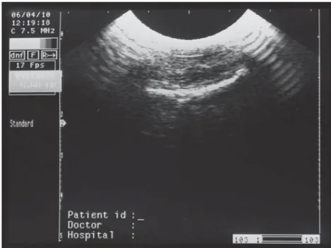
Research material included fore and hind feet claws of 4 Holstein heifers and 6 cows which gave birth 1, 2, 3 times or more in each case. All were slaughtered in an abattoir in Konya. Claws were harvested immediately after slaughter, routine hoof trimming was performed and at the junction of the heel and sole digital cushion thicknesses were determined ultrasonographically with a curved array dual-frequency probe set at 7.5 MHz
(Fig. 2).
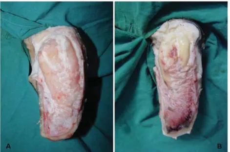
Later on, the claw horn of the sole was dissected carefully, solar corium and digital cushion segments were exposed (Fig. 3A-B). Tissue samples were obtained
from digital cushion segments. Lipid extraction was made in tissue samples according to the Folch17 method
and their fatty acid composition was determined by gas chromatography (HP6890, Agilent Technologies, USA) with an automatic injector. Peaks in chromatograms were obtained with three repetitions and their percentage areas’ arithmetic means and standard deviati ons were calculated.
RESULTS
It was determined that, according to the ultra sonographic evaluations of the heifer’s front right lateral and medial claws had approximately 0.85±0.04 cm and
Fig 2. Determination of the digital cushion thickness by ultrasound
Şekil 2. Ökçe yastığı’nın kalınlığının ultra sonografik olarak belirlenmesi
0.87±0.04 cm digital cushion thickness, respectively. Right hind lateral and medial claws had approximately 0.73±0.08 cm and 0.81±0.01 cm digital cushion thickness, respectively. In an old cow (approximately 12 years old) front left lateral and medial claws both had digital cushion thicknesses of approximately 0.65±0.04 cm and 0.65±0.04 cm. Gas chromatography analysis showed that heifers and cows that gave birth more than 3 times had a lower unsaturated fatty acid rate than the other cows (Fig. 4).
Fig 3.A-B. Appearence of the digital cushion’s segments after the sole dissection
Şekil 3.A-B. Tabanın diseksiyonundan sonra ökçe yastığı segmentlerinin görünümü
segment had been longer than the other ones. For heifers and other cows it may be stated that data obtained from the study of digital cushion thickness of the lateral and medial claws of fore and hind feet is parallel to literature data 10.
Thickness of the digital cushion can be measured by ultrasonography 10,19. In this study, the thickness of the
digital cushion was measured ultrasonographically from the junction of the heel and sole where a typical sole
Fig 4. The result of gas chromatography analysis. (SFA: Saturated Fatty Acid, M: Monounsaturated Fatty Acid, P: Polyunsaturated Fatty Acid) Şekil 4. Gaz kromatografi analiz sonuçları. (SFA: Doymuş Yağ Asidi, M: Doymamış Yağ Asidi, P: Aşırı Doymamış Yağ Asidi)
DISCUSSION
It is not wrong to claim that there is not much known about the structure of the digital cushion dairy cows have. It is known that each digital cushion has three cylindrical parallel segments (axial, medial and abaxial) containing connective tissue capsules filled by fatty tissue. Among these segments, axial one is the longest (Fig. 1-3.AB) 6,12,15,18. The macroscopic appearance of a
heifer’s digital cushion is whiter than that of cows which gave birth 2 or 3 times. It was determined that the axial
ulcer is located. Digital cushions of heifers have a lower percentage of fat than those of cows. Heifers also have a higher rate of saturated fatty acid in their digital cushion. After first birth and lactation the percentage of unsaturated fatty acids increases. In this study gas chromatography analysis showed that heifers’ digital cushion fatty acid rates are lower than of the cows which gave birth 2-3 ti mes. Physiologic and metabolic changes occur during gestation and birth period which decreases strength of connective tissues 20-22. As a result of aging,
162
A Study About Determining ...
the third lactation fat rates decrease, also 14. In this
study it was found that cows that gave birth more than three times had higher saturated fatty acid rates than the other cows. These results support the previous studies 10,12,14,15 that state traumas and contusions on
the connective ti ssues inside the claw have an effect on the development of sole ulcer and white line disease. According to these findings, it can be emphasized that thickness of the digital cushion can be measured by ultrasonography and, also digital cushions of heifers have a lower percentage of fat than cows have and a higher rate of saturated fatty acid in their digital cushion. It can be asserted that these periodical changes of digital cushion increase traumati c effects inside the claw which may cause sole ulcer and white line disease.
In conclusion, decreasing digital cushion thickness and structural changes with the effect of factors like stand and walk on hard flooring surfaces and unwillingness to lie down increase traumati c effect inside the claw and predisposes to claw horn lesions such as sole ulcer and white line disease. Based on this, it can be suggested that taking precautions to protect digital cushion’s content and structural characteristics may be a new approach to decrease claw horn lesions.
REFERENCES
1. Kossaibati MA, Esslemont RJ: The costs of producti on diseases
in dairy herds in England. Vet J, 154, 41-51, 1997.
2. Warnick LD, Janssen D, Guard CL, Grohn YT: The Eff ect of
Lameness on Milk Production in Dairy Cows. J Dairy Sci, 84, 1988 1997, 2001.
3. Bicalho RC, Vokey F, Erb HN, Guard CL, Grohn YT: Visual
locomotion scoring in the first seventy days in milk: Impact onpregnancy and survival. J Dairy Sci, 90, 4586-4591, 2007. 4. Baran V: Sığırlarda Tırnak Bozuklukları ve Bunların Neden Olduğu
Taban Ulkuslarının Sağaltımında Anti biyotik ve Enzim Uygulamaları. Kafkas Univ Vet Fak Derg, 3 (2): 201-210, 1996.
5. Ossent P, Greenough PR, Vermunt JJ: Laminitis. In, Greenough
PR, Weaver AD (Eds): Lameness in Cattle. pp. 277-289, John Wright and Sons Ltd, Britol, 1997.
6. Greenough PR: Bovine Laminitis and Lameness. pp. 84-106.
Saunders Elsevier, Edinburg, 2007.
7.Logue DN, Offer JE, McGovern RD: The bovine digital
cushion-how crucial is it to contusions on the bearing surface of the claw of the cow? Vet J, 167 (3): 220-221, 2004.
8. Enevoldsen C, Grohn YT, Thysen I: Sole ulcer in catt le. Association
with season, cow characterstics, disease and production. J Dairy Sci, 74, 1284-1298, 1991.
9. Smilie RH, Hoblet KH, Eastridge ML, Weiss WP, Schnitkey GL, Moeschberger ML: Subclinical laminitis in dairy cows: Use of
severity of hoof lesions to rank and evaluate herds. Vet Rec, 144, 17-21, 1999.
10. Bicalho RC , Machadon VS, Caixeta LS: Lameness in dairy
cattle: A debilitating disease or a disease of debilitated catt le? A cross-sectional study of lameness prevalence and thickness of the digital cushion. J Dairy Sci, 92, 3175-3184, 2009.
11. Alsleben B, Russke A, Wrede J, Hamann H, Distl O: Messung
der Druckverteilung unter den Klauen bei Rindern der Rasse Deutsches Braunvieh in den ersten-zwei Lebensjahren. Tierarztl Umsch, 57, 657-666, 2002.
12. Räber M, Lischer ChJ, Geyer H, Ossent P: The bovine digital
cushion-a descriptive anatomical study. Vet J, 167, 258-64, 2004.
13. Fürst A: Die anatomie der rinderklaue. Dissertati on,
Veterinar-Medizinische Fakültaet, University of Zürich, 1993.
14. Lischer CH, Ossent P, Raber M, Geyer H: Suspensory structures
and supporting tissues of the third phalanx of cows and their relevance to the development of suspensory structures and supporting tissues of the third phalanx of cows and their relevance to the development of tpical sole ulcers (Rusterholz ulcer). Vet Rec,151, 694-698, 2002.
15. Räber M, Scheeder MR, Ossent P, Lischer ChJ, Geyer H: The
content and composition of lipids in the digital cushion of the bovine claw with respect to age and location - A preliminary report. Vet J, 172, 173-177, 2006.
16. Baird LG, Dawson LER, Young IS, O’Connell NE: Lipid content
and fatt y acid composition of the digital cushion of bulls off ered different amounts of linseed. J Anim Sci, 88, 2403-2409, 2010.
17. Folch J, Lees M, Sloane-Stanley GH: A simple method for the
isolation and purification of total lipids from animal tissues. J Biol Chem, 226, 497-509, 1957.
18. van Amstel S, Shearer J: Manual For Treatment and Control of
Lameness in Cattle. pp. 22-30, Blackwell Publishing, Ames, USA, 2006.
19. van Amstel SR, Shearer J, Palin FL: Moisture content, thickness,
and lesions of sole horn associated with thin soles in dairy cattle. J Dairy Sci, 87, 757-763, 2004.
20. Leach KA, Logue DN, Kempson SA, Offer JE, Ternent HE, Randalls JM: Claw lesions in dairy cattle: development of sole and
white line haemorrhages during the fi rst lactation. Vet J, 154, 215 225, 1997.
21. Ossent, P: Subclinical bovine laminitis. Catt le Practice, 7, 193
195, 1999.
22. Tarlton JF, Holah DE, Evans KM, Jones S, Pearson GR, Webster AJ: Biomechanical and histopathological changes in the support
structures of bovine hooves around the ti me of fi rst calving. Vet J, 163, 196-204, 2002.