Summary
In this study, clinical and laboratory findings along with serum IgE, IgA, IgM and IgG concentrations were determined in stray dogs (healthy and with dermatological problems). It was determined that skin lesions were localized in the rate of 69.8% (n=44) and generalized in the rate of 30.2% (n=19) of sick animals. Serum IgE concentration in stray dogs with dermatologic problems was significantly higher (P<0.001) than that of healthy dogs whereas serum IgM and G concentrations were significantly lower (P<0.05). It was evaluated that, serum total immunoglobulin concentrations could be useful in making contribution to differential diagnosis of skin diseases in stray dogs.
Keywords: Skin disease, Immunoglobulin E, A, G and M, Dog
Sağlıklı ve Dermatolojik Problemli Başıboş Köpeklerde Total
Serum İmmunoglobulin E, A, G ve M Konsantrasyonları
Özet
Bu araştırmada sağlıklı ve deri problemleri bulunan başıboş köpeklerde serum IgE, IgA, IgM ve IgG konsantrasyonları ile birlikte klinik ve laboratuar bulgular belirlendi. Köpeklerde gözlenen tüm deri lezyonlarının %69.8 (n=44) oranında lokalize, %30.2 (n=19) oranında generalize lezyonlardan oluştuğu belirlendi. Deri problemleri bulunan köpeklerde sağlıklı gruba göre IgE konsantrasyonunun önemli düzeyde (P<0.001) yüksek, IgG ve IgM konsantrasyonlarının ise önemli düzeyde (P<0.05) düşük olduğu tespit edildi. Sonuç olarak, serum total immunoglobulin konsantrasyonlarının deri hastalıklarının ayırıcı tanısına katkı sağlayabileceği değerlendirildi.
Anahtar sözcükler: Deri hastalığı, İmmunoglobulin E, A, G ve M, Köpek
Concentrations of Total Serum Immunoglobulin E, A, G and M in
Stray Dogs with Healthy and Dermatological Problems
Mehmet MADEN
1 Fatih Mehmet BİRDANE
2Uçkun Sait UÇAN
3Vahdettin ALTUNOK
41 2 3 4
Selçuk Üniversitesi, Veteriner Fakültesi, İç Hastalıkları Anabilim Dalı, TR-42031 Konya - TÜRKİYE
Afyon Kocatepe Üniversitesi, Veteriner Fakültesi, İç Hastalıkları Anabilim Dalı, TR-03200 Afyon - TÜRKİYE Selçuk Üniversitesi, Veteriner Fakültesi, Mikrobiyoloji Anabilim Dalı, TR-42031 Konya - TÜRKİYE
Selçuk Üniversitesi, Veteriner Fakültesi Biyokimya Anabilim Dalı, TR-42031 Konya - TÜRKİYE
Makale Kodu (Article Code): KVFD-2012-7161
Most skin diseases are diagnosed by detailed anamnesis, physical examination and able to be treated 1.
Immunoglobulin E (IgE)-mediated hypersensitivity against environmental allergens is associated with atopic diseases in both humans and dogs 2. Significant increase in serum
IgE concentration has been observed in flea-bite allergic and atopic dermatitis. IgE is also known to be principal antibody class in food allergy 3. Fadok 4, has suggested that
measurement of IgE with ELISA was a useful tool in the diagnosis of food allergy. Similarly, it has been suggested
that measurement of IgE using ELISA was a reliable method (93-97%) in the diagnosis of atopic dermatitis in dogs 5. Day 6,
has suggested that markedly low serum IgA and subnormal serum IgG concentrations was found in Rottweiler pups with inflammatory skin diseases. In the majority of canine cases of bacterial diseases, hypersensitivity disorders, endocrine dermatosis, dermatomycosis, parasitic disease, cutaneous Leishmaniasis and non-specific dermatopathies of uncertain aetiology were found to show intercellular deposits of IgG, M and A in skin sections 7.
INTRODUCTION
İletişim (Correspondence)
+90 332 2233596
[email protected]Journal Home-Page: http://vetdergi.kafkas.edu.tr online SubmiSSion: http://vetdergikafkas.org
SHORT
COMMUNICATION
Kafkas Univ Vet Fak Derg19 (2): 347-350, 2013
348
Concentrations of Total Serum ...
Stray dogs are susceptible to many diseases due to life conditions. Stray dogs live in dirty environments, eat spoiled food remains in dump, sleep in dirty areas and are highly susceptible to a wide variety of fatal diseases. In a study, low body condition score (70%), and skin problems (69%) were the most common health problems in stray dog population 8. Therefore, this study was carried out in
the stray dogs.
The aim of this study was to determine IgE, A, G and M concentrations, and to evaluate their clinical value in the differential diagnosis of skin disease in stray dogs by comparing healthy and sick individuals.
MATERIAL and METHODS
A total of 84 stray dogs [21 healthy (control group) and 63 with dermatologic problem (experimental group)] of both sexes, and different ages (from 1 to 3 years old) were used as materials. All dogs admitted to the Animal Hospital of Faculty of Veterinary Medicine for treatment from the Animal Facilities of Konya Municipality. The study was approved by the Ethic Committee of Selcuk University Faculty of Veterinary Medicine (code: 2012/052).
All dogs were examined for the presence of dermato-logical disorders including pruritis, erythema, papula, pustule, seborrhoea, crusted lesions, hyperpigmentation, erosion, ulcer and complications (pyoderma, dermatitis etc.) were evaluated in the dogs. Endo- and ecto-parasitic examinations were performed in all dogs.
Blood samples were collected from each dog by cephalic venipuncture into plain and anticoagulated Vacutainer tubes (Venoject® - Terumo Corp. Belgium) in the morning following 12 h of fasting. Blood samples with anticoagulant were kept for complete blood count. Serum was separated by centrifugation at 3.000 rpm for 15 min and stored at -20°C until biochemical analyses. Serum Alanine aminotransferase (ALT), Aspartate aminotransferase (AST), Alkaline phosphatase (ALP), Creatinine phosphokinase (CPK) enzyme activities, total protein (TP), albumin (ALB), triglyceride (TRI), cholesterol (COL), urea nitrogen (BUN) and creatinine (CR) concentrations were determined using a commercial enzyme immunoassay kit (Biocon®) on a Schimadzu spectrophotometer (UV-Vis 2100 Model). Serum total immunoglobulin concentrations (IgE, A, M and G) were measured by use of a commercially available Sandwich ELISA kit (E40-125, E40-104, E40-118 ve E40-116; Bethyl Lab. Inc., Montgomery, TX). Complete blood count (erythrocyte (RBC), leukocyte (WBC), haemoglobin (Hb) and packed cell volume (PCV) was performed in all dogs.
Data were analyzed by the two-sample t test in order to detect statistically significant differences between the results of control and experimental groups (SPSS 10, Statistical Package of Social Science, SPSS Inc., USA).
RESULTS
The local and general skin lesions were determined in 69.8% (n=44) and 30.2% (n=19) dogs, respectively. Local lesions were observed in neck, thorax, extremities, tail, gluteal areas, periocular and perioral areas of the face, abdomen, inguinal areas and ears. Table 1 shows the extent and rates of these lesions. Non of the dermatologic lesions were observed in healthy dogs.
Serum IgE concentration was significantly higher (P<0.001) while serum IgG and M concentrations were significantly lower (P<0.05) in dogs with dermatological problems (Table 2). The biochemical and haematological parameters measured for each group and the results of statistical analyses are summarized in Table 3 and Table 4
respectively.
DISCUSSION
In this study, clinical and laboratory findings along with serum IgE, IgA, IgM and IgG concentrations were determined and their contribution to differential diagnosis of skin diseases in healthy and dermatologically sick stray dogs. Serum IgE concentration in stray dogs with dermatologic problem was significantly higher (P<0.001) than that of
Table 1. The extent and rates of skin lesions in stray dogs with
dermatological problems (n=63)
Tablo 1. Deri problemleri bulunan başıboş köpeklerde deri lezyonlarının
kapsamı ve oranları Lesions % Lesions % Pruritis 75.7 Hyperkeratosis 35.1 Hyperpigmentation 45.9 Seborrhoea 21.6 Papula 24.3 Erosion 18.9 Pustule 18.9 Ulceration 10.8 Alopecia 86.5 Pyoderma 18.9 Crustacea 45.9
Table 2. Total serum concentrations of IgE, A, G and M (g/L) in healthy stray
dogs and stray dogs with dermatological problems
Tablo 2. Sağlıklı ve deri problemleri bulunan başıboş köpeklerde total
serum IgE, A, G ve M (g/L) konsantrasyonları
Parameters Healthy DogsMean±SD (n: 21)
Dogs with Dermatological Problems Mean±SD (n: 63) P IgE (0.005-0.122)0.029±0.028 (0.027-0.203)0.092±0.056 0.001*** IgA (0.26-1.45)0.78±0.38 (0.22-1.58)0.62±0.33 0.127 -IgG (10.57-19.98)16.35±2.74 (10.29-19.09)14.33±2.67 0.015* IgM 1.45±0.53(0.79-2.6) (0.59-2.08)1.10±0.39 0.015* - : P>0.05, * P<0.05 ** P<0.01 *** P<0.001
349 MADEN, BİRDANE UÇAN, ALTUNOK healthy dogs whereas serum IgM and G concentrations were
significantly lower (P<0.05).
The significant increases of IgE concentrations were observed in the cases of flea-bite dermatitis, atopic dermatitis and food allergy 6,9. Taszkun 10 found polysensitization in
98.6% of dogs with atopic dermatitis in intradermal skin tests. Hence, the differential diagnosis of dermatologic disease is difficult problem for veterinary clinician. Hill et al.11 have found that increases of total serum IgE and IgG
concentration and decrease of total serum IgA concentration in atopic and parasitized dogs. In a study 12, IgE and IgG
response to different food antigens have found indicating significant variation between dogs with atopic dermatitis (78.0-92.3 μg/ml, 2.3-4.01 μg/ml), gastrointestinal diseases (21.9-98.4 μg/ml, 2.6-4.09 μg/ml), and healthy (55-100 μg/ml, 1.9-4.12 μg/ml). A variety of factors may influence antigen-specific serum IgE production. These include possible genetic variation within and between breeds, the type of antigen, and the dose, route, frequency and interval of administration. Previous or concurrent gastrointestinal, parasitic and microbial infections may influence immune responses 11. Even age and gender may be associated
with variation in total serum IgE concentrations 13. In our
study, mean total serum IgE concentrations significantly
increased (P<0.001) in stray dogs with dermatological problems. So, it could be said that increases observed in the serum IgE concentrations may be indicator of any allergic diseases in stray dogs.
Day 6, has reported that markedly low serum IgA and
subnormal serum IgG concentration was found in Rottweiler pups with inflammatory skin disease. IgA concentrations in serum and skin washings have been compared in a study 14
and showed that there was no significantly difference in the mean serum IgA concentration in normal dogs versus atopic animals. When skin washings from all sites in both groups were compared, atopic dogs had significantly greater concentrations of IgA in their skin washings than normal dogs. In a study 14 further investigations that need
to determine whether the greater concentrations were caused by nonspecific inflammation or by secretion of allergen-specific IgA onto the skin surface were emphasized. Similarly, skin sections from 71 dogs and 10 cats with bullous autoimmune skin diseases and various non-auto- immune dermatopathies have been studied for the presence of immunoglobulins (canine IgG, IgM, IgA; feline IgG) using the direct immunofluorescence and indirect immunoperoxidase methods. Positive reactions have been found in canine cases with hypersensitivity disorders,
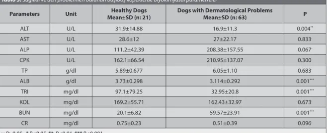
Table 3. Biochemical parameters in healthy stray dogs and stray dogs with dermatological problems Tablo 3. Sağlıklı ve deri problemleri bulunan başıboş köpeklerde biyokimyasal parametreler
Parameters Unit Mean±SD (n: 21)Healthy Dogs Dogs with Dermatological Problems Mean±SD (n: 63) P
ALT U/L 31.9±14.88 16.9±11.3 0.004** AST U/L 28.6±12 27±22.17 0.833 -ALP U/L 111.2±42.39 208.38±157.55 0.067 -CPK U/L 162.1±66.54 210.95±137.07 0.300 -TP g/dl 5.89±0.677 6.05±1.10 0.683 -ALB g/dl 3.73±0.298 3.114±0.292 0.001*** TRI mg/dl 97.1±79.25 32.95±20.8 0.001*** KOL mg/dl 169.2±55.71 162.43±32.97 0.673 -BUN mg/dl 20.1±6.82 59.57±23.91 0.001*** CR mg/dl 0.75±0.23 0.51±0.39 0.096 -- : P>0.05, * P<0.05 ** P<0.01 *** P<0.001
Table 4. Hematological parameters in healthy stray dogs and stray dogs with dermatological problems Tablo 4. Sağlıklı ve deri problemleri bulunan başıboş köpeklerde hematolojik parametreler
Parameters Unit Mean±SD (n: 21)Healthy Dogs Dogs with Dermatological ProblemsMean±SD (n: 63) P
WBC x103 /µl 19.42±9.49 13.44±2.11 0.115-RBC x106 /µl 5.52±1.4 7.12±0.721 0.010* HGB g/dl 11.59±2.66 15.23±1.26 0.002** HCT % 35.06±8.566 46.34±4.74 0.003** MCV fL 64.51±5.11 65.57±4.44 0.634 -MCHC g/dl 33.36±1.65 33.91±0.96 0.417 -- : P>0.05, * P<0.05 ** P<0.01
350
Concentrations of Total Serum ...
endocrine dermatosis, dermatomycosis, parasitic disease, cutaneous Leishmaniasis and in cases with non-specific dermatopathies of uncertain aetiology 15. Furthermore,
it was suggested that the determination of anti-canine IgG, IgM and IgA using an immunoperoxidase method in skin sections from dogs with autoimmune skin disease were useful 9. In our study, it could be considered that
insignificant decreases (P>0.05) in serum IgA concentrations and significant increases (P<0.05) in serum IgG and IgM concentrations might be resulted from local accumulation in skin surface.
Serum ALT, ALB and TRI concentrations in dogs with dermatologic disease significantly decreased (P<0.01) when compared with healthy dogs in our study. The BUN concentration significantly increased (P<0.01) in dogs with dermatologic disease (Table 3). It was determined that these parameters were including reference range except BUN concentration which was interpreted as prerenal azotemia and dehydration considered along with haematological parameters. The significant increase of RBC, Hb and PCV concentrations were found in dogs with dermatologic disease in this study (Table 4). This might be attributed to hemoconcentration and dehydration on the basis of clinical and laboratory findings. When management and feeding conditions for these dogs were evaluated, it was concluded that these dogs were fed insufficiently by an inappropriate diet.
Skin disease of uncertain aetiology is common in the stray dogs and generally complicated by allergic, parasitic, and inflammatory conditions. Therefore clinical diagnosis of stray dogs with dermatologic disease is challenging cases. In our study, it was also concluded that serum total immunoglobulin concentrations (IgE, A, G and M) could be useful in making contribution to differential diagnosis of allergic or inflammatory skin diseases in stray dogs. However, the measurement of specific immunoglobulin formations could be recommended in the definitive diagnosis of allergic skin disease in stray dogs.
REFERENCES
1. Mueller RS, Cannon A, Reubl GH: Serum and skin IgA concentrations
in normal and atopic dogs. Austr Vet J, 75 (12): 906-909, 1997.
2. Roque JB, O’Leary CA, Duffy DL, Kyaw-Tanner M, Latter M, Mason K, Vogelnest L, Shipstone M: IgE Responsiveness to Dermatophagoides
farinae in West Highland white terrier dogs is associated with region on
CFA35. J Heredity, 102 (1): 74-80, 2011.
3. Hill PB, Griffin CE: The ACVD task force on canine atopic dermatitis
(X): Is there a relationship between canine atopic dermatitis and cutaneus adverse food reactions? Vet Immunol Immunopathol, 81, 227-231, 2001.
4. Fadok VA: Diagnosing and managing the food-allergic dog. Compend
Contin Educ, 16, 12, 1541-1544, 1994.
5. Chalmers SA, Medleau L: An update on atopic dermatitis in dogs. Vet
Med, 4, 326-341. 1994.
6. Day MJ: Possible immunodeficiency in related rottweiler dogs. J Small
Anim Pract, 40, 12,561-568, 1999.
7. Hewicker-Trautwein M, Trautwein G: Demonstration of immunoglobulins
and complement in canine and feline autoimmune and non-autoimmune skin diseases with the direct immunofluorescence and indirect immuno-peroxidase method. Zentralblatt Fur Veterinarmedizin. Reihe A, 39 (7): 494-501, 1992.
8. Totton SC, Wandeler AI, Ribble CS, Rosatte RC, McEwen SA: Stray
dog population health in Jodhpur, India in the wake of an animal birth control (ABC) program. Prev Vet Med, 98 (2-3): 215-20, 2011.
9. Bradley GA, Mays MB: Immunoperoxidase staining for the detection
of autoantibodies in canine autoimmune skin disease: Comparison to immunofluorescence results. Vet Immunol Immunopathol, 26 (2): 105-113, 1990.
10. Taszkun I: The results of intradermal skin tests (IDST) in dogs with
atopic dermatitis from the Lublin voivodeship. Polish J Vet Sci, 14 (1): 95-101, 2011.
11. Hill PB, Moriello KA, Deboer DJ: Concentrations of total serum IgE,
IgA, and IgG in atopic and parasitized dogs. Vet Immunol Immunopathol, 44 (2): 105-113, 1995.
12. Foster AP, Knowles TG, Hotston Moore A, Cousins PDG, Day MJ, Hall EJ: Serum IgE and IgG responses to food antigens in normal and atopic
dogs, and dogs with gastrointestinal disease. Vet Immunol Immunopathol, 92, 113-124, 2003.
13. Racine BP, Marti E, Busato A, Weilenmann R, Lazary S, Griot-Wenk ME:
Influence of sex and age on serum total immunoglobulin E concentration in Beagles. Am J Vet Res, 60, 93-97, 1999.
14. Kuhl KA: Newly reported skin diseases. Vet Med, 11, 1007-1020, 1996. 15. Rosser EJ: Diagnosis of food allergy in dogs. JAVMA, 203 (2): 259-262,