Summary
In previous reports, it was indicated that measurement of activity of Interferon-tau Stimulated Genes (ISGs) in Peripheral Blood Leucocytes (PBLs) may be used as an alternative early pregnancy detection method in dairy cows. However, there are no data showing the expression profiles of ISGs in other body fluids containing leucocytes such as milk. In the present study, it was hypothesized that leucocytes in milk samples may reflect the increases in expression profiles of ISGs as shown in PBLs. For this purpose, nine pregnant lactating Holstein cows were used. Insemination day was accepted as day zero (day 0). Blood and milk samples were collected on day 0 and 18 after insemination for cell isolation. Total RNA was extracted from isolated cells and converted to cDNA. Steady state levels of Interferon-tau Stimulated Gene 15 (ISG15), Myxovirus (influenza virus) resistance 1 (MX1) and 2 (MX2) mRNA transcripts were assayed by using real-time reverse transcriptase PCR. Relative Expression Software Tool (REST2009) was used for statistical analyses. There was no statistical significant difference for expression levels of ISG15, MX1 and MX2 mRNAs between days 0 and 18 in milk samples. However, when compared to day 0, levels of ISG15 and MX2 transcripts were increased 6.97±0.68 fold and 5.84±1.27 fold on day 18 in PBLs in pregnant cows, respectively (P<0.05). According to this result, it may be suggested that milk cells are not suitable measurement of expression profiles of ISGs to detect early pregnancy in lactating dairy cows.
Keywords: Early pregnancy detection, ISGs, qPCR, Milk, Cow
Sütçü Gebe İneklerde Periferal Kan Lökositleri (PBLs) ve
Süt Hücrelerindeki Interferon-Tau Stimulated Genlerinin (ISGs)
Ekspresyon Profili
Özet
Önceki çalışmalarda sütçü ineklerde periferal kan lökositlerindeki interferon-tau stimulated genlerindeki (ISGs) aktivite ölçümünün alternatif erken gebelik teşhis yöntemi olarak kullanılabileceği gösterilmiştir. Bununla birlikte süt gibi lökosit içeren diğer vücut sıvılarında ISGs’lerin ekspresyon profilindeki değişimleri inceleyen bir veri bulunmamaktadır. Sunulan çalışmada hipotez olarak süt örneklerindeki lökositlerde de, PBLs’lerde gösterildiği gibi ISGs’lerin ekspresyon profilini arttırabileceği düşünüldü. Bu amaç için dokuz (n=9) sağmal Holştayn ırkı gebe inek kullanıldı. Tohumlama günü sıfırıncı gün (0. gün) olarak kabul edildi. Kan ve süt örnekleri hücre izolasyonu için 0. ve 18. gün alındı. İzole edilen hücrelerden total RNA elde edildi ve cDNA’ya dönüştürüldü. Interferon-tau Stimulated Gene 15 (ISG15), Myxovirus (influenza virus) resistance 1 (MX1) and 2 (MX2) genlerinin mRNA transkriptlerinin seviyeleri real-time reverse transcriptase PCR tekniği ile ölçüldü. İstatistiksel analizler için Relative Expression Software Tool (REST2009) programı kullanıldı. Süt örneklerinin 0 ve 18. günleri arasında ISG15, MX1 ve MX2 mRNA’larının ekspresyon seviyelerindeki farklılık istatistiki olarak önemli olmadı. Bununla birlikte, gebe ineklerin 18. gün PBLs’deki ISG15 ve MX2’nin transkript seviyeleri 0. gün ile karşılaştırıldığında sırasıyla 6.97±0.68 ve 5.84±1.27 kat arttı (P<0.05). Bu sonuçlara göre sağmal ineklerde süt hücrelerindeki ISGs ekspresyon seviyelerinin ölçülmesinin erken gebelik teşhisi için uygun olmadığı önerilebilir.
Anahtar sözcükler: Erken gebelik teşhisi, ISGs, qPCR, Süt, İnek
Expression Profiles of Interferon-Tau Stimulated Genes (ISGs) in
Peripheral Blood Leucocytes (PBLs) and Milk Cells in
Pregnant Dairy Cows
[1]Mehmet KÖSE
1Murat GÖRGÜLÜ
2Mehmet Salih KAYA
3Nurettin AYDİLEK
3Faruk BOZKAYA
4Tahir BAYRIL
5Ercan KURAR
6Zekeriya KIYMA
7Aydın GÜZELOĞLU
6Mehmet Osman ATLI
1
[1] 1 2 3 4 5 6 7
This study was supported by Dicle University DUBAP - (10-VF-43)
Dicle University, Faculty of Veterinary Medicine, Department of Obstetrics and Gynaecology, TR-21280 Diyarbakır - TÜRKİYE
Cukurova University, Faculty of Agriculture, Department of Animal Science, TR-01330 Adana - TÜRKİYE Dicle University, Faculty of Veterinary Medicine, Department of Physiology, TR-21280 Diyarbakır - TÜRKİYE Harran University, Faculty of Veterinary Medicine, Department of Genetics, TR-63100 Şanlıurfa - TÜRKİYE Dicle University, Faculty of Veterinary Medicine, Department of Animal Science, TR-21280 Diyarbakır - TÜRKİYE Selcuk University, Faculty of Veterinary Medicine, Department of Genetics, TR-42031 Konya - TÜRKİYE
Eskisehir Osman Gazi University, Faculty of Agriculture, Department of Animal Science, TR-26160 Eskişehir - TÜRKİYE
Makale Kodu (Article Code): KVFD-2013-9776
İletişim (Correspondence)
+90 412 2488020/8660INTRODUCTION
Detection of pregnancy as early as possible in dairy cows is an important factor to sustain the farm management. In practice, the bovine pregnancy can be easily detected around day 30 after ovulation by using transrectal ultrasonography [1]. However, day 30 means
one missed valuable cycle to reschedule non pregnant cows for re-insemination. Furthermore, one missed cycle for each day/cow costs about 5 USD for dairy farms in TURKEY [2]. Besides economical effect of late detection of
pregnancy, using ultrasonography requires skilled large animal veterinarians [3]. All these factors put pressure
on the scientists to develop alternative early pregnancy detection methods in cows. Nowadays, molecular biology techniques such as PCR, RIA and ELISA are major candidates as alternative pregnancy detection methods in cow. Early pregnancy factor on day 2, expression profile of ISGs after day 18, remaining elevated progesterone level (>1 ng/ ml) between day 18 and day 21 of pregnancy, pregnancy associated glycoproteins (PAGs) on day 28 are accepted as alternative pregnancy detection markers for cows [1,3-5].
Studies have shown that the bovine embryo signals its presence to the dam by a glycoprotein, interferon -tau, which is secreted by embryonic trophoblastic cells [6]. During the maternal recognition of pregnancy,
embryonic interferon-tau abolishes endometrial luteolytic mechanism by specifically causing down regulation of endometrial estrogen receptor alpha and oxytocin receptor expressions [7]. Without an embryo, pulsatile releasing
of endometrial PGF results in functional and structural demise of the CL (corpus luteum) in cows [8]. Therefore,
elongated tropho-blastic cells of the embryo must secrete enough amounts of interferon-tau in a timely manner to maintain the CL, source of progesterone [9]. Embryonic
interferon-tau not only causes inhibition of luteolytic mechanism but also stimulates expression of its target genes both in intra-uterine and extra-uterine tissues [10].
Those genes are named as Interferon-tau Stimulated Genes (ISGs) [11,12]. Being a cytokine, interferon-tau can
stimulate leukocyte activity. Expression profiles of ISGs in PBLs have been studied in detail and a significant increase in some ISGs in PBLs of early pregnant cows compared to those of non-pregnant cows were reported [12,13].
We hypothesize that immune cells present in milk may also reflect the changes in expression profiles of ISGs as shown in PBLs and milk would be an easily collectable candidate sample for a possible use of early pregnancy detection in cows. For this purpose, we sought to elucidate expression profiles of ISG15, MX1, MX2 mRNAs in milk cells collected from lactating cows.
MATERIAL and METHODS
All animal experimental procedures were approved
by ethical committee of Dicle University, Diyarbakır (#2012-51), Turkey and the experimental procedures were performed in the Dairy Farm of Cukurova University, Adana, Turkey. Twenty lactating Holstein dairy cows (3-5 years old, 137±7 DIM, 35 kg/day milk) were synchronized using standard ovsynch protocol (day -10 GnRH, day -3 PGF2 alpha, day -1 GnRH, day 0 Timed AI). Cows were free of any health problems, specifically of mastitis. Evidence of mastitis was eliminated by indirect Somatic cells count (Califronia Mastitis Test, CMT) and direct Somatic cells count measurements (DeLaval Cell counter DCC, DeLaval, Tumba, Sweden). Blood (10 mL) and milk (50 mL) samples were collected from cows on days of 0 and 18. Nine cows were diagnosed pregnant on day 35 by using transrectal ultrasonography. PBLs were isolated according to Kurar et al.[14]. Cells from milk samples were isolated according
to the following protocol. Fifty mL milk sample was centrifuged at 4.000 RPM (+4°C) for 10 min. Cell pellet was washed three times with chilled PBS. After each washing step cell suspension was centrifuged at 4.000 RPM (+4°C) for 5 min. Immediately after washing, the isolated cells were snap frozen and stored at -80°C until RNA isolation.
RNA isolation kit (RNeasy Mini Kit, Qiagen) was used for total RNA isolation from milk samples. Kit protocol was followed accordingly. Briefly, frozen milk cells were re-suspended in Lysis buffer passed through a 25 G needle for homogenization. Following 10 min incubation at room temperature (RT), the homogenized sample was centrifuged at 10.000 x g for 5 min at RT. Upper phase of the solution was carefully transferred into a new sterile tube and was mixed with 70% ethanol. The mixture was loaded on a filter cartilage and centrifuged for 15 sec at 8.000 x g at RT. Subsequently, the filter cartilage was washed once with Buffer RW1 and twice with Buffer RPE. Following each washing step, the filter cartilage was centrifuged for 1 min at 8.000 x g, and for one min to remove residual fluid from the filter during the last wash. Finally, filter was transferred to a new collection tube and 50 µL RNAse, and DNase free water was applied to the center of the filter and centrifuged for 30 sec at max speed to recover total RNA.
For total mRNA isolation from PBLs, a protocol described by Kurar et al.[14] was followed. Concentrations and purity
of total RNA were determined by using NanoDrop-1000 spectrophotometer (NanoDrop Technologies, Palo Alto, CA). Total RNA from PBLs and milk cells were treated with DNAse I to clean gDNA contamination and was then reverse transcribed in the presence of both random hexamer and oligo dT primers in equal volume by using Revert Aid First Strand cDNA Synthesis Kit (Fermentas Life Science, USA) according to the manufacturer’s protocol.
Primers for MX1, MX2, and ISG15 mRNAs were derived from Gifford et al.[12] and Boerboom et al.[15]. The primer
pair sequences and product sizes are shown in Table 1. qPCR reactions were set up as follows: 5 µL SYBR Green Master Mix (2X, Fermentas Life Science, USA), 2.5 pMol
of each primer, 0.5 µL cDNA, and ddH2O to bring final
volume to 10 µL. Thermal cycling was done by initially incubating the mixture at 50°C for 2 min with subsequent denaturation at 95°C for 10 min. This was followed by 40 cycles of denaturation, annealing, and amplification (95°C 30 sec, 60°C 30 sec, 72°C 30 sec). All reactions were done on the Real-Time PCR System (Applied Bioscience Stepone plus, Foster City, CA). In each run, negative controls with no cDNA template and RT negative controls were included. All samples were evaluated in duplicate for each cDNA. Glyceraldehyde-3 phosphate dehydrogenase (GAPDH) mRNA primer was derived from Boerboom et al.[15] and
used as a housekeeping gene to normalize the expression of ISG15, MX1 and MX2. Mean threshold of cycle values from day of 0 was used as a reference point and Ct values for each days (day 0 and day 18) were used to calculate the fold change from this reference point using reference point Ct values according to 2-∆∆Ct method described by
Livak and Schmittgen[16]. Expression levels of ISG15, MX1
and MX2 transcripts were compared by using algorithm (Relative Expression Software Tool 2009) in which the
mathematical model is based on the PCR efficiency of each gene investigated and the mean deviation in Ct between groups [17]. The expression ratios were considered
statistically significant at P<0.05.
RESULTS
Total amounts of RNA isolated from 50 ml milk and 10 ml blood samples were 148.6±9.6 ng and 30±8 µg, respectively. Optical densities of 260/280 UV in Nanodrop measurement were 1.7±0.1 for milk isolated total RNA, 2.0±0.1 for PBLs isolated total RNA. Amplification products were verified by separation on a 2% agarose gel (Fig. 1).
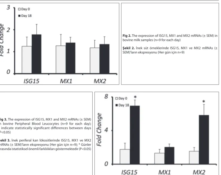
Expression levels of ISG15, MX1 and MX2 mRNA were not statistically significant between comparison days (days 0 and 18) for milk cells (Fig. 2). However, when compared to day 0, steady state levels of ISG15 and MX2 transcripts were up regulated as 6.97±0.68 fold and 5.84±1.27 fold on day 18 in PBLs, respectively (Fig. 3, P<0.05). MX1 mRNA expression did not change between days 0 and 18 in PBLs (Fig. 3).
Table 1. Primers for Interferone-tau stimulated genes (ISGs) used in Real-time PCR Tablo 1. Real-time PCR’da kullanılan İnterferon-tau tarafından uyarılan genlerin primerleri
Locus Primer Sequence Reference Access Code
ISG15 F 5’- ggtatccgagctgaagcagtt -3’
R 5’- acctccctgctgtcaaggt -3’ Gifford et al.[12] NM_174366 MX1 F 5’- gtacgagccgagttctccaa -3’
R 5’- atgtccacagcaggctcttc -3’ Gifford et al.[12] AF047692 MX2 F 5’- cttcagagacgcctcagtcg -3’
R 5’- tgaagcagccaggaatagtg -3’ Gifford et al.[12] NM_173941 GAPDH F 5’- atcaccatcttccaggagcgaga-3’
R 5’- gtcttctgggtggcagtgatgg-3’ Boerboom et al.
[15] XM_001502360
Fig 1. Agarose gel electrophoresis of GAPDH (341 bp), ISG15
(87 bp), MX1 (197 bp) and MX2 (232 bp) PCR Amplification products
Şekil 1. GAPDH (341 bp), ISG15 (87 bp), MX1 (197 bp) ve MX2
DISCUSSION
Apart from the endometrium, the CL and PBLs in which the effect of interferon-tau were very well documented [10,18,19], there is no available data that
demonstrates presence and profile of ISGs expression in the other body fluids containing leucocytes such as milk. Therefore, the present study aimed to detect the presence of ISGs in milk cells and their expression profile in search of any possible change in expression levels due to pregnancy.
In the present study, expression profile of ISGs in lactating pregnant cow blood and milk cells on day 18 was demonstrated by qPCR. MX1, MX2 and ISG15 mRNA transcripts showed an up regulated profiles due to early pregnancy in both pregnant ewes and cows in previous studies [12,20] Therefore, these genes were chosen as
candidate pregnancy detection genes for milk samples from among ISGs. As internal control for ISG expression on day 18, day 0 samples collected from each individual animal were used. Fold changes on day 18 for each ISGs were calculated according to day 0 values. This model allows us to eliminate individual animal effect on day 18
ISGs expression. Compared to isolated PBLs from 10 mL blood, very small amount of cells were isolated from the 50 mL milk sample. As expected, isolated total RNA from milk cells were also less abundant than that of PBLs. This may be explained by the number of cells from blood (7-10x 106/mL) and milk (1-2x10⁵/mL) from a healthy cow [21].
Yankey et al.[20] reported an increased MX1 expression
in PBLs of pregnant ewes and Gifford et al.[12] also indicated
that PBLs produces ISGs due to embryonic interferon- tau during the early pregnancy in cow. This effect was also confirmed by intrauterine interferon-tau infusion studies [22]. Despite a significant increase in ISGs transcripts
(MX2 and ISG15 mRNAs) [12] on day 18 in PBLs in the present
study, we could not see any significant changes for ISGs mRNA expression in milk cells. This might indicate that INF- tau is not responsible for activation of ISGs in milk immune cells or the amount and effect of INF-tau is not enough to detect any expression changes for ISGs.
Milk cells are composed mostly of leukocytes (95%) and udder epithelial cells (5%) and these are called as somatic cells [23]. The number of somatic cells is accepted as a gold
standart for udder health [24]. Milk progesterone level is
Fig 2. The expression of ISG15, MX1 and MX2 mRNAs (± SEM) in
bovine milk samples (n=9 for each day)
Şekil 2. İnek süt örneklerinde ISG15, MX1 ve MX2 mRNAs (±
SEM)’ların ekspresyonu (Her gün için n=9)
Fig 3. The expression of ISG15, MX1 and MX2 mRNAs (± SEM)
in bovine Peripheral Blood Leucocytes (n=9 for each day); * indicate statistically significant differences between days (P<0.05)
Şekil 3. İnek periferal kan lökositlerinde ISG15, MX1 ve MX2
mRNAs (± SEM)’ların ekspresyonu (Her gün için n=9); * Günler arasında istatistiksel önemli farklılıkları göstermektedir (P<0.05)
also used for detection of open-cow [25]. Previous reports
were shown that the effect of IFN-tau can be monitored by ISGs expression in endometrium and CL and PBLs [10,20].
Interferon-tau, like other type I interferons, is a chemokine and has immunomodulatory activities [26]. Both in vivo and
in vitro studies were clearly demonstrated that interferon-tau proliferates leucocytes [12,13]. Interferon-tau mediated
expression of more than 100 ISGs as has been reported by [27] but only a few increase in PBLs due to pregnancy [28,29].
The measurement of ISGs in PBLs provides an alternative method to follow early pregnancy in cows. However, according to present study result, milk cells may not be suitable for monitoring early bovine pregnancy.
Moreover, healthy milk mostly contains macrophages (70%) and they are accepted as resident cell population. Compared to macrophages, number of neutrophil comprise only 5-20% of total cells in healthy milk [23].
However, if inflammation occurs, neutrophiles invade into the udder from blood and cell composition changes in favor of the neutrophils (70-80%) [30]. Furthermore it was
demonstrated that the neutrophils are more sensitive to INF-tau compared to other peripheral blood mononuclear cells [13]. We suppose that our method does not reflect
the same changes in ISGs mRNA expression as in PBLs, since the expression was assessed in healthy pregnant animal milk cells. In the latter, the predominant cells are macrophages.
ISGs expression (MX1, MX2 and ISG15) profiles in PBLs and milk cells were followed to detect any changes due to early pregnancy by the present study. Although expression of MX2 and ISG15 mRNA transcripts showed a significantly increased expression profile in PBLs on day 18 compared to day 0, MX1, MX2 and ISG15 did not change in milk samples during early pregnancy. According to this result, we may suggest that milk cells are not suitable for following ISGs expression profiles to detect early pregnancy in lactating dairy cow.
REFERENCES
1. Romano JE, Thompson JA, Forrest DW, Westhusin ME, Tomaszweski MA, Kraemer DC: Early pregnancy diagnosis by transrectal
ultrasonography in dairy cattle. Theriogenology, 66, 1034-1041, 2006.
2. Yalcın C: Süt sığırcılığında infertiliteden kaynaklanan mali kayıplar. Lalahan Hay Arast Enst Derg, 40, 39-47, 2000.
3. Lucy M, Green J, Poock S: Pregnancy determination in cattle: A review
of available alternatives. In, Proceedings Applied Reproductive Strategies in Beef Cattle, Joplin/Missouri, USA August 31-September 31, pp.367-376, 2011.
4. Nancarrow CD, Wallace AL, Grewal AS: The early pregnancy factor of
sheep and cattle. J Reprod Fertil (Suppl): 30, 191-199, 1981.
5. Szenci O, Beckers JF, Humblot P, Sulon J, Sasser G, Taverne MA, Varga J, Baltusen R, Schekk G: Comparison of ultrasonography,
bovine pregnancy-specific protein B, and bovine pregnancy-associated glycoprotein 1 tests for pregnancy detection in dairy cows. Theriogenology, 50, 77-88, 1998.
6. Roberts RM, Leaman DW, Cross JC: Role of interferons in maternal
recognition of pregnancy in ruminants. Proc Soc Exp Biol Med, 200, 7-18, 1992.
7. Guzeloglu A, Bilby TR, Meikle A, Kamimura S, Kowalski A, Michel F, MacLaren LA, Thatcher WW: Pregnancy and bovine somatotropin
in nonlactating dairy cows: II. Endometrial gene expression related to maintenance of pregnancy. J Dairy Sci, 87, 3268-3279, 2004.
8. Atli MO, Bender RW, Mehta V, Bastos MR, Luo W, Vezina CM, Wiltbank MC: Patterns of gene expression in the bovine corpus luteum
following repeated intrauterine infusions of low doses of prostaglandin F2alpha. Biol Reprod, 86, 1-13, 2012.
9. Bazer FW, Burghardt RC, Johnson GA, Spencer TE, Wu G: Interferons
and progesterone for establishment and maintenance of pregnancy: interactions among novel cell signaling pathways. Reprod Biol, 8, 179-211, 2008.
10. Oliveira JF, Henkes LE, Ashley RL, Purcell SH, Smirnova NP, Veeramachaneni DN, Anthony RV, Hansen TR: Expression of interferon
(IFN)-stimulated genes in extrauterine tissues during early pregnancy in sheep is the consequence of endocrine IFN-tau release from the uterine vein. Endocrinology, 149, 1252-1259, 2008.
11. Gray CA, Abbey CA, Beremand PD, Choi Y, Farmer JL, Adelson DL, Thomas TL, Bazer FW, Spencer TE: Identification of endometrial genes
regulated by early pregnancy, progesterone, and interferon tau in the ovine uterus. Biol Reprod, 74, 383-394, 2006.
12. Gifford CA, Racicot K, Clark DS, Austin KJ, Hansen TR, Lucy MC, Davies CJ, Ott TL: Regulation of interferon-stimulated genes in
peripheral blood leukocytes in pregnant and bred, nonpregnant dairy cows. J Dairy Sci, 90, 274-280, 2007.
13. Shirasuna K, Matsumoto H, Kobayashi E, Nitta A, Haneda S, Matsui M, Kawashima C, Kida K, Shimizu T, Miyamoto A: Upregulation
of interferon-stimulated genes and interleukin-10 in peripheral blood immune cells during early pregnancy in dairy cows. J Reprod Dev, 58, 84-90, 2012.
14. Kurar E, Atli MO, Guzeloglu A, Semacan A: POMC, iNOS, PGES, IL-4,
IL-5 and IL-10 gene expression in peripheral blood mononuclear cells of cyclic and pregnant mares. Kafkas Univ Vet Fak Derg, 17 (2): 319-323, 2011.
15. Boerboom D, Brown KA, Vaillancourt D, Poitras P, Goff AK, Watanabe K, Dore M, Sirois J: Expression of key prostaglandin synthases
in equine endometrium during late diestrus and early pregnancy. Biol Reprod, 70, 391-399, 2004.
16. Livak KJ, Schmittgen TD: Analysis of relative gene expression data
using real-time quantitative PCR and the 2(-Delta Delta C(T)) method. Methods, 25, 402-408, 2001.
17. Pfaffl MW, Horgan GW, Dempfle L: Relative expression software
tool (REST) for group-wise comparison and statistical analysis of relative expression results in real-time PCR. Nucleic Acids Res, 30, e36, 2002.
18. Choi Y, Johnson GA, Burghardt RC, Berghman LR, Joyce MM, Taylor KM, Stewart MD, Bazer FW, Spencer TE: Interferon regulatory
factor-two restricts expression of interferon-stimulated genes to the endometrial stroma and glandular epithelium of the ovine uterus. Biol Reprod, 65, 1038-1049, 2001.
19. Gifford CA, Assiri AM, Satterfield MC, Spencer TE, Ott TL: Receptor
transporter protein 4 (RTP4) in endometrium, ovary, and peripheral blood leukocytes of pregnant and cyclic ewes. Biol Reprod, 79, 518-524, 2008.
20. Yankey SJ, Hicks BA, Carnahan KG, Assiri AM, Sinor SJ, Kodali K, Stellflug JN, Ott TL: Expression of the antiviral protein Mx in peripheral
blood mononuclear cells of pregnant and bred, non-pregnant ewes. J Endocrinol, 170, 7-11, 2001.
21. Bradley A, Green M: Use and interpretation of somatic cell count
data in dairy cows. In Practice, 27, 310-315, 2005.
22. Spencer TE, Gray A, Johnson GA, Taylor KM, Gertler A, Gootwine E, Ott TL, Bazer FW: Effects of recombinant ovine interferon tau, placental
lactogen, and growth hormone on the ovine uterus. Biol Reprod, 61, 1409-1418, 1999.
23. Boutinaud M, Jammes H: Potential uses of milk epithelial cells: A
Gonzalez RN: Monitoring udder health and milk quality using somatic
cell counts. Vet Res, 34, 579-596, 2003.
25. Nebel RL: On-farm milk progesterone tests. J Dairy Sci, 71,
1682-1690, 1988.
26. Chelmonskasoyta A: Interferon tau and its immunobiological
role in ruminant reproduction. Arch Immunol Ther Exp, 50, 47-52, 2002.
27. Garcia-Sastre A, Biron CA: Type 1 interferons and the virus-host
relationship: A lesson in detente. Science, 312, 879-882, 2006.
SJ, Kodali K, Johnson GA, Hansen TR, Mirando MA, Woods GL, Vanderwall DK, Ott TL: Expression of the uterine Mx protein in cyclic
and pregnant cows, gilts, and mares. J Anim Sci, 81, 1552-1561, 2003.
29. Austin KJ, Carr AL, Pru JK, Hearne CE, George EL, Belden EL, Hansen TR: Localization of ISG15 and conjugated proteins in bovine
endometrium using immunohistochemistry and electron microscopy. Endocrinology, 145, 967-975, 2004.
30. Rainard P, Riollet C: Mobilization of neutrophils and defense of the