490 Ufuk Ozer1,2, Karen Wood Barbour2 Abstract Objectives: Cancer cells require reactive oxygen species (ROS) in order to keep up with growth rate. The accumulation of ROS induced by anticancer drugs can promote cell death through oxidative damage. A potential source of ROS is the family of NADPH oxidase (NOX) enzyme that produces ROS as their sole function. In this study, we aimed to investigate expression of NOX1 and NOX2 subunits in response to fluoropyrimidines in human colon cancer cell line, HCT116. Methods: We used fluoropyrimidines, 5-fluorouracil (FUra) and 5’-fluoro-2’-deoxyuridine (FdUrd) as anticancer drugs, and measured mRNA levels of NOX1 and NOX2 with semi-quantitative polymerase chain reaction (PCR), quantitative PCR (qPCR) and microarray assays in order. Results: We found that expression of none of enzyme subunits was altered in response to FUra or FdUrd, except expression of p67phox. Expression of p67phox was induced by drugs approximately 25-fold relative to basal level. Conclusion: p67phox subunit may be a key subunit in NOX-mediated ROS production following exposure to drugs. Key words: NADPH Oxidase, p67phox, ROS, colon cancer, FUra 1 Department of Molecular Biology and Genetics, Faculty of Science, Dicle University, Diyarbakir, Turkey 2 Department of Biological Sciences, University of South Carolina, Columbia, Usa Corresponding author : Ufuk Ozer, Ph.D., Department of Molecular Biology and Genetics, Faculty of Science, Dicle University, Diyarbakir, 21280, Turkey, e-mail: [email protected] Overexpression of p67phox in Response to Fluoropyrimidines in HCT116 Cells
ORIGINAL ARTICLE
Özet Giriş: Kanser hücreleri büyüme hızı ile başa çıkmak için reaktif oksijen türlerine (ROS) ihtiyaç duyarlar. Antikanser ilaçları tarafından indüklenen ROS’un akümülasyonu oksidatif hasarla hücre ölümünü kolaylaştırabilir. Potansiyel bir ROS kaynağı tek fonksiyonu ROS üretmek olan NADPH oksidaz (NOX) enzimidir. Bu çalışmada insan kolon kanseri hücre hattı olan HCT116 da floropirimidinlere karşı NOX1 ve NOX2’nin ekspresyonunu araştırmayı amaçladık. Yöntemler: Antikanser ilaçlar olarak floropirimidinleri; 5-florourasil (FUra) ve 5’-floro-2’-deoksiuridin (FdUrd) kullandık ve sırasıyla semi-kantitatif polimeraz zincir reaksiyonu (PCR), kantitatif PCR (qPCR) ve mikroarray metodları ile NOX1 ve NOX2’nin mRNA seviyelerini ölçtük. Bulgular: p67phox hariç enzimin hiçbir alt ünitesinin ekspresyonunun FUra ve FdUrd’ye karşı değişmediğini bulduk. p67phox ekspresyonu ilaçlarla bazal seviyelere nazaran yaklaşık 25 kat indüklendi. Sonuç: p67phox alt ünitesi ilaçlarla muamele sonrasında NOX ile üretilen ROS’da önemli bir alt ünite olabilir. Anahtar kelimeler: NADPH Oksidaz, p67phox, ROS, kolon kanseri, FUra HCT116 Hücrelerinde Floropirimidinlere Karşı p67phox’un Fazla Ekspresyonu Received:30 Jan 2016; Revised:28 Sept 2016 Accepted:30 Sept 201643 (4) 2016 www.diclemedj.org
491
INTRODUCTION
The generation and accumulation of reactive oxygen species (ROS) can physiologically occur as a by-product of functioning or damaged mitochondria, by enzyme systems such as peroxisomal oxidases and lipoxygenases, and in response to ROS-producing environmental exposures or inflammatory conditions [1,2]. In contrast, the family of NADPH oxidases (NOX) produce ROS as their primary and sole function [3,4]. This enzyme family occurs as multi-protein complexes consisting of a membrane-spanning catalytic NOX subunit and regulatory subunits that are localized in the cytosol and the membrane [3-5]. Among them, NOX1 and NOX2 subunits are the best characterized. Catalytic NOX1 and NOX2 subunits bind to a common protein, p22phox in the membrane, forming a membrane complex termed flavocytochrome b558. The flavocytochrome b558 complex of NOX1 binds to NOX activator 1 (NOXA1) and NOX organizer 1 (NOXO1), while the complex of NOX2 binds the respective subunits p67phox and p47phox and p40phox. Rac, a small GTPase, binds to NOXA1 and p67phox upon activation of enzymes [5-7]. Assembly of all subunits brings about the activation of NOX catalytic function, involving the transportation of electrons from cytoplasmic NADPH to FAD, first and second heme groups, respectively and finally to extracellular or phagosomal oxygen to produce superoxide (O2.-) [3,4,6].
The fate of cancer cells is thus bound up by the level of ROS induced by anticancer drugs [8]. It has been suggested that human tumor cells generate their characteristically elevated ROS levels by NOX, indicating the contribution of this enzyme to carcinogenesis for various types of cancer [4,9-13]. Expression and regulation of NOX isoforms vary in tissues and are differentially localized in subcellular compartments [5,14]. NOX1 and NOX2 are potentially important targets of fluoropyrimidines, 5-fluorouracil (FUra) and its
nucleoside analog, 5’-fluoro-2’-deoxyuridine (FdUrd), that induce ROS formation, since they are highly expressed in colon tissue [15,16]. For decades, fluoropyrimidines have been widely used in chemotherapy of colorectal cancer because of their strong cytotoxic impacts. It is known that the activity of ROS generating enzymes like NADPH oxidases is increased to kill cancer cells [4,9]. Greater efficacy in causing apoptotic programmed cell death may be achieved by activating ROS-producing systems. Therefore, the NOX enzyme family by virtue of its ability to produce the excessive ROS in colon cancer cells may be an attractive target of fluoropyrimidines.
Here, in 3 different methods, we demonstrate expression of NOX1 and NOX2 subunits in human colon cancer cell, HCT116. Among all NOX regulatory subunits, p67phox is the only one whose expression is increased by drugs, thereby indicating that it may be a key subunit in activation of NOX2 in HCT116 cells following exposure to drugs. METHODS Cell culture
Human colon cancer cell line, HCT116 was obtained from Dr. Michael G. Brattain. Cells were grown in RPMI-1640 medium (Cellgro, Corning, Manassas, VA, USA) supplemented with 10% heat-activated fetal bovine serum (FBS, Atlanta Biologicals) at 37 °C in a humidified 5% CO2 atmosphere. Cells were
treated with fluoropyrimidines; FUra and FdUrd (Sigma-Aldrich Co., St. Louis, MO, USA) at concentration of 10 µM for 24 hours.
Semi-quantitative PCR
In order to isolate total RNA, RNeasy Mini Kit (Qiagen, Maryland, USA) was used and RNase Free DNase Set (Qiagen, Hilden, Germany) as applied to eliminate contaminating genomic DNA. Each samples’ RNA levels of samples were spectrophotometrically measured at 260 and 280 nm absorbance (NanoDrop ND-1000 Spectrophotometer, Thermo Fisher Scientific,
43 (4) 2016 www.diclemedj.org
492
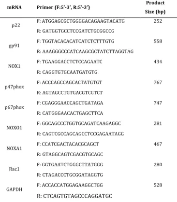
USA). According to manufacturer instructions, 1 µg RNA was reversely transcribed by iscript cDNA synthesis kit (Biorad, Hercules, CA, USA) for each reaction in a Mycycler Thermal Cycler (Biorad, Hercules, CA). The samples were incubated for 5 min in 25 °C, 30 min in 42 °C, heated for 5 min in 85 °C and hold at 4 °C. Indicated PCR cycles were used to determine the relative expression of each gene. In order to eliminate contamination, negative controls including no reverse transcriptase enzyme and no template RNA were used for each gene. The different PCR tubes within each series were set up due to increasing numbers of amplification cycles for each gene. Each reaction was run with 1 μl of cDNA and 250 ng primers (Table 1, Integrated DNA Technologies Inc., Coralville, IA, USA) in a total volume of 25 μl, using GoTaq Hot Start Green Master Mix (Promega, Madison, WI, USA).
Table 1: Primers used for semi-quantitative PCR
mRNA Primer (F:5’-3’, R:5’-3’) Product Size (bp) p22 F: ATGGAGCGCTGGGGACAGAAGTACATG 252 R: GATGGTGCCTCCGATCTGCGGCCG gp91 F: TGGTACACACATCATCTCTTTGTG 558 R: AAAGGGCCCATCAAGCGCTATCTTAGGTAG NOX1 F: TGAAGGACCTCTCCAGAATC 434 R: CAGGTGTGCAATGATGTG p47phox F: ACCCAGCCAGCACTATGTGT 767 R: AGTAGCCTGTGACGTCGTCT p67phox F: CGAGGGAACCAGCTGATAGA 747 R: CATGGGAACACTGAGCTTCA NOXO1 F: GGCAGCCCTGGTGCAGATCAAGAGGC 281 R: CAGTCGCCAGCAGCCTCCGAGAATAGG NOXA1 F: CCATCGACTACACGCAGCT 467 R: GTAGGCAGTCGACGTGCAGC Rac1 F: GGTGAATCTGGGCTTATGGG 280 R: CTAGACCCTGCGGATAGGTG GAPDH F: ACCACCATGGAGAAGGCTGG 528 R: CTCAGTGTAGCCCAGGATGC
All PCR reactions consisted of an initial denaturation step for 2 min at 95 °C and followed by denaturation (30 s at 95 °C), annealing step (30 s
at various temperature), extension (30 s at 72 °C) and final extension step (5 min at 72 °C). Annealing step was 30 s at 50 °C for GAPDH, Rac1, NOXA1, gp91 and p67phox; at 49.5 °C for p47phox; at 60 °C for NOXO1 and at 52 °C for NOX1. Amplification cycles for GAPDH and Rac1 were from 18 to 33 and 32 to 44 for other genes. 1.5% (w/v) agarose gel electrophoresis was used to resolve the amplified cDNA products which were visualized by staining with ethidium bromide (Sigma-Aldrich Co., St. Louis, MO, USA).
Quantitative PCR
Power SYBR Green PCR Master Mix (Applied Biosystems, Foster City, CA) was used for amplifying cDNA (1 μl) according to the manufacturer’s procedures. PCR reactions were set up for one cycle at 95 °C for 10 min, and 40 cycles of 95 °C for 15 s, 50 °C for 15 s and 72 °C for 40 s utilizing an Applied Biosystems 7300 Real Time PCR System. By detecting SYBR green incorporation during quantitative PCR (qPCR), all mRNA levels were calculated relative to GAPDH as a reference gene. Calculations and statistical analyses were done as described in the manufacturer’s protocol. Relative changes in each gene levels between drug-treated and control samples are expressed as fold induction relative to the basal level of expression in control samples. Table 2 shows primers (Integrated DNA Technologies Inc., Coralville, IA, USA) used for qPCR. Microarray Analysis RNA was isolated from HCT116 cells treated in quadruplicate with 10 µM of FUra for 24 hours. RNeasy Mini Kit (Qiagen, Maryland, USA) was used for the isolation from 6 x 10⁶ cells and RNase Free DNase Set (Qiagen, Hilden, Germany) was performed to eliminate contaminating genomic DNA according to the recommendations of the manufacturer. Utilizing the RNA 6000 Nano LabChip, 2100 Bioanalyzer (Agilent Biotechnologies) was used to measure the integrity and concentration of the total RNA. The RNA integrity was checked to make sure the quality of RNA and it ranged from 9.8 to 10.0. RNA samples were amplified and labeled using Agilent’s Low Input Quick
43 (4) 2016 www.diclemedj.org 493 Amp Labeling Kit (Cat. # 5190-2306) according to the manufacturer suggestions. Table 2: Primers used for qPCR
mRNA Primer (F:5’-3’, R:5’-3’) Size (bp) Product
p22 F: ATGGAGCGCTGGGGACAGAAGTACATG 77 R: GATGGTGCCTCCGATCTGCGGCCG gp91 F: TGGTACACACATCATCTCTTTGTG 94 R: AAAGGGCCCATCAAGCGCTATCTTAGGTAG NOX1 F: CTCCCTTGCCTCCATTCTC 149 R: AGGCTATTGTCATGATCACTCC p40phox F: AAAGTCAAGAGCGTGTCCC 132 R: GAGGAAGATCACATCTCCAGC p47phox F: ACACCTTCATCCGTCACATC 143 R: GAACTCGTAGATCTCGGTGAAG p67phox F: CGAGGGAACCAGCTGATAGA 131 R: CATGGGAACACTGAGCTTCA NOXO1 F: AGATCAAGAGGCTCCAAACG 117 R: AGGTCTCCTTGAGGGTCTTC NOXA1 F: CAGGCTGTGGATCGTGG 150 R: CACGGCTTGGTCAAATGC Rac1 F: GGTGAATCTGGGCTTATGGG 82 R: CTAGACCCTGCGGATAGGTG GAPDH F: ACCACCATGGAGAAGGCTGG 218 R: CTCAGTGTAGCCCAGGATGC By using a poly-dT primer consisting of the T7 RNA polymerase promoter sequence, cDNA was produced by mRNA from 200 ng of total RNA and. Then, cDNA samples were amplified by T7 RNA polymerase and were simultaneously incorporated by cyanine 3- or cyanine 5-labeled CTP (cRNA). As an experimental quality control, Agilent RNA spike-in controls (Cat. # 5188-5279) were added to samples before cDNA synthesis. After amplification and incorporation steps, Qiagen’s RNeasy Mini Kit (Cat. # 74104) was used for purification of labeled RNA molecules. Samples were spectrophotometrically assessed in terms of dye incorporation and cRNA yield and then stored at -80 °C until hybridization. According to the manufacturer’s recommendations, Agilent’s Gene Expression Hybridization Kit (Cat. # 5188-5242) was used for hybridization
of labeled cRNA samples to Agilent Human GE 4 x 44K v2 Microarrays (Cat. # G4845A) at 65 °C for 17 hours. We hybridized four control sample replicates against four FUra treated sample replicates in a dye swap design. After washes, Agilent DNA Microarray Scanner System (Cat. # G2565CA) was utilized to scan arrays. Images from Feature Extractor Software version 10.7.3.1 (Agilent) extracted data by correcting background utilizing additive and multiplicative detrending algorithms. Additionally, dye normalization was performed by linear and LOWESS methods. Final data was uploaded into GeneSpring GX version 11.5.1 for analysis. This data was log2 transformed, quantile normalized and base line transformed using the median of all samples. Then, data was filtered by flags in a way that 100% of the samples in at least one of the two treatment groups have a “detected” flag. Analysis of the data was done with an unpaired t-test statistics to determine differentially expressed genes and this was corrected for multiple testing using the Benjamini-Hochberg algorithm. A cutoff for p-value was 0.005. To filter data, 1.5 was used as a fold change cutoff value.
Statistical Analysis
All data were analyzed as the mean ± SEM. Student’s t-test were performed to determine statistical significance of the mean for each groups. Differences with P ≤ 0.05 were considered statistically significant.
RESULTS
Indication of drug-induced p67phox expression by semi-quantitative PCR and qPCR
NOX isoforms are expressed in various tissues and have different localizations in subcellular compartments [5,14]. Previous studies had indicated that NOX enzyme contributes elevation of ROS levels in various types of cancer, implicating its strong cytotoxic effects on cancer chemotherapy [4,9-13]. In order to induce ROS, fluoropyrimidines target NOX1 and
43 (4) 2016 www.diclemedj.org
494
NOX2, which are highly expressed in colon tissue [15,16]. As such, these drugs may induce expression of their subunits.
To test whether mRNA expressions of all NOX1 and NOX2 subunits―p22phox, NOX1, NOXA1, NOXO1, NOX2, p67phox, p47phox, Rac, altered by FdUrd in HCT116 cells, we utilized semi-quantitative PCR method. mRNA levels were detected semi-quantitatively in absence and presence of FdUrd and GAPDH was used as an experimental control. None of subunits, except p67phox, were altered by FdUrd treatment (Figure 1). HCT116 cells were treated with FdUrd and qPCR was done to verify expression of p67phox mRNA levels induced by FdUrd. In this assay, mRNA expression of another NOX2 regulatory subunit―p40phox was determined as well. Similarly, only mRNA levels of p67phox was induced by FdUrd about 28-fold (p<0.01) among all subunits (Figure 2). Figure 1. mRNA levels of NOX1 and NOX2 subunits in response to FdUrd. HCT116 cells were treated with ± 10 µM FdUrd for 24 h. Indicated subunits were examined to show relative levels of mRNAs in untreated and FdUrd treated cells by semi-quantitative PCR. GAPDH was tested as a loading control. Indication of FUra-induced p67phox expression by microarray analysis
In order to examine if mRNA expressions of various NOX1 and NOX2 subunits is changed by another fluoropyrimidine, FUra was used in HCT116 cells. FUra treated to cells and microarray analysis was done to measure expression. Four sample replicates for both control and FUra treatment were averaged and represented by black color. Any sample below average was indicated as green while sample
over it was shown as red. With the exception of p67phox, expression of all NOX1 and NOX2 subunits were unchanged by FUra (Figure 3). Thus, p67phox may be a key regulator of NOX enzyme in response to FdUrd.
DISCUSSION
Primary and sole function of NOX family is producing O2.- by transporting electrons (e-)
across the membrane to reduce oxygen (O2)
[3,4]. They were expressed and localized in subcellular compartments in diverse tissues [5,14]. It has been suggested that NOX induces ROS levels in human tumor cells and contributes to cytotoxicity [10-13] and ROS generation in colorectal cancer is increased by NOX1 and NOX2 enzymes [17,18]. Increase in ROS can contribute to cell signaling and proliferation, but the extent of ROS induction alters the fate of cells. The excessive amount of ROS induced by NOX1 and NOX2 switches scales of a balance between oxidative stress and defense capability in favor of the stress. Fluoropyrimidines have been widely used in chemotherapy of colorectal cancer for decades due to their powerful cytotoxic impacts. NOX1 and NOX2 are potentially important targets of these drugs that induce ROS formation, since they are highly expressed in colon tissue [15,16]. Activation of ROS-producing systems may provide convenience to cancer cell death. Thus, the NOX enzyme family may be a considerable target of drugs as unique ROS producers.
In this study, we examined gene expression of NOX1 and NOX2 subunits in response to fluoropyrimidines, FUra and FdUrd. We found that except p67phox, expressions of all subunits are not changed in treatment of FdUrd. Induction in p67phox expression by FdUrd was confirmed by semi-quantitative PCR and qPCR (Figure 1 and 2). Similarly, in microarray assay, only p67phox expression induced in treatment of FUra (Figure 3). This shows these drugs potentially induce NOX2 via p67phox, thereby indicating a possible target
43 (4) 2016 www.diclemedj.org
495
for fluoropyrimidine-directed therapy. Despite requirement of future studies, NOX2 may be a new target for chemotherapy in colorectal cancer. Figure 2. Only p67phox mRNA is induced in treatment of FdUrd. HCT116 cells were treated with ± 10 µM FdUrd for 24 h. mRNA levels of NOX1 and NOX2 subunits were assayed by qPCR using GAPDH as a loading control. Bars represent an average of fold increase ± SEM from 2 separate experiments (*p<0.01).
Figure 3. Gene expression of NOX1 and NOX2 subunits in
response to FUra. HCT116 cells were treated in quadruplicate with ± 10 µM FUra for 24 h. cDNAs are synthesized from isolated mRNA samples. T7 RNA polymerase was added to cDNA samples to amplify original mRNA molecules and was added to cDNA samples and to simultaneously incorporate cyanine 3- or cyanine 5-labeled CTP (cRNA) into the amplification product. Labeled cRNA samples were hybridized to Agilent Human GE 4 x 44K v2 Microarrays at 65 °C for 17 hours. Arrays were scanned using an Agilent DNA Microarray Scanner System. Acknowledgements We acknowledge Dr. Franklin G. Berger for experimental guidance and thank to Dr. Diego Altomare for help with microarrays experiment. Declaration of Conflicting Interests: The authors declare that they have no conflict of interest. Financial Disclosure: This work was supported by the National Cancer Institute [Grant CA44013] and the National Institute of General Medical Sciences [Grant GM103336]. REFERENCES 1. Balaban RS, Nemoto S, Finkel T. Mitochondria, oxidants, and aging. Cell 2005;120:483–95. 2. Schrader M, Fahimi HD. Mammalian peroxisomes and reactive oxygen species. Histochem Cell Biol. 2004;122:383–93. 3. Bedard K, Krause KH. The NOX family of ROS-generating NADPH oxidases: physiology and pathophysiology. Physiol Rev. 2007;87:245-313. 4. Lambeth JD. NOX enzymes and the biology of reactive oxygen. Nat Rev Immunol. 2004;4:181-9. 5. Altenhöfer S, Kleikers PW, Radermacher KA. The NOX toolbox: validating the role of NADPH oxidases in physiology and disease. Cell Mol Life Sci. 2012;69:2327-43. 6. Hayes P, Knaus UG. Balancing Reactive Oxygen Species in the Epigenome: NADPH Oxidases as Target and Perpetrator. Antioxid Redox Signal. 2013;18:1937-45. 7. Wingler K, Hermans JJ, Schiffers P. NOX1, 2, 4, 5: counting out oxidative stress. Br J Pharmacol. 2011;164:866-83. 8. Zhou BB, Elledge SJ. The DNA damage response: putting checkpoints in perspective. Nature. 2000;408:433-9. 9. Kumar B, Koul S, Khandrika L, et al. Oxidative stress is inherent in prostate cancer cells and is required for aggressive phenotype. Cancer Res. 2008;68:1777–85. 10. Kamata T. Roles of Nox1 and other Nox isoforms in cancer development. Cancer Sci. 2009;100:1382-8. 11. Laurent E, 3rd McCoy JW, Macina RA, et al. Nox1 is over-expressed in human colon cancers and correlates with activating mutations in K-Ras. Int J Cancer. 2008;123:100-7. 12. Lassègue B, Griendling KK. NADPH oxidases: functions and pathologies in the vasculature. Arterioscler Thromb Vasc Biol. 2010;30:653-61. 13. Bánfi B, Maturana A, Jaconi S, et al. A mammalian H+ channel generated through alternative splicing of the NADPH oxidase homolog NOH-1. Science. 2000;287:138-42. 14. Brown DI, Griendling KK. Nox proteins in signal transduction. Free Radic Biol Med. 2009;47:1239-53. 15. Hwang PM, Bunz F, Yu J, et al. Ferredoxin reductase affects p53-dependent, 5-fluorouracil–induced apoptosis in colorectal cancer cells. Nat Med. 2001;7:1111-7. 16. Juhasz A, Ge Y, Markel S, et al. Expression of NADPH oxidase homologues and accessory genes in human cancer cell lines, tumours and adjacent normal tissues. Free Radic Res. 2009;43:523-32.
43 (4) 2016 www.diclemedj.org 496 17. Kikuchi H, Hikage M, Miyashita H, et al. NADPH oxidase subunit, gp91(phox) homologue, preferentially expressed in human colon epithelial cells. Gene. 2000;254:237-43. 18. Perner A, Andresen L, Pedersen G, et al. Superoxide production and expression of NAD(P)H oxidases by transformed and primary human colonic epithelial cells. Gut. 2003;52:231-6.