CLINICAL QUIZ
Proteinuria and progressive kidney failure due to an inborn error
of metabolism: Answers
Özlem Ünal Uzun1 &Nurcan Cengiz2&Büşra Çavdarlı3&Umut Bayrakçı4&Saba Kiremitçi5& Aynur Küçükçongar Yavaş4
Received: 11 November 2020 / Accepted: 3 December 2020 # IPNA 2021
Keywords Child . Dysostosis multiplex . Kidney disease . Cherry-red spot . Nephrosialidosis . Lysosomal storage disorder . NEU1 mutation
Answers
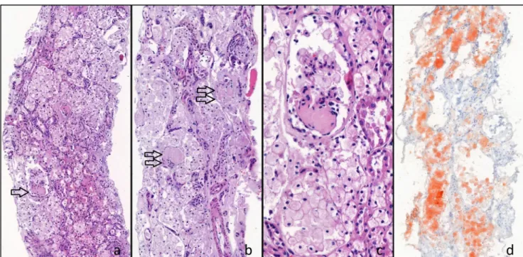
1. What would be the next diagnostics for the diagnosis? A. Kidney biopsy: Kidney biopsy showed diffuse and severe vacuolization of glomerular and tubular epi-thelial cells. Light microscopic examination of kid-ney biopsy revealed 19 glomeruli showing either segmental sclerosis or global sclerosis. In nonsclerotic loops of the glomeruli, swollen podocytes with cytoplasmic vacuolization were re-markable. Most of the tubules were dilated and the lumens were filled with swollen tubular epithelial cells showing cytoplasmic enlargement and foamy appearance. Oil Red O stain, applied to the frozen section of the biopsy, showed diffuse lipid deposits in tubular epithelial cells. Immunofluorescence ex-amination showed no deposits of immune com-plexes (Fig.1a, b, c, d).
B. Molecular genetic analysis: A lysosomal storage dis-order was suspected in the patient; however, whole exome sequencing (WES) was performed due to the lack of specific pre-diagnosis. Analysis of the data revealed that the patient had two heterozygous path-ogenic variants (c.1109A>G (p.Tyr370Cys)/ c.689G>A (p.Cys230Tyr)) of the NEU1 gene that explained the clinical findings with a diagnosis of nephrosialidosis. Mutations were proven to be in trans position by confirming that each parent was a carrier for one allele via Sanger sequencing (Fig.2). 2. What is your diagnosis?
Nephrosialidosis: Inborn errors of metabolism (IEMs) may involve almost all organs and systems, either as a part of a systemic clinical picture or individually. The kidney is one of the target organs for inherited metabolic disor-ders and a variety of IEMs present with different types of kidney disorders. Fanconi syndrome, proteinuria, renal tubular acidosis, nephrolithiasis, kidney cysts, acute kid-ney injury, and chronic kidkid-ney disease can be clinical manifestations of IEMs. Specific patterns of kidney in-volvement could be indicative of an underlying IEM. Kidney failure is a well-defined severe morbidity in Fabry disease, primary hyperoxaluria, cystinosis, and methylmalonic acidemia. Nephrosialidosis is a very rare subgroup of the lysosomal storage disorders leading to proteinuria and kidney failure [1].
3. The patient has a life-limiting inherited metabolic disease and kidney failure due to this disorder. Genetic counsel-ling and information about the condition should be given to the parents. To date, the age of symptom onset in all cases is under 2 years of age, and most patients have died under age 10. Our patient became dialysis-dependent at 8 years of age. In comparison with other cases reported This refers to the article that can be found athttps://doi.org/10.1007/
s00467-020-04891-y. * Özlem Ünal Uzun
Kocaeli University, Pediatric Metabolism, Umuttepe Yerleşkesi, İzmit, Kocaeli, Turkey
2
Muğla Sıtkı Koçman University, Pediatric Nephrology, Muğla, Turkey
3 Ankara City Hospital, Medical Genetics, Ankara, Turkey 4
Ankara City Hospital, Pediatric Nephrology, Pediatric Metabolism, Ankara, Turkey
5 Ankara University, Pathology, Ankara, Turkey Pediatric Nephrology
https://doi.org/10.1007/s00467-020-04901-z
before, the mild clinical phenotype in our patient may be associated with compound heterozygosity. It is difficult to say anything definite about the prognosis in this case.
Discussion
Sialidosis is a rare lysosomal storage disorder characterized by neuraminidase deficiency. It is caused by mutations in the NEU1 gene on chromosome 6p21 [2]. Neuraminidase defi-ciency affects glycoprotein degradation and leads to abnormal accumulation of sialyl oligosaccharides and glycoproteins. Sialidosis is divided into two groups: type 1 and type 2. Clinical severity is heterogeneous depending on the residual enzyme activity. Type 1 sialidosis is the milder form and usually presents in the second decade with neurological find-i n g s . T y p e 2 p r e s e n t s w find-i t h e a r l find-i e r o n s e t a n d mucopolysaccharide-like phenotype with dysostosis multi-plex and organomegaly. Rarely, type 2 may affect the kidney and lead to proteinuria progressing to nephrotic syndrome and chronic kidney disease.
Maroteaux et al. proposed the term ‘nephrosialidosis’ for patients with sialidosis and kidney involvement [3]. Firstly, in 1977, Kelly and Graetz reported an 8-month-old female with neuraminidase deficiency [4]. Roth et al. presented a follow-up on the patient, with the fulminant nature of nephrotic syndrome an unusual
feature of the disease. Pathological examination of the kidneys revealed kidney epithelial cell damage, most marked in the membranes of the glomeruli and proximal tubules [5]. Since these first reports on patients with sialidosis and kidney involvement, only 16 patients have been described in the literature before our patient, and two of these have been genetically confirmed [1].
C h e n e t a l . r e p o r t e d a 2 - y e a r - o l d b o y w i t h nephrosialidosis who had steroid-resistant nephrotic syn-drome. Kidney biopsy showed diffuse and severe vacuolization of glomerular and tubular epithelial cells [6]. Kidney pathology was described in 11 patients with nephrosialidosis, and vacuolated cells and appearance of focal segmental glomerulosclerosis were common find-ings, as in our patient [1]. Because treatment success has not been reported in the literature, we did not initiate ste-roid therapy in our patient.
Recently, Maroofian et.al. reviewed the literature on 16 patients with nephrosialidosis and reported an Iranian female infant who presented with nephrotic syndrome at 9 months old. The patient was diagnosed with nephrosialidosis with parental WES analysis after the patient’s death. Her parents were heterozygous for c.1109A>G mutation [1]. Molecular genetic analysis of our patient revealed compound heterozygosity and he was also found carry-ing this same mutation in one allele.
Fig. 1 a–d Diffuse cytoplasmic vacuolization of tubular epithelial cells and glomerular sclerosis are seen. Segmental sclerosis (single arrow) and global sclerosis (double arrow). Hematoxylin & Eosin × 143. c Higher magnification of a segmental sclerotic glomerulus shows swollen
podocytes having vacuolated foamy cytoplasm. H&E × 403. e Extensive red oil droplets are seen in tubular epithelial cells by Oil Red O stain in a frozen section of the biopsy. Oil Red O × 129
Pediatr Nephrol
Appyling the criteria of the American College of Medical Genetics and Genomics (moderate pathogenic criteria: PM1-6, supporting pathogenic criteria: PP1-5) [7], these two vari-ants were classified as likely pathogenic varivari-ants that describe the clinical findings of the patient. The variants are demon-strated in Fig.2. The c.1109A>G (rs1310267862) variant is located in the“helical” transmembrane region of the gene that is known to be a functional domain (PM1) and its frequency is very low in the healthy population (PM2). The rate of benign missense variants in the NEU1 gene is very low, and the missense variants formed in this gene are responsible for the disease mechanism (PP2). In silico analyses predict a patho-genic effect of the variant in terms of evolutionary conserva-tion, effects on the protein level, and coding dysfunctions (PP3). And finally, the variant was reported as causal for nephrosialidosis in the literature before (PP5). In our patient, the other variant, c.689G>A, was also a rare (PM2), missense variant (PP2) with pathogenic predictions (PP3) and it was determined to be in trans position with the other likely patho-genic variant (c.1109A>G) (PP3).
The clincal findings of our patient were milder and progression rate was slower compared with most of the patients reported in the literature. The other molecular genetically confirmed patient was reported by Caciotti et al. nearly 10 years ago. She was found to carry a ho-mozygous c.807+1G>A splicing defect and exhibited the congenital lethal form of the disease. Fourteen of the 16 previously reported cases were no longer alive at the time of the literature review conducted by Maroofian et al. 2 years ago. Hepatomegaly was present in nearly all cases, whereas corneal clouding and cherry-red spot was observed in only 50% of cases. In our patient, ophthalmologic evaluation was initally nor-mal, but cherry-red spot was observed 6 months after
first examination. This finding shows that clinical fea-tures may become prominent in the follow-up period, and long-term follow-up and investigating the clinical course of older patients is very important for these very rare and often lethal disorders.
Until now, the age of onset of symptoms in all patients was under 2 years of age, and most patients died aged under 10. In our patient, mild coarse face, organomegaly, and proteinuria were noticed incidentally during an infec-tious disease at 6 years old, and he became dialysis de-pendent at 8 years. In comparison with other patients re-ported before, mild clinical phenotype in our patient may be associated with compound heterozygosity.
In conclusion, here we report the clinical features of a Turkish boy who presented with proteinuria and rapidly progressed to kidney failure at the age of 7. Consideration of nephrosialidosis and evaluation for lysosomal storage findings are important in the differential diagnosis of proteinuria and kidney failure.
Compliance with ethical standards
Conflict of interest The authors declare that they have no conflict of interest.
Informed consent statement Informed consent was obtained from the parents of the patient.
References
1. Maroofian R, Schuele I, Najafi M, Bakey Z, Rad A, Antony D, Habibi H, Miriam Schmidts M (2018) Parental whole exome se-quencing enables sialidosis type II diagnosis due to an NEU1 Fig. 2 Demonstration of the two variants in the NEU1 gene. The c.1109A>G variant was in exon 6 of the maternal allele of the NEU1 gene while the c.689G>A variant was in exon 4 of the paternal allele
Pediatr Nephrol p21.33 Maternal 3 S'UTR 3'UTR Paternal 3 6 c.689G>A heterozygous ~ Springer
missense mutation as an underlying cause of nephrotic syndrome in the child. Kidney Int Rep 3:1454–1463
2. Khan A, Sergi C (2018) Sialidosis: a review of morphology and molecular biology of a rare pediatric disorder. Diagnostics (Basel) 8:E29
3. Maroteaux P, Humbel R, Strecker G, Michalski JC, Mande R (1978) A new type of sialidosis with kidney disease: nephrosialidosis. I. Clinical, radiological and nosological study. Arch Fr Pediatr 35: 819–829
4. Kelly TE, Graetz G (1977) Isolated acid neuraminidase deficiency: a distinct lysosomal storage disease. Am J Med Genet 1:31–46 5. Roth KS, Chan JC, Ghatak NR, Mamunes P, Miller WW, O'Brien JS
(1988) Acid alpha-neuraminidase deficiency: a nephropathic pheno-type? Clin Genet 34:185–194
6. Chen W, Yang S, Shi H, Guan W, Dong Y, Wang Y, Wang L (2011) Histological studies of renal biopsy in a boy with nephrosialidosis. Ultrastruct Pathol 35:168–171
7. Richards S, Aziz N, Bale S, Bick D, Das S, Gastier-Foster J, Grody WW, Hegde M, Lyon E, Spector E, Voelkerding K, Rehm HL, ACMG Laboratory Quality Assurance Committee (2015) Standards and guidelines for the interpretation of sequence variants: a joint consensus recommendation of the American College of Medical Genetics and Genomics and the Association for Molecular Pathology. Genet Med 17:405–424
Publisher’s note Springer Nature remains neutral with regard to jurisdic-tional claims in published maps and institujurisdic-tional affiliations.
Pediatr Nephrol