The effect of different levels of teat-end hyperkeratosis on mammary
infrared thermograph and mastitis in dairy cows
Vida JUOZAITIENE
1, Arunas JUOZAITIS
1, Judita ZYMANTIENE
2, Vaidas OBERAUSKAS
2,
Albina ANIULIENĖ
3, Lina KAJOKIENĖ
4, Ayhan YILMAZ
5, Aistė SIMOKAITIENĖ
11Department of Animal Breeding and Nutrition, Faculty of Animal Husbandry Technology, Lithuanian University of Health
Sciences, Kaunas, Lithuania;2Department of Anatomy and Physiology, Faculty of Veterinary Medicine, University of Health
Sciences, Kaunas, Lithuania;3Department of Veterinary Pathology, University of Health Sciences, Kaunas, Lithuania;4Institute of
Biology Systems and Genetics, University of Health Sciences, Kaunas, Lithuania, 5Department of Animal Science, Agricultural
Faculty, Siirt University, Siirt, Turkey.
Summary: The object of this study was to assay different levels of teat hyperkeratosis and to determine the connections between
teat thermographic characteristics, somatic cells count (SCC) and mastitis in dairy cows. A total of 920 teats of 230 Lithuanian Black and White cows were evaluated to assess teat-end conditions and the thermographic characteristics were determined before evening milking. Teats of the animals were grouped into four different classes of hyperkeratosis. Additionally, quarters of udder were divided into three different classes based on the California mastitis test (CMT) and clinical signs: group 1 (healthy), group 2 (subclinical mastitis), and group 3 (clinical mastitis). The 44.3% of the teats were given a score of N (No ring), 41.1% of the teats were scored S (smooth ring), 11.8% of teats were given a score R (rough ring) and 2.9% of teats were given a score VR (very rough skin). The N and S groups had more healthy udder quarters than R and VR groups (P<0.001). Analysis of thermographic images at the teat sinuses showed that group 1 had lower teat temperature (0.93-1.32 0C) than group 2 and group 3 (P<0.01). There was a significant positive
correlation between milk SCC and temperature of the teats evaluated by hyperkeratosis scores N, S, and R. The results of the present study clearly showed that there was a significant connection between different levels of hyperkeratosis and teat temperature in all groups, indicating a greater risk to mastitis.
Keywords: Dairy cows, infrared thermography, mastitis, somatic cell count, teat-end hyperkeratosis
Süt sığırlarında farklı seviyelerdeki meme başı hiperkeratozun meme infrared termografi ve mastitis
üzerine etkisi
Özet: Bu çalışmanın amacı süt sığırlarında meme başı hiperkeratozun farklı seviyeleri ile mastitis, somatic hücre sayısı ve meme
başı infrared thermografi özellikleri arasındaki bağlantıları belirlemektir. 230 Litvanya Siyah Beyaz sığırında toplam 920 meme başı değerlendirildi ve akşam sağımından önce thermografik özellikleri belirlendi. Hayvanların meme başı özelliklerine göre hiperkeratozları dört seviyede gruplandırıldı. Ek olarak meme lobları Kaliforniya Mastitis Testi (CMT) ve klinik belirtilerine göre Grup 1 (sağlıklı), Grup 2 (subklinik mastitis) ve Grup 3 (klinik mastitis) olarak üç farklı sınıfa ayrıldı. Araştırmada, değerlendirilen meme başlarının %44.3`üne N (halka olmayan), %41.1`ine S (pürüzsüz halka), %11.8`ine R (keratinleşmiş halka) ve %2.9`una VR (çok keratinleşmiş halka) puanı verildi. Araştırmada, N ve S grupların R ve VR gruplarına oranla daha sağlıklı meme loblarına sahip olduğu gözlenmiştir (P<0.01). Meme başı sinüs infrared thermografi analizlerinde 1. Grubun, 2. ve 3. gruplara oranla daha düşük meme başı sıcaklığına (0.93-1.32) sahip olduğunu göstermiştir (P<0.001). Hiperkeratoz N, S ve R seviyelerindeki gruplarda süt somatik hücre sayısı ve meme başı sıcaklıkları arasında önemli pozitif korelasyon saptanmıştır (P<0.001). Bu çalışmanın sonuçları bütün gruplarda hiperkeratozun farklı seviyeleri ile meme başı sıcaklığı arasında önemli bir bağlantı olduğunu açık bir şekilde göstermektedir.
Anahtar sözcükler: Infrared termografi, mastitis, meme başı hiperkeratoz, somatik hücre sayısı, süt sığırcılığı.
Introduction
Bovine mastitis is one of the most frequent diseases in dairy farms and has an important economic effect on the dairy farming sector (9). Particularly, because of reduction in milk production, subclinical mastitis leads to more significant economic losses compared to other mastitis types (9,19). In Italy, the cost of mastitis was reported
around 318 euros/head and it was an essential criterion in cow culling (30).
The anatomical characteristics of teats for a healthy milking process are a crucial issue and especially teat canal is the main barrier against the invasion of mastitis pathogens into udder (14,24). The integrity of teat orifice is needed to protect against bacterial colonization of the
quarter (4). Guarín et al (12) indicated that there were
significant relationships between teat anatomical
characteristics, teat dimensions, hyperkeratosis, and subclinical mastitis at quarter level. The hyperkeratosis is associated with clinical mastitis (13), reduced milk production (8), increased somatic cell count (8,11) and new intramammary infections (29). Additionally, the infrared thermography is a novel and non-invasive tool to measure the temperature of cow`s udder in veterinary studies. As known, there is a heat circulation over bloodstream in mammary gland and other active organs (1). Hovinen et al. (15) and Çolak et al. (6) found that infrared thermography has an important capacity in the detection of temperature increases (>1°C) in cow’s udder with the clinical mastitis.
The purpose of this study was to assay different levels of teat hyperkeratosis and to determine the relationships between teat thermographic characteristics, SCC and mastitis in Lithuanian Black and White cows.
Materials and Methods
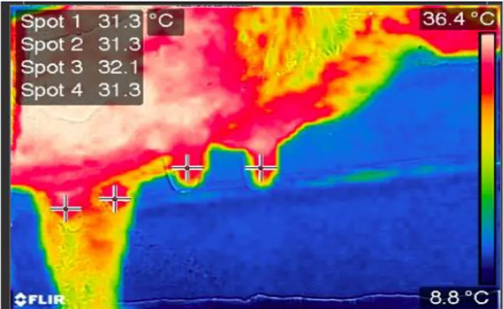
This study was carried out in the herd of Black and White cattle breeders’ association in Southern Lithuania. The experiment was completed in February 2017. A total of 920 teats of 230 Lithuanian Black and White cows (73 cows of first parity, 61 of the second parity and 96 cows of the third parity) were used in this study. Before the study, the cows were examined according to a general clinical examination plan (an average rectal temperature of 38.8 °C, without signs of lameness, metritis, digestive and respiratory disease). The cows were kept in a loose housing system and fed with a balanced feed ration according to their physiological needs throughout the year. Feeding scheduled to 06:00 AM and 18:00 PM every day. The teat-end conditions and thermographic traits were evaluated on each animal and teat-end conditions were measured based on Teat Club International guideline (18). The four different levels (score) of hyperkeratosis were named: no ring, where the teat- end is smooth with a small even orifice (N); smooth ring, where a raised ring encircles the orifice (S), rough ring, where a raised roughened ring with isolated fronds of old keratin extending 1-3 mm from the teat canal orifice (R), and very rough, where a raised ring with rough fronds of old keratin (VR). The quarters of udders were divided into three groups according to the CMT test and clinical signs: group 1 (healthy), group 2 (subclinical mastitis), and group 3 (clinical mastitis). All the measurements were performed at the same time for all analyzed animals. Thermographic measurements of udder quarters of cows, milk SCC, and CMT tests were carried out before morning milking and, clinical signs of udder quarters were evaluated during the morning milking. Thermal images were taken with an infrared thermographic camera (FLIR Systems E4)
positioned 1 m from the lateral side of the udder before milking. Assessment of the teats was carried out at a 90° angle. All cows were acclimated to environmental temperature for 10 minutes before thermal testing. Moreover, the temperatures of mammary in cows were measured at teat sinuses. After colorimetric thermal mapping with software, the elements (pixels) were
converted to give the surface temperatures (o C).
Thermographic images were analyzed using the FLIR software (FLIR Tools version 2.1). The temperature in teat sinuses was rated on four different points: point 1 (left front), point 2 (right front), point 3 (left rear) and point 4 (right rear) (Figure 1).
Figure 1. Multispectral (MSX) image Şekil 1. Multispectral (MSX) görüntü
The SCC in milk was determined using “Somascope” device (CA-3A4, 2004; Delta Instruments, the Netherlands), which uses on flow cytometry technology in State enterprise “Pieno tyrimai”. Besides, CMT was used in addition to physical examination of cows.
Data were analyzed using SPSS (Statistical Package for the Social Sciences, 13.0). Pearson’s Chi-square test was used to identify statistical differences among groups. Value of P<0.05 was considered significant in statistical evaluations.
Results
Teat hyperkeratosis: According to results, only
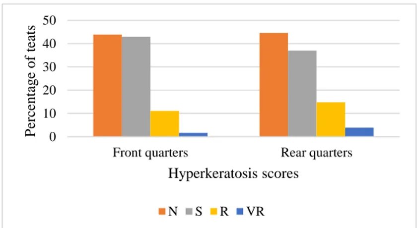
11.8% of the teats were found as rough (R) and 2.9% as very rough for teat hyperkeratosis (Figure 2), while 44.3% of the teats were classified as no ring (N), 41.1% were classified as a smooth ring (S).
The percentages of hyperkeratosis for front and rear quarters are presented in Figure 2. The prevalence of the hyperkeratosis score was significantly (χ2=40.377: DF=3, P<0.001) higher for rear quarters than for front quarters. The percentage of teats classified in the worst classes of hyperkeratosis (R and VR) for front and rear quarters were 12.8% and 18.7%, respectively (Figure 2).
Figure 2. The incidence of teat hyperkeratosis for front and rear quarters Şekil 2. Ön ve arka meme loblarında hiperkeratoz yoğunluğu
Table 1. Teat hyperkeratosis in dairy cows with different lactation levels (%) Tablo 1. Farklı laktasyon seviyelerindeki süt sığırlarında meme başı hiperkeratozu
Teat end score Lactation 1 Lactation 2 Lactation 3>
N 52.25a 37.40b 42.60c
S 42.56a 48.37b 32.73c
R 3.81a 12.60b 20.00c
VR 1.38a 1.63b 4.68c
a, b, c: The differences among values in the same line were significant (X2, P<0.001)
The effect of lactation number on teat hyperkeratosis is presented in Table 1. Multiparous cows had increased degrees of hyperkeratosis when compared with the younger ones (P<0.001). Regarding the teat-end condition of primiparous cows, only 5.19% of the teats were classified as rough (R) and as very rough (VR). The percentage of teats assigned to R and VR classes in the second parity and multiparous cows had 2.74-4.75 times than primiparous R and VR classes (Table 1).
There were significant differences between front and rear teats for the prevalence of hyperkeratosis in first, second and third (≥3 lactations) lactation cows (P<0.001). The prevalence of the hyperkeratosis score was significantly (χ2=8.423; DF =3, P<0.05) higher for rear quarters than for front quarters. The percentage of teats classified in the worst classes of hyperkeratosis (R and VR) equaled 12.8% in front quarters and 18.7% in rear quarters. Rear teats of first lactation cows showed more prevalence of hyperkeratosis compared to front quarters (χ2=9.692, DF=3, P<0.05); in second lactation cows we did not establish a statistically significant difference between hyperkeratosis scores of rear and front teats (χ2=1,752, DF=3, P>0.05); rear quarters of the multiparous cows were more likely to hyperkeratosis (χ2=40.029, DF=3, P<0.0001) than front quarters.
Mastitis and teat hyperkeratosis: Regarding quarters
of the udder, which was divided into three groups according to the CMT test and clinical signs, the percent of udder quarters with healthy, subclinical, and clinical mastitis were 74.1% (682 of quarters), 8.7% (80 of
quarters) and 17.0% (158 of quarters), respectively. There were significant (P<0.05) differences between front and rear udder quarters. Rear quarters had more subclinical and clinical mastitis compared to front quarters (1.28 and 1.40 times). These results clearly showed that there were significant relationships between teat condition and udder quarter cases (χ2=258.072, DF=6, P<0.001). Teat condition was significantly (χ2=84,239, DF=3, P<0.001) better on healthy udder quarters of group 1 when compared to group 3.
The number of clinical mastitis cases was higher for the udder quarters in which hyperkeratosis scores were very high. The hyperkeratosis scores of N and S teats had 2.17 times more healthy udder quarters (P<0.001) than the hyperkeratosis scores of R and VR teats (Figure 3).
Mastitis and thermographic characteristics:
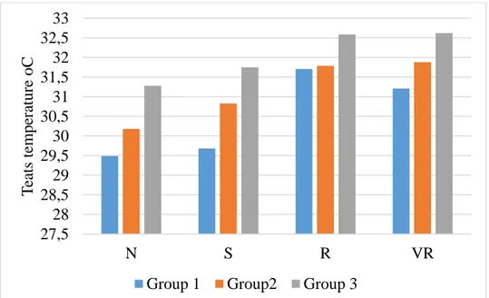
According to the analysis of thermographic images at the teat sinuses, the average temperature of teats was
30.22±0.123oC (temperature of rear quarters was higher
than the front quarters of the udder, 0.08oC). The average
temperature of teats in group 1 was 29.72±0.118oC, while
it was 0.93oC and 1.32oC higher in group 2 and group 3,
respectively, when compared with group 1 (P<0.01). Figure 4 shows different levels of hyperkeratosis that lead to an increase in teat temperature in each group, which was divided based on CMT and clinical signs (from
1.34oC in group 3 to 1.72oC in group 1; P<0.01). The
average milk SCC for group 1, group 2, and group 3 was
137.76±4.40, 298.53±12.718 and 526.50±44.78 thousand/ml, respectively. 0 10 20 30 40 50
Front quarters Rear quarters
P er ce n tag e o f teats Hyperkeratosis scores N S R VR
Figure 3. Relationships between teat hyperkeratosis and prevalence of mastitis Şekil 3. Mastitis yaygınlığı ile hiperkeratoz arasındaki ilişki
Figure 4. Teat temperature based on the groups of mastitis Şekil 4. Mastitis gruplarına göre meme başı sıcaklığı
Additionally, the correlation between teat
temperature and milk SCC were analyzed. Hyperkeratosis of score VR had no effect on milk SCC depending on thermographic characteristics of teats, while the correlation in this group was the highest. However, a significant correlation (P<0.05) was seen between milk SCC and temperature of teats evaluated by hyperkeratosis scores N, S, and R.
Discussion and Conclusion
The results of the present study demonstrate that increased risks of mastitis in dairy herds are associated with different levels of teat hyperkeratosis. A significant connection in many studies was reported between hyperkeratosis and SCC. On the contrary, such a
connection was not observed in some studies. It is generally thought that a severe degree of hyperkeratosis is most likely associated with a mastitis infection rather than mild hyperkeratosis (5,8,11,17). Neijenhuis (20) reported significant associations between severe degree of teat hyperkeratosis and increased risk of clinical mastitis with a different scale they used. The same author et al (21) has also shown that cows with clinical mastitis had worst teat condition (rough rings) when compared with non-infected animals, especially for animals with clinical mastitis between the second and fifth months of lactation. Breen et al (2) found that only 7% of TEC (greater degree of teat-end callosity) scores were classified as N (no ring). In that study, it was also reported that the majority of TEC scores were score 1A and 1B (thin and moderate smooth callosity
N S R VR Group 3 26,3 4,6 22,7 26,9 Group 2 2,9 3,8 37 38,5 Group 1 70,8 91,6 40,3 34,6 0% 10% 20% 30% 40% 50% 60% 70% 80% 90% 100%
27,5
28
28,5
29
29,5
30
30,5
31
31,5
32
32,5
33
N
S
R
VR
T
ea
ts
tempe
ra
ture
oC
ring); score 2D (extreme thickening. severe HK) was present in 1% of all teats scored during the study period.
The development of hyperkeratosis varies based on a number of lactation. It was found that multiparous cows had increased degrees of hyperkeratosis compared to younger ones (P<0.01). Some studies have indicated that a number of lactations have no effect on hyperkeratosis development (5,8). Emre and Alaçam (8) reported that the development of hyperkeratosis was more prevalent in the fifth lactation and above.
In our study, we detected significantly (P<0.05) higher teat hyperkeratosis in cows for rear quarters than for front quarters of the udder. Trajcev and Nakov (27) reported 60.61% and 39.39% subclinical mastitis for the rear and front quarters, respectively. It was reported that the rear udder quarters had a higher risk of CM incidence when compared to the front udder quarters (19). Our results showed that rear udder quarters had 1.28-1.40 times higher (P<0.01) frequency of subclinical and clinical mastitis cases.
Additionally, we detected new data about thermographic parameters in cases of mentioned pathologies. In fact, several diagnostic tests exist for detection and prediction of mastitis such as milk color, pH test, electrical conductivity (10,22), CMT (16), SCC (28), culture test, biomarkers, proteomic technique, and immunoassay method (3,7,26). Infrared thermography (IRT) is an informative method to determine the temperature changes in the udder and in teats within a distance. In the present study, however, there was not any significant difference between the healthy (evaluated as N) teats and teats with a weak degree of hyperkeratosis (evaluated as S) for the temperature of teats. On the other hand, the temperature of udder quarters was significantly lower in healthy quarters (P<0.01) and was related with increased milk SCC (P<0.05), and with the prevalence of subclinical and clinical mastitis in dairy cows (P<0.01). The IRT is a simple, effective, on-site, and noninvasive method that detects surface heat which is emitted as infrared radiation and generates pictorial images without causing radiation exposure (1,23). Our study shows that the level of hyperkeratosis is associated with increased teat temperature and greater risk of mastitis. This can be useful to extend the knowledge on the risk factors, and on the prevention of this disease (25). According to Porcionato et al. (23), the minimum and maximum temperature in the upper area of the udder was 31.17°C and 35.83°C, respectively. The minimum temperature at the end of the teat in the same study was reported as 29.00°C, while the maximum temperature was 34.80°C.
We investigated the relationships between
anatomical characteristics of teats and the prevalence of subclinical mastitis at the quarter level. The results highlight that hyperkeratosis should be considered as an
important problem in the dairy farm we evaluated. Anatomical characteristics of teats in quarter level can be easy and quick criterion in mastitis prevention programs of dairy herds. Additionally, it was concluded that the level of hyperkeratosis is associated with an increase in teat temperature and greater risk of mastitis. This may help to extend our knowledge on the risk factors and on added prevention tools for this disease.
References
1. Bhattacharya A, Mahajan RL (2003): Temperature
dependence of thermal conductivity of biological tissues.
Physiological Measurement, 24, 69–783.
2. Breen JE, Green MJ, Bradley AJ (2009): Quarter and
cow risk factors associated with the occurrence of clinical mastitis in dairy cows in the United Kingdom. J Dairy Sci,
92, 6, 2551–2561.
3. Bu RE, Wang JL, Wu JH, et al (2017): Indirect
enzyme-linked immunosorbent assay method based on Streptococcus agalactiae rSip-Pgk-FbsA fusion protein for detection of bovine mastitis. Pol J Vet Sci, 20, 2, 355-362.
4. Capuco AV, Bright SA, Pankey JW, et al (1992):
Increased susceptibility to intramammary infection following removal of teat canal keratin. J Dairy Sci, 75,
2126–2130.
5. Chrystal MA, Seykora AJ, Hansen LB (1999):
Heritabilities of teat end shape and teat diameter and their relationships with somatic cell score. J Dairy Sci., 82 9,
2017-22.
6. Colak A, Polat B, Okumus Z, et al (2008): Short
communication: early detection of mastitis using infrared thermography in dairy cows. J Dairy Sci., 91, 4244–4248.
7. Culina M, Hahne J, Vorlop KD (2006): Design of an
online sensor array for an early detection of udder affections in automatic milking systems. World Congress of
Agricultural Engineering for a Better World: Book of Abstracts. VDI Verlag GmbH, Germany 453–454. 8. Emre B, Alaçam E (2015): The Occurrence of teat
hyperkeratosis in cows and its effect on milk somatic cell counts. Türkiye Klinikleri J Vet Sci, 6, 1, 1–6.
9. Espeche M, Pellegrino M, Frola I, et al (2012): Lactic
acid bacteria from raw milk as potentially beneficial strains to prevent bovine mastitis. Anaerobe, 18, 103–109.
10. Gáspárdy A; Ismach G, Bajcsy AC, et al (2012):
Evaluation of the on-line electrical conductivity of milk in mastitic dairy cows. Acta Vet Hung, 60, 1, 145–55.
11. Gleeson DE, Meaney WJ, O’Callaghan EJ, et al (2004):
Effect of teat hyperkeratosis on somatic cell counts of dairy cows. J App Res Vet Med, 2, 115–122.
12. Guarín JF, Paixão MG, Ruegg PL (2017): Association of
anatomical characteristics of teats with quarter-level somatic cell count. J. Dairy Sci, 100, 643–652.
13. Haghkhah M, Ahmadi MR, GheisarI HR, et al (2011):
Preliminary bacterial study on subclinical mastitis and teat condition in dairy herds around Shiraz. Turk J Vet Anim
Sci, 35, 6, 387–394.
14. Hamann J (1987): Machine Milking and Mastitis Section
3: Effect of Machine Milking on Teat End Condition - A
15. Hovinen, MJ, Siivonen J, Taponen S, et al (2008):
Detection of clinical mastitis with the help of a thermal camera. J Dairy Sci, 91, 12, 4592-8.
16. Kaşıkçı G, Çetin Ö, Bingöl EB, et al (2012): Relations
between electrical conductivity, somatic cell count, California mastitis test and some quality parameters in the diagnosis of subclinical mastitis in dairy cows. Turk J Vet
Anim Sci, 36, 1, 49–55.
17. Lewis S, Cockroft PD, Bramley RA, et al (2000): The
likelihood of subclinical mastitis in quarters with different types of teat lesion in dairy cow. Cattle Pract, 8, 3, 293-9.
18. Mein GA, Neijenhuis F; Morgan WF, et al (2001):
Evaluation of bovine teat condition in commercial dairy herds. 1. Non-infectious factors. In: Proceedings of the
AABP-NMC International Symposium on Mastitis and Milk Quality. Vancouver. BC. Canada, 347–351.
19. Nakov D, Hristov S, Andonov S, et al (2014):
Udder-related risk factors for clinical mastitis in dairy cows.
Veterinarski archiv, 84, 2, 111–127.
20. Neijenhuis F. (1998): Teat End Callosity Classification
System. Proc Intern Dairy Housing Conf., 4, 17–123.
21. Neijenhuis F, Barkema HW, Hogeveen H, et al (2001):
Relationship between teat-end callosity and occurrence of clinical mastitis. J Dairy Sci, 84, 2664–2672.
22. Norberg E (2005): Electrical conductivity of milk as a
phenotypic and genetic indicator of bovine mastitis: A review. Livest. Prod Sci, 96, 129–139.
23. Porcionato MAF, Canata TF, De Oliveira Cel, et al (2009): Udder thermography of Gyr cows for subclinical
mastitis detection. BioEng. Campinas. Set/Dez, 3, 251–257.
24. Sandrucci A, Bava L, Zucali M, et al (2014): Management
factors and cow traits influencing milk somatic cell counts and teat hyperkeratosis during different seasons. R Bras
Zootec, 43, 9, 505–511.
25. Sathiyabarathi M, Jeyakumar S, Manimaran A, et al (2016): Infrared thermography: A potential noninvasive
tool to monitor udder health status in dairy cows. Veterinary
World. EISSN: 2231-0916 Available at www.veterinaryworld.org/Vol.9/October-2016/7.pdf. 26. Špakauskas V, Klimienė I, Matusevičius AA (2006):
Comparison of indirect methods for diagnosis of subclinical mastitis in lactatting dairy cows. Veterinarski Archiv, 76, 2,
101–109.
27. Trajcev M, Nakov D (2010): Distribution of abnormal
secretion and subclinical mastitis among the udder quarters in dairy cows. Yearbook of the Faculty of agricultural
science and food, 55, 129–138.
28. Yarabbi H, Mortazavi A, Mehraban M, et al (2014):
Effect of somatic cells on the physic-chemical and microbial properties of raw milk in different seasons. IJPAES, 4, 3,
289–298.
29. Zecconi A, Piccinini R, Casirani G, et al (2003): Effects
of automatic milking system on teat tissues, intramammary infections and somatic cell counts. Italian J of Anim Sci, 2,
275-282.
30. Zecconi A, Di Bella L (2013): La mastite costa piu di 300 euro a capo. Informatore agrario 69, 23-26.
Geliş tarihi : 29.10.2017 / Kabul Tarihi : 10.04.2018
Address for correspondence:
Ayhan YILMAZ
Siirt University, Agricultural Faculty, Department of Animal Science Siirt, Turkey e-mail: [email protected]