steoma is a slowly-growing, asymptomatic and benign bone tumor.1Although its pathogenesis is not clearly understood, there
are several theories have been proposed. Some authors consider that the development of osteoma is associated with stimulation of os-teogenic cells by muscle contraction due to trauma.2As in the study of
Kashima et al., muscle traction may play a role in development of osteoma.3
Periosteal ossifying fibroma, exostosis, chondroma, osteosarcoma, fibrosis dysplasia, Paget’s disease and chronic osteomyelitis should be considered for the differential diagnosis of osteomas. Osteomas are most often con-fused with exostosis.4The lesions were considered to be osteomas due to
the presence of polypozis coli. Although osteomas are generally diagnosed in the early period of Gardner Syndrome; they may also be detected in the late period, as in this case. Therefore, long-term follow-up is recom-mended.
Gardner syndrome should be considered for the patients presenting with osteoma, in accordance with family history and the patient should be referred to colonoscopic examination for the presence of intestinal polyps. Likewise, patients diagnosed with intestinal polyp should be referred to the department of oral and maxillofacial surgery considering the presence of osteoma in the mandible. The aim of this study was to describe the diagno-sis and treatment of Gardner syndrome.
Gardner Syndrome Associated with
Late Mandibular Osteoma
AABBSS TTRRAACCTT Osteomas are well-differentiated, slowly-growing and asymptomatic benign tumors of mature bone. Osteomas are usually observed on the jaw and classified into two types as central and peripheral osteoma. Diagnosis is made by observing radio-opacities on computed tomography. While follow-up is recommended for minor cases, surgical excision should be performed for major cases. Gardner syndrome is an autosomal dominant disease characterized by intestinal polyps and multiple osteomas. Due to the malignancy potential of intestinal polyps, early diagnosis and treat-ment are important. In this case report, diagnosis, surgical treattreat-ment and two years follow-up of a patient with Gardner syndrome are presented.
KKeeyywwoorrddss:: Gardner syndrome; familial adenomatosis polyposis; protocolectomy; late osteoma
Orkhan ISMAYILOVa, Ali EKEMENa, Haydar CELASİNb, Hakan Alpay KARASUa, İbrahim Ethem GEÇİMc aDepartment of Oral and Maxillofacial Surgery,
Ankara University Faculty of Densitry, bDepartment of General Surgery, Lokman Hekim University Faculty of Medicine,
cDepartment of General Surgery, Ankara University Faculty of Medicine, Ankara, TURKEY
Re ce i ved: 14 Nov 2018
Received in revised form: 07 Mar 2019 Ac cep ted: 11 Mar 2019
Available online: 15 Mar 2019 Cor res pon den ce:
Orkhan ISMAYILOV
Ankara University Faculty of Dentistry, Department of Oral and
Maxillofacial Surgery, Ankara, TURKEY
Cop yright © 2019 by Tür ki ye Kli nik le ri
DOI: 10.5336/caserep.2018-63701
CASE REPORT
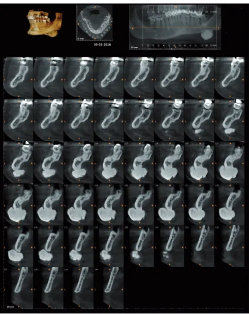
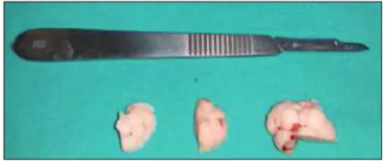
The mother of the patient was learned to have been diagnosed with familial adenomatosis polyposis (FAP) and had been, treated with total colectomy and ileorectal anastomosis and a secondary liver cancer had been developed thereafter. During this treatment, patients family was invited for screening and her son was diagnosed with FAP at the age of 20. Restorative proctocolectomy was performed due to multiple adenomatous polyps detected on colonoscopy. During follow-up, millimetric polyps were detected in the anal transition zone (ATZ) and repeated polypectomies were performed dur-ing follow-up periods. Durdur-ing the last physical ex-amination, after 16 years from the initial surgery, a solid mass was detected on the lower part of the mandible and he was referred to the Department of Oral and Maxillofacial Surgery in the Faculty of Dentistry of Ankara University for the evaluation of the palpable mass and asymmetry on the angle of the mandible. After the physical examination, computerized tomography scan revealed four ra-diopaque and oval bone lesions in the mandible; three small lesions on the right side and one large lesion on the lower part of the left side (Figure 1). The patient underwent surgery under general anes-thesia and local anesthetic with vasoconstrictor was administered to incision line. Subsequently, cuta-neous and subcutacuta-neous tissues were incised to platysma muscle layer in parallel with the lower edge of the mandible and 2 cm distant from the bone with no 10 scalpel. The deep cervical fascia was exposed by blunt dissection through superior-inferior fibers of platysma muscle. By preserving the marginal mandibular branch of the facial nerve, an incision measuring the same with the skin inci-sion was performed on the platysma muscle. The superficial and deep layers of cervical fascia were dissected and periosteum was exposed (Figure 2). The periosteum was dissected with no 15 scalpel blade to reveal the mass. Osteoma was completely excised with drill and osteotomes (Figure 3). The lower edge of the mandible was corrected with round drills and the layers were sutured anatomi-cally (periosteum, fascia, muscle, subcutaneous and
cutaneous tissues in order). In order not to lead the pathologist to the diagnosis of osteoma, the removed specimen was sent to the laboratory without inform of pre-diagnosis of Gardner syndrome. The pathol-ogy result of the specimen was reported as osteoma.
The patient is under being followed up for 24 months and no intestinal recurrence was detected. An X-ray was performed during follow-up after 2 years and the incision site was detected to have anatomic borders and no recurrence was detected (Figure 4).
DISCUSSION
Isolated osteomas are very rare in the stomatog-nathic system and they most commonly occur at the angle and body of the mandible. On the other hand, multiple lesions are associated with Gardner syndrome.5
According to a study conducted in 2002, os-teomas do not vary in accordance with age and gender.6Nevertheless, Kaplan et al. showed that
os-teoma has a higher incidence in males. They are frequently observed between 16 and 74 years and occur at 3rdor 5thdecade of life.7Our patient's being
an adult is consistent with literature.
In clinical practice, osteomas may be observed as well-demarcated oval shaped tumors with pedi-cles or unilateral, slowly growing asymptomatic ad-herent tumors and externally adad-herent to the bone surface. Osteoma may be observed as a radiopaque mass with bone density and with a thin radiolucent line around. Three-dimensional computed tomog-raphy was shown to be the best radiographic method for diagnosis of osteomas.8The presented
case is clinically and radiographically consistent with literature data.
Gardner syndrome is considered as a version of familial adenomatous polyposis (FAP); however, the presence of osteomas should be excluded. FAP is characterized by the presence of hundreds of in-testinal polyps. Gardner syndrome is the associa-tion of polyposis, osteomas and soft tissue tumors (desmoid tumors, fibromas, epidermoid cysts). As the malignancy risk of intestinal polyps is very high, early diagnosis has vital importance. The
dis-ease is autosomal dominant and colorectal adeno-carcinoma may develop in untreated patients. Al-though pathological lesions, such as osteomas, skin cysts, etc., may be asymptomatic for a long time; polyposis lesions may lead to symptoms, such as rectal bleeding, diarrhea, and abdominal pain.9Also
Gardner’s syndrome (Familial adenomatous poly-posis) is associated with papillary thyroid cancer in 89% of the cases.10,11In the present case, symptoms
of Gardner syndrome were noted.
If osteoma is diagnosed during head and neck examination, upper and lower endoscopic exami-nation should be recommended. In patients with
FIGURE 1: Preoperative CBCT.
colonic or duodenal polyposis, the investigation of osteoma and papillary thyroid cancer by per-forming head and neck examination will be use-ful.
I
Innffoorrmmeedd CCoonnsseenntt
Informed consent was obtained from the patient prior to the operation.
S
Soouurrccee ooff FFiinnaannccee
During this study, no financial or spiritual support was received neither from any pharmaceutical company that has a direct connection with the research subject, nor from a company that provides or produces medical instruments and materials which may negatively affect the evaluation process of this study.
C
Coonnfflliicctt ooff IInntteerreesstt
No conflicts of interest between the authors and / or family members of the scientific and medical committee members or
FIGURE 3: Removed Lesion.
members of the potential conflicts of interest, counseling, ex-pertise, working conditions, share holding and similar situa-tions in any firm.
A
Auutthhoorrsshhiipp CCoonnttrriibbuuttiioonnss
I
Iddeeaa//CCoonncceepptt:: Ibrahim Ethem Gecim, Hakan Alpay Karasu, Orkhan Ismayilov; DDeessiiggnn:: Haydar Celasin, Ali Ekemen; CCoonn--t
trrooll//SSuuppeerrvviissiioonn:: Hakan Alpay Karasu, Ali Ekemen, Orkhan Is-mayilov; DDaattaa CCoolllleeccttiioonn aanndd//oorr PPrroocceessssiinngg:: Haydar Celasin,
Ibrahim Ethem Gecim, Ali Ekemen; AAnnaallyyssiiss aanndd//oorr IInntteerrpprree--t
taattiioonn:: Orkhan Ismayilov, Ali Ekemen; LLiitteerraattuurree RReevviieeww:: Ibrahim Ethem Gecim,, Hakan Alpay Karasu, Haydar Celasin;WWrriittiinngg tthhee AArrttiiccllee:: Orkhan Ismayilov, Ibrahim Ethem Gecim, Hakan Alpay Karasu; CCrriittiiccaall RReevviieeww:: Hakan Alpay Karasu, Ali Ekemen; RReeffeerreenncceess aanndd FFuunnddiinnggss:: Haydar Celasin, Orkhan Ismayilov; MMaatteerriiaallss:: Ibrahim Ethem Gecim, Hakan Alpay Karasu, Orkhan Ismayilov.
1. Kerckhaert A, Wolvius E, van der Wal K, Oost-erhuis JW. A giant osteoma of the mandible: case report. J Craniomaxillofac Surg. 2005;33(4):282-5. [Crossref] [PubMed]
2. Kaplan I, Nicolaou Z, Hatuel D, Calderon S. Solitary central osteoma of the jaws: a diag-nostic dilemma. Oral Surg Oral Med Oral Pathol Oral Radiol Endod. 2008;106(3):e22-9. [Crossref] [PubMed]
3. Kashima K, Rahman OI, Sakoda S, Shiba R. Unusual peripheral osteoma of the mandible: report of 2 cases. J Oral Maxillofac Surg. 2000;58(8):911-3. [Crossref] [PubMed]
4. Guimarães KB, Cavalcante JR, Ferraro-Bez-erra M, Silva DN, de Holanda Vasconcellos RJ, do Egito Vasconcelos BC. Peripheral os-teoma bilateral of the mandible without
as-sociation with Gardner syndrome. J Cranio-fac Surg. 2012;23(2):e83-6. [Crossref] [PubMed]
5. Zouloumis L, Lazaridis N, Maria P, Epiva-tianos A. Osteoma of the ethmoidal sinus: a rare case of recurrence. Br J Oral Maxillofac Surg. 2005;43(6):520-2. [Crossref] [PubMed]
6. Sayan NB, Uçok C, Karasu HA, Günhan O. Peripheral osteoma of the oral and maxillofa-cial region: a study of 35 new cases. J Oral Maxillofac Surg. 2002;60(11):1299-301.
[Crossref] [PubMed]
7. Kaplan I, Calderon S, Buchner A. Peripheral osteoma of the mandible: a study of 10 new cases and analysis of the literature. J Oral Maxillofac Surg. 1994;52(5):467-70. [Cross-ref]
8. Ogbureke KU, Nashed MN, Ayoub AF. Huge peripheral osteoma of the mandible: a case report and review of the literature. Pathol Res Pract. 2007;203(3):185-8. [Crossref] [PubMed]
9. Waller A, Findeis S, Lee MJ. Familial adeno-matous polyposis. J Pediatr Genet. 2016;5(2): 78-83. [Crossref] [PubMed] [PMC]
10. Bell B, Mazzaferri EL. Thyroid cancer in famil-ial polyposis coli: case report and literature re-view. Dig Dis Sci. 1993; 38(1):185-90.
[Crossref] [PubMed]
11. Isik A, Firat D, Yilmaz I, Peker K, Idiz O, Yil-maz B, et al. A survey of current approaches to thyroid nodules and thyroid operations. Int J Surg. 2018;54(Pt A):100-4. [Crossref] [PubMed]