Ankara Üniv Vet Fak Derg, 58, 61-64, 2011
Short Communication / Kısa Bilimsel Çalışma
Immunohistochemical distribution of ghrelin positive cells in the
abomasum of sheep
Nesrin ÖZFİLİZ, Şerife TÜTÜNCÜ, Hatice ERDOST
Department of Histology and Embryology, Faculty of Veterinary Medicine, University of Uludağ, Bursa, Türkiye.
Summary: Ghrelin is a novel 28-amino-acid-peptide isolated from the rat and human stomach. This peptide is predominantly produced by the stomach, in addition lower amounts derive from small and large intestines, pancreas, pituitary gland, kidney, placenta, immun system organs and cells, testis, ovary and arcuate nucleus of hypothalamus. In this study, distrubition of ghrelin immunoreactive cells in the abomasum of sheep was investigated. Tissue samples were taken from cardia, fundus , the proximal region of pylorus, main/distal pylorus regions of abomasum. The results have revealed that ghrelin immunopositive cells distrubuted throughout the mucosal layer of abomasum of sheep, in other mammalian species.
Key words: Abomasum, Ghrelin, immunohistochemistry, sheep,
Koyun abomasumunda ghrelin pozitif hücrelerin immunohistokimyasal dağılımı
Özet: Ghrelin rat ve insan midesinden izole edilen 28 amino asitli bir peptiddir. Bu peptid öncelikle mide tarafından üretilmektedir, ayrıca ince ve kalın barsaklardan, pankreas, hipofiz, böbrek, plasenta, immun sistem, testis, ovaryum ve hipotalamusun arkuat nukleusundan küçük miktarlarda elde edilmiştir. Çalışmamızda, koyun abomasumunda Ghrelin immun pozitif hücrelerin dağılımı incelendi. Abomasum cardia, fundus, proksimal ve distal pilorus olmak üzere 4 bölüme ayrılarak incelendi. Sonuç olarak, Ghrelin immunpozitif hücrelerin diğer memeli hayvanlarda olduğu gibi abomasumun mukozal katmanına dağıldığı görüldü.
Anahtar sözcükler: abomasum, Ghrelin, immunohistokimya, koyun.
Introduction
Ghrelin is an endogenous ligand for the growth hormone secretagogue receptor (GHS-R) and stimulates growth hormone release from the pituitary gland(6, 7). This peptide was reported to function not only in the control of growth hormone secretion but also in the regulation of food intake and energy metabolism (9, 14, 16). Ghrelin is predominantly produced in the stomach mucosa. In addition, lower amounts derive from small and large intestines, pancreas, pituitary gland, kidney, placenta, immun system cells and organs, testis, ovary and arcuate nucleus of hypothalamus. At the histological level ghrelin producing cells were immunohistochemically identified, in the gastrointestinal tract of humans same of the mammalian species (2, 6, 10, 13). Hayashida et al. (3) examined distribution of the ghrelin immunoreactive cells in the stomachs of domestic animals and rats, using a polyclonal antibody for the N-terminal fragment of rat ghrelin. Wada et al. (15) demonstrated that ghrelin exsisted in the hatching and adult chicken proventrikulus. Sakata et al. (11) demonstrated that ghrelin producing cells of the rat located in the mucosal layer of the
stomach, fundus, duodenum, ileum, cecum and colon. Ahmed and Harvey (1) showed that ghrelin immunopositive cells existed in the chicken hypotalamus but not in the chicken proventriculus. Huang et al. (5) investigated the distrubition of ghrelin mRNA of different tissues in sheep. Ghrelin mRNA expression was significantly higher in the abomasum than other tissues by RT-PCR and real-time PCR. In another study, Ghrelin mRNA was predominantly expressed in abomasum of the digestive tract of beef cattle(8).
However, the distrubition of ghrelin immunoreactive cells in the abomasum of sheep, has not been described yet. In this study, the distrubition of ghrelin immunoreactive cells in the abomasum of sheep was investigated.
Awassi sheep were used in the study. The abomasum pieces were removed from 4 sheep after slaughtering and fixed with Bouin solution for 24-36 hours (12). The abomasum were divided into four regions: cardia, fundus, the proximal region of pylorus and main/distal pylorus. The fixed tissues were dehydrated in alcohol, cleared in xylene and embedded in parafin blocks. Immunostaining was performed with a labelled
Nesrin Özfiliz - Şerife Tütüncü - Hatice Erdost 62
avidin- biotin (LAB) technique (4) using polyclonal ghrelin antibody (Phoenix Pharma. Inc.H- 031-31) as a primary antibody and using Histostain-Plus Bulk Kits (Zymed 2nd generation LAB-SA Detection System). The
sections were incubated for 30 minutes at 37ºC with 1:2500 dilution of the ghrelin antibodies. Finally, 3.3’-diaminobenzidine (DAB) was used for colour development
and haematoxylin was used for counterstaining. Negative control slides processed without primary antibodies were included for each staining. Section were examined using a Nikon eclipse 80i Microscope-Ds Camera Control Unit DS-L1.
The present study showed that ghrelin immunpositive cells scattered throughout the mucosal layer of
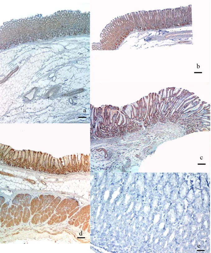
Figure 1. a, b, c, d. General view of cardia, fundus, proximal pylorus, and distal pylorus regions, Bar 350 µm, e. Negative control, Bar 140µm
Şekil 1. a, b, c, d. Cardia, Fundus, Pilorisin proksimal ve distal bölümlerinden genel görünüm, skala 350 µm, e. Negatif kontrol, Skala 140 µm
Ankara Üniv Vet Fak Derg, 58, 2011 63
abomasum. The abomasum were examined by dividing into four regions:
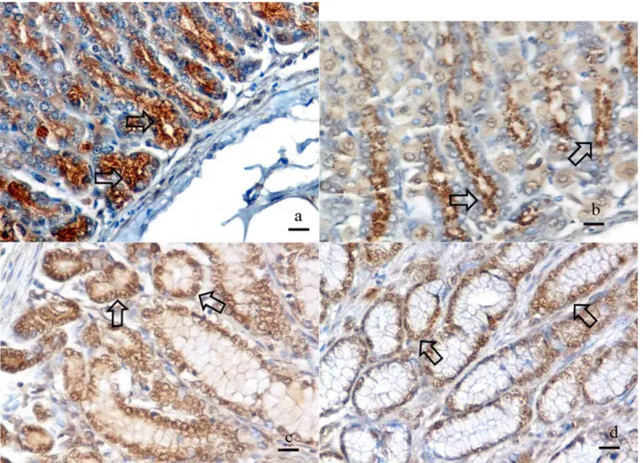
The cardia Region: Ghrelin immunopositive reaction was granular and located in intracytoplasmic and peculiar to perinuclear region of the glandular cells(Fig.1a, 2a).
The fundus Region: Ghrelin immunopositive reaction was granular and located in the cytoplasm, specifically in the apical region of the glandular cells(Fig.1b, 2b).
The proximal region of pylorus: Ghrelin immunopositive reaction was granular and located in the cell nucleus and paranuclear cytoplasm of the glandular cells (Fig.1c, 2c).
The main/distal pylorus: Ghrelin immunopositive reaction was granular and located in the cell nucleus and paranuclear cytoplasm of both glandular and surface epithelial cells (Fig.1d, 2d).
Negative controls did not give any spesific immunostaining for Ghrelin(Fig. 1e).
Many investigations, most of which were biochemical and physiological have been published since then ghrelin was identified in 1999 (5, 7-9, 14, 16). In only a few
histological studies ghrelin producing cells were immunohistochemically identified in the gastrointestinal tract (2, 6, 10, 13). The present study focused on, to determine the distrubition of ghrelin immunoreactive cells throughout the abomasum of sheep.
Wada et al. (15) reported that ghrelin immunopositive cells were found in the mucosal layer of proventriculus in the hatching and adult chicken. Rindi et al. (10) demonstrated that ghrelin immunoreactive cells, in the human, canine and rodent stomach. In addition, Tanaka and Watanabe (13) found ghrelin immunoreactive cells in all regions of human gastric mucosa, but were distrubuted mainly in the fundic region. Sakata et al. (11) observed ghrelin producing cells the mucosal layer of the stomach, fundus, duodenum, ileum, cecum, colon of the rat and they could be classified into two different cell types. Hayashida et al. (3) reported that ghrelin immunopositive cells were widely distributed from the neck to the base of oxyntic glands in the cow, sheep, pig and horse stomach. Previous papers reported that ghrelin immunopositive cells were abundant in oxyntic glands of the glandular stomach. Our results were consistent with those reports. Ghrelin immunopositive cells scattered
Figure 2. a, b ,c, d. Ghrelin immunopositive reaction in cardia, fundus, proximal pylorus and distal pylorus regions, Arrows, Bar 35µm
Nesrin Özfiliz - Şerife Tütüncü - Hatice Erdost 64
troughout the mucosal layer of abomasum in the glandular stomach. Results of the previous studies on the ghrelin mRNA distrubition and ghrelin gene expression support our immunohistochemical results (2, 5, 8). As a contrast to our findings, Ahmed and Harvey(1) did not observe ghrelin immunoreactivity both in the chicken proventriculus and in the remaning parts of the gastrointestinal tract but the reactivity was found in the chicken hypothalamus. A possible explanation, for this inconsistency might be the difference in the recognition sites of the antiserum.
In conclusion, we have demonstrated that ghrelin immunopositive cells scattered throughout the mucosal layer of the sheep abomasum.
References
1. Ahmed S., Harvey S (2002): Ghrelin a Hypotalamic
GH-releasing factor in Domestic fowl (Gallus Domesticus). J. Endocrinol. 172:117-25.
2. Date Y, Kojima M, Hosoda H, Sawaguchi A, Mondal MS, Suganuma T, Matsukura S, Kangawa K, Nakazato M (2000): Ghrelin, a novel growth hormone-releasing
acylated peptide, is synthesized in a distinct endocrine cell type in the gastrointestinal tracts of rats and humans. Endocrinology. 141:4255–4261.
3. Hayashida T, Murakami K, Mogi K, Nishihara M, Nakazato M, Mondal MS, Horii Y, Kojima M, Kangawa K, Murakami N (2001): Ghrelin in domestic
animals: distribution in stomach and its possible role. Domest Anim Endocrinol. 21:17–24
4. Hsu S. M., Raiıne L.,Fanger H (1981): Use of
Avidin-Biotin-Peroxidase Complex (ABC) in immunperoxidase techniques. The Journal of Histochemistry and Cytochemistry,
29: 577-580,.
5. Huang Z.G., Xiong L., Liu Z. S., Qiao Y., Dai R., Xie Z., Liu s.R.,Shi G.Q., Liu G.Q (2006): The tissue
distribution and developmental changes of ghrelin mRNA expression in sheep.Yi Chuan Xue Bao. Sep; 33(9):808-13.
6. Kojima M.,Hosoda H., Date Y, Nakazato M, Matsuo H, Kangawa K (1999): Ghrelin is a Growth-
Hormone-releasing acylated peptide from stomach, Nature.
402:656-60.
7. Kojima M., Kangawa K (2001): Ghrelin: a novel growth
hormone releasing peptide (in japanese). Nippon Rinsho
59:1400-7.
8. Leaflet A. S (2004): Effect of Feed Intake on Plasma
Ghrelin Concentration in Beef Cattle. Iowa state University Animal Industry Report, 1881,.
9. Nakazato M, Murakami N, Date Y, Kojima M, Matsuo H, Kangawa K, et al. (2001): A role for ghrelin in the
central regulation feeding. Nature. 409:194-8
10. Rindi G., Necchi V., Savio A., Torsello A., Zoli M., Locatelli V., Raimondo F., Cocchi D., Solcia E (2002):
Characterisation of Gastric Ghrelin Cells in Man and Other Mammals: Studies in Adult and Fetal Tissues, Histochem Cell Bio. 117:511-519.
11. Sakata I., Nakamura K., Yamazaki M., Matsubara M., Hayashi Y., Kangawa K., Sakai T (2002):
Ghrelin-Producing cells exist as two types of cells, closed and opened type cells, in the rat gastrointestinal tract. Peptides. 23:531-6.
12. Smith A., Bruton J (1978): A Colour Atlas of Histological
Staining Techniques. 2nd Edition ,Wolfe Medical
Publications Ltd., London 181-184,
13. Tanaka M., Watanabe M (2005): Distrubition of
Ghrelin-İmmunreactive Cells in Human Gastric Mucosa: Comparision with that of Parietal Cells, J. Gastroenterol.
40: 345-349
14. Tschöp M, Smiley DL, Heiman ML (2000): Ghrelin induces adiposity in rodents, Nature. 407:908-13.
15. Wada R., Sakata I., Kaiya H., Nakamura K., Hayashi Y., Kangawa K., Sakai T (2003): Existence of
ghrelin-immunpositive and expressing cells in the proventriculus of the hatching and adult chicken, Peptides. 111;123-128.
16. Wren AM, Small CJ, Ward HL, Murphy KG, Dakin CL, Taheri S, et al. (2000): The novel hypotalamic
peptide ghrelin stimulates food intake and growth hormone secretion, Endocrinology. 141: 4325-8.
Geliş tarihi: 15.05.2009 / Kabul tarihi: 22.01.2010
Corresonding author:
Nesrin Özfiliz
Uludağ Üniversitesi Veteriner Fakültesi Histoloji Embriyoloji Anabilim Dalı 16059 Bursa, Türkiye
Tel: 224 2941263,