https://doi.org/10.1007/s00464-019-06856-1
A surgical and anatomo‑histological study on Transoral Endoscopic
Thyroidectomy Vestibular Approach (TOETVA)
Servet Celik1 · Ozer Makay2 · Mustafa Deniz Yoruk3 · Ilke Bayzit Kocer1 · Murat Ozdemir2 · Kubilay Dogan Kilic4 ·
Canberk Tomruk4 · Okan Bilge1 · Yigit Uyanikgil4,5 · Gianlorenzo Dionigi6
Received: 6 March 2019 / Accepted: 18 May 2019 / Published online: 30 May 2019 © Springer Science+Business Media, LLC, part of Springer Nature 2019
Abstract
Background The number of TOETVA surgeries has increased worldwide but the anatomical passage of trocars is not clearly defined. We aimed to define detailed surgical anatomical passage of the trocars in cadavers. The incisions in oral vestibule, anatomical pathways of trocars, affected mimetic muscles, neurovascular relations of trocars and histological correlation of surgical anatomy were investigated.
Methods Four cadavers and 6 six patient oral vestibules were used. The locations of optimised vestibular incisions were measured photogrammetrically. Initial steps of TOETVA surgery were performed on cadavers according to those optimal incisions. TOETVA preformed cadavers dissected to determine anatomical passages of the trocars. Afterwards, flap of lower lip and chin were zoned by software appropriate to the trocars routes. Histological analyses of the zones were made in cor-relation with dissections.
Results Mimetic muscles associated with median (MT) and lateral trocars (LT) are orbicularis oris, mentalis, depressor anguli oris, depressor labii inferioris and platysma muscles. Trocars affect mimetic muscles in the perioral, chin and submental regions in different ways. The risk of mental nerve injury by MT is low. LT pass through the DLI muscle. The transmission of LT to the subplatysmal plane in the submental regions can be in two different ways. The arterial injury risk is higher with LT than the MT.
Conclusions The surgical anatomy of the perioral, chin and submental regions for the initial TOETVA steps has been defined. Detailed surgical anatomical passages of the MT and LT were determined. Anatomical pattern to reach subplatysmal plane are presented. Mimetic muscles effected by trocars were determined. Endocrine surgeons should know the anatomical pas-sage of TOETVA trocars.
Keywords TOETVA · Surgical anatomy · Histological preparation · Oral vestibule · Perioral and chin region · Mimetic muscles
The scarless thyroidectomy approach has gained popular-ity in recent years. As a result of the efforts to develop new thyroidectomy techniques, surgery through the oral vesti-bule, called the Natural Orifice Transluminal Endoscopic Surgery (NOTES), has been described [1–3]. An entirely transoral endoscopic approach, the transoral video-assisted thyroidectomy (TOVAT), has been reported in experiments on cadavers [4]. The first transoral thyroid and parathyroid surgeries using a lateral sublingual approach were executed on living pigs [5]. Wang et al. reported a completely oral
vestibular approach with 3 trocars on 12 patients [6]. The struggle to evaluate the NOTES techniques finally led to the Transoral Endoscopic Thyroidectomy Vestibular Approach (TOETVA), which is considered to be a novel technique [7].
In the NOTES progress, two lateral trocars (LT) and one median trocar (MT) were initially inserted through the oral vestibule and the mouth floor in different combinations [2,
8, 9]. Due to the difficulties with and complications in the application of MT through the mouth floor, it was subse-quently applied through the oral vestibule instead [10, 11]. The first fully oral vestibular application of trocars was the Endoscopic Thyroidectomy Using the Oral Vestibular Approach (ETOVA), which represents almost the same technique as TOETVA. In the ETOVA, one median incision
* Servet Celik
[email protected]; [email protected]
Extended author information available on the last page of the article
(MI) and two lateral incisions (LI) are made transversely for trocar insertion close to the buccogingival (BG) sulcus on the inferior oral vestibule. After the insertion through the oral vestibule, the trocars make their way down through the perioral, chin and submental regions and the subplatysmal plane, respectively, to reach the thyroid gland [6]. Those initial steps of the operation are made blindly and, since the surgeon can consequently proceed only manually, a detailed anatomical description of the trocars’ passages, not currently found in literature, is needed.
During blind placement of the trocars, it is possible to injure the mental nerve (MnN) when using different NOTES techniques [8]. Different locations and positions have been tried at the oral vestibules of patients to prevent MnN injury during trocar placement. Recently, Anuwong described ver-tical orientation LI for TOETVA, which is unlike the previ-ous techniques [12]. In his first 60 cases, Anuwong made a 1-cm MI close to the frenulum of the lower lip and two 0.5 cm LIs close to the BG sulcus, opposite to the junc-tion between the incisor and canine teeth on both sides. Afterwards, Anuwong refined LIs and MI to avoid MnN injury by making the incisions more anterolateral. Accord-ing to his most recent description, the 1 cm MI should be made opposite the incisor teeth, at the mid-level of the oral vestibule, and the 0.5 cm LI should be made near the ver-milion in a vertical orientation opposite the first or second premolar teeth. This repositioning resulted in a lower MnN injury rate (1.5%) [13–15]. Nowadays, TOETVA continues gaining popularity around the world according to the above-standardised definition and steps [16–18].
Although the number of TOETVA surgeries has increased worldwide, obscure issues exist regarding surgical anatomy and trocar relations. How the trocars pass downward from the oral vestibule to the subplatysmal plane is not clear. Dur-ing the initial steps of TOETVA, the trocar transmissions from the vestibular incisions to the subplatysmal plane are not seen by the surgeon. A tunnel is created using a tunnel-ling probe until the subplatysmal plane is reached and after-wards the trocars are pushed downafter-wards using gentle hand manoeuvres while the neck is extended [13, 19]. The MT passage through the mentalis (Mn) muscle was previously reported [10, 19, 20]. However, the relations and effects of the median and lateral trocars on other mimetic muscles in the perioral, chin and submental regions are not well defined. The neurovascular and muscular anatomical relations of the trocars during their passage to the subplatysmal plane are not described. Detailed knowledge about the anatomical passage of the trocars and their relations with the mimetic muscles, as well as mental and facial neurovascular structures, may provide information that would increase the understanding of the initial steps of the technique better. Surgeons must attain perfect knowledge of the anatomical course and effects of the trocars on their pathway. Further descriptions of the
TOETVA surgical anatomy would be useful for educating experts about this specific technique.
Due to restricted literature about the TOETVA surgical anatomy, we aimed towards a better understanding of the ini-tial steps of this technique in this paper. TOETVA applied to cadaver dissection has not been previously reported. Cadaver dissections are essential for increasing the understanding of the trocar anatomical passages in the initial steps of TOE-TVA at the perioral, chin and submental regions.
The locations and proportions of MIs and LIs on the oral vestibule, the detailed anatomical trocar pathways, the affected mimetic muscles, the neurovascular relations of trocars and the histological correlation of surgical anatomy were the targets of this study.
Materials and methods
To determine the optimal locations for MIs and LIs, six clinical cases during operation and two simulated surgery cadaver vestibules (eight in total) were analysed photogram-metrically. The cadavers used for simulations and subsequent descriptive dissections were embalmed with a modified Larssen solution that provides protection for life-like prop-erties and is suitable for modern surgical instrumentation [21]. Another cadaver, on which surgery was not performed, was dissected for comparison with the two surgical simu-lation cadavers, looking at muscle fibre changes resulting from the TOETVA trocar applications. To determine the microanatomy of the surgical area and correlate it with the gross anatomy of the trocar tunnels, sagittal-sliced samples from another 10% formalin-fixed cadaver were examined histologically.
All the cadavers used in this study were donated to the Ege University Department of Anatomy in accordance with the organ and tissue donation law of the Republic of Tur-key. The study was designed according to the Declaration of Helsinki (2010). The signed approval was received from the Head and Academic Committee of the Department of Anatomy and the Institute Review Board (IRB) of the Ege University Medical Faculty. The signed consent form of each patient is obtained.
TOETVA steps application
Previously described TOETVA surgery steps were per-formed by experienced endocrine senior surgeons (OM and GD) [7, 12, 19, 20]. The surgeons marked the MI and LI using a blue-coloured pen on both the patients’ and the cadavers’ oral vestibules, taking into consideration that the incisions should be made as close as possible to the vermil-ion to avoid MnN injury [19, 20]. The MI was transverse ori-ented and 10 mm in length. The LI was oriori-ented vertically,
5 mm in length and carried out on both sides, respectively, opposite to the second premolar teeth (Fig. 1A).
Before photographing, a millimetre sensitive scale bar was placed in the oral vestibule as the lower lip was half-pulled forward to see the oral vestibule, BG sulcus and teeth. Photographs were taken with the Nikon D7100, Sigma 17-50 f2.8 lens.
After making the incision in the oral vestibular mucosa through the transverse MI mark, a downward blunt dissec-tion with a Kelly clamp was carried out (Fig. 1B, C). A tunnel to the subplatysmal plane from the transverse MI was created using a tunnelling probe. A Veress needle was applied for hydrodissection from the transverse MI and 25 to 50 ml of saline solution was injected into the subplatysmal plane. Additionally, 50 ml of saline solution was injected into the subplatysmal plane of the submental and neck regions through the skin (Fig. 1D, E). After making inci-sions through the vertical LI marks, about 20 ml of saline solution was injected per side. One MT 12 mm in diameter and two LTs 5 mm in diameter were inserted, respectively, through the incisions (Fig. 1F, G). Further subplatysmal flap preparation to create an air pocket in the neck was not car-ried out on the cadaver samples.
Measuring
Graphic analysis software (Image J version 1.51e, supplied by the National Institute of Health) was used for photogram-metry. The lengths measured included MI to the vermilion mucosal border (VM), MI to the BG sulcus, the superior tip of LI to VM, the inferior tip of LI to the BG sulcus and the distance between MI and LI on both sides, respectively (Fig. 2).
Dissection of the cadavers
After applying the above-described initial steps of the TOE-TVA procedure, the dissections were carried out in layers by anatomists (SC, MDY, IBK, OB) under 2×–8× dissection microscopes (HS Möller-Wedel Int. Universa 300 Operating Microscope) and head loupes (H&S® Illuminated LED Light
Head Loupe Magnifier) without removing the trocars. The skin was dissected from the dermis plane to separate it from the superficial fascia following the median vertical incision and the transverse incision that passed along the vermilion cutaneous border (Fig. 1H). After the removal of the superfi-cial fascia, the mimetic muscles and neurovascular structures in the surgical field were carefully determined.
Afterwards, the trocars were removed to determine the structures left behind them and the roots of the neurovascular structures (Fig. 4B). To show the structures behind MT, the plane was vertically incised down to the mandible. To describe the posterior LT structures, the inferior mandibular attachment
of the depressor anguli oris (DAO) muscle was detached and the muscle was elevated (Fig. 5G). The dissection went on to separate the platysma lateral from the labial commissure. Lateralisation of the platysma was made to determine the mar-ginal mandibular branch (MMB) of the facial nerve (FN) and facial vessels. Finally, the inferior mandibular attachment of the depressor labii inferioris (DLI) muscle was separated and was elevated laterally to localise the neurovascular structures below it (Fig. 4D).
Histological sampling
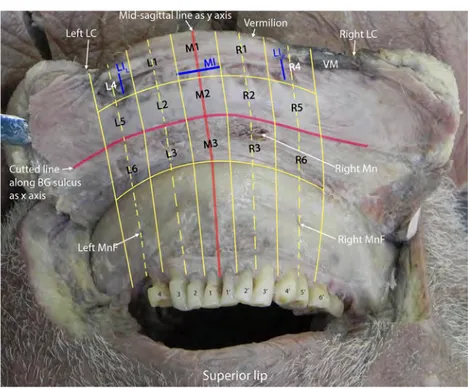
The lower lip and chin were harvested as a flap from a 10% formalin-fixed cadaver (Fig. 3). In order to do so, a circular incision along the BG sulcus towards the mandible was made and a part of the flap under the BG sulcus level, including the periosteum, was separated from the mandible using an elevator. The flap was wider than the TOETVA-related area. The anatomical structures on the harvested flap were identi-fied and photographed together with a metric scale bar. The photographs were transported to a computer and a grid was created photogrammetrically.
The median plane of the grid was accepted as the y-axis and the line that passed along the incision of the BG sulcus was accepted as the x-axis. Nine zones were created with lines parallel to the y- and x-axes. The lines parallel to the y-axis were compatible with the interdental gingival pulps (Fig. 3). The zones on the flap were identified as follows: the M1-3 zones were in the median position on the flap and on the MT route; the R1–6 zones and L1–6 zones, respectively, were on the right and left sides of the flap and the R4–6 zones and L4–6 zones were on the LTs routes. These zones are well described in Fig. 3.
Trimming for a histological sampling of the flap was made according to the grid shown in Fig. 3. Haematoxylin and eosin and Masson’s trichrome staining methods were carried out for microscopic examination.
Results
The findings are organised as follows—the locations and proportions of MIs and LIs in the oral vestibule, the affected relative muscles, the relative vessels and nerves in the pas-sage of trocars and the histological findings and anatomical correlation.
Anatomical results
The locations and proportions of incisions
The length of the MI drawn by surgeons was 1 cm in the transverse direction. In the half-pulled position of the lower
lip, the MI–VM border length was 1.24 cm and the MI–BG sulcus length was 1.36 cm. The MI was almost in the mid-dle of the VM–BG length or on the internal surface of the lower lip (Fig. 2).
The length of LI was 0.5 cm in the vertical direction, opposite to the second premolar tooth. In the half-pulled position of the lower lip, the length of the superior tip of the LI–VM border was 0.5 cm and the length of the inferior tip of the LI–BG sulcus was 1.49 cm. Namely the distance above the LI to VM border was nearly one-third of the dis-tance below the LI to BG sulcus. The disdis-tance between the MI and LI marks on both sides in the half-pulled position of the lower lip was 2.3 cm (Fig. 2).
The anatomical course of the trocars and relations
The mimetic muscles determined to be in close relation to MT and LT in the perioral, chin and submental regions are the orbicularis oris (OO), the mentalis (Mn), the depressor anguli oris (DAO), the depressor labii inferioris (DLI) and the platysma. The relations and effects of the trocars to each muscle were different (Figs. 4 and 5).
Muscle relations and effects of the median trocar
The relations of MT with the mimetic muscles are as follows:
Orbicularis oris (OO) muscle After it is inserted through the mucosal incision, MT is passed down below the lower border of OO (Figs. 1B and 4). The fibres of the OO muscle are pushed forward and stretched by MT.
Mentalis (Mn) muscle As the trocar advances further, it passes between the fibres of the Mn muscle. The trocar separates the muscle in both superficial and deep fibres. The superficial fibres pass from the lateral side of the tro-car to reach the skin above. These fibres are stretched and some of them are lacerated by the trocar. Deep fibres lie between the trocar and the mandible. After MT removal, a thin connective tissue was found on the midline in front of
the periosteum to which some deep fibres of the Mn muscle are attached (Fig. 4A–C).
Depressor labii inferioris (DLI) muscle The most medial fibres of the DLI muscle are laid over the superficial fibres of the Mn muscle. MT stretches these medial fibres without tearing by pushing forward.
Depressor anguli oris (DAO) muscle Only a few infero-medial overcrossing fibres of the DAO muscle are stretched by MT.
Platysma muscle Below the lower edge of the mandible, the inferior MT tip is located in the fatty tissue over the platysma (Fig. 4A, B). That means that MT does not pen-etrate the platysma, neither below the lower border of the mandible nor in the submental region during its transmission (Fig. 4A, B). Further below, MT penetrates the superficial fascia of the neck, where the medial borders of the right and left platysma muscles are away from each other and became in the intended subplatysmal plane (Fig. 7A).
Muscle relations and effects of the lateral trocars
The relations of LT with the mimetic muscles are as follows: Orbicularis oris (OO) muscle: Near the labial commis-sure, LT passes downward behind the posterior surface of the OO muscle and below its lower border (Fig. 5A, B). LT pushes the OO muscle fibres forward without tearing. On the cadaver’s left side, LT had separated a small inferior bundle posteriorly (Fig. 5F).
Depressor labii inferioris (DLI) muscle: This lies at an angle between OO and DAO muscles and is covered only by superficial fascia and skin. LT passes through the DLI fibres from inside to outside (Fig. 5A–C). LT separates the fibres of the DLI muscle and some of the fibres were lacerated on both sides of all cadavers (Fig. 5F).
Depressor anguli oris (DAO) muscle: Some of the upper medial fibres of the DAO muscle extend from the lateral to the medial side over LT without laceration (Fig. 5C). Lit-tle tension of those fibres was observed at that point in the lower part of the mandible. LT passes over the DAO fibres after it exits from the front surface of the DLI muscle. The lowermost medial fibres of the DAO muscle lie under the LT without damage (Fig. 5A–C).
Platysma muscle: The transmission of LT to the sub-platysmal plane in the submental region was observed in two different ways (Fig. 7B, C). In the first, LT reaches the subplatysmal plane between the fibres of the platysma near its medial border. There was no tear on the fibres of the platysma; LT only separated the fibres to reach the plane (Figs. 5C and 7C). In the second, LT passes medially the medial border of the platysma to reach the subplatysmal plane. Here, the LT penetrates the superficial fascia, not the platysma muscle (Figs. 5I and 7B).
Fig. 1 A The marks of median and lateral incisions in the oral ves-tibule. B The orbicularis oris muscle is seen through the median transverse incision. C Blunt dissection with Kelly clamp through the median transverse incision. D Hydrodissection through the median transverse incision. E Hydrodissection through subcutaneous injec-tion. F After insertion of all trocars. G Incision lines marked with a blue pen for skin dissection. H Dissection of the skin and expose the superficial layer. BG, buccogingival sulcus; FrL, frenulum of lower lip; iLT, inferior tip of lateral trocar; iMT, inferior tip of median tro-car; LC, labial commissure; LIL, left lateral incision; LIR, right lateral
incision; LT lateral trocar; MI, median incision; MT median trocar; SF, superficial fascia; VM, vermilion mucosa border; 1–4, numbers of teeth from median to lateral on the left side; 1′-5′, numbers of teeth from median to lateral on the right side
Fig. 2 Anatomical positions and vestibular distances of median and lateral incisions of TOE-TVA surgery. BG, buccogingi-val sulcus; iIL, inferior tip of lateral incision; LI, lateral inci-sion; MI, median inciinci-sion; sIL, superior tip of lateral incision; V, vermilion; VM vermilion mucosa border; 1–4, numbers of teeth from median to lateral on the left side; 1′–4′, numbers of teeth from median to lateral on the right side
Fig. 3 Dissection of the flap from a 10% formalin-fixed cadaver and graphic software prepared zones for histological trimming. M1 zone: Median upper zone includes transverse MI mark of TOETVA proce-dure. M2 zone: Median middle zone is below the level of all incision marks and above the BG sulcus (x-axis). M3 zone: Median lower zone is below the level of BG sulcus (x-axis). R1 zone: Right upper intermediate zone, vertically between MI and LI marks. R2 zone: Right middle intermediate zone, vertically between MI and LI, hori-zontally below the level of all incision marks and above the BG sul-cus. R3 zone: Right lower intermediate zone vertically between MI and LI marks, below BG sulcus. R4 zone: Right upper lateral zone includes a right lateral incision mark. R5 zone: Right middle lateral zone, below the level of all incision marks and above BG sulcus. R6 zone: Right lower lateral zone is below the BG sulcus level. L1 zone:
Left upper intermediate zone, vertically between MI and LI marks. L2 zone: Left middle intermediate zone vertically between MI and LI, horizontally below the level of all incision marks and above the BG sulcus. L3 zone: Left lower intermediate zone vertically between MI and LI marks, below BG sulcus. L4 zone: Left upper lateral zone includes right LI mark. L5 zone: Left middle lateral zone, below the level of all incision marks and above BG sulcus. L6 zone: Left lower lateral zone is below the BG sulcus level. BG, buccogingival sulcus; LC, labial commissure; MI, median incision; Mn, mentalis muscle, MnF, mental foramen; LIL, left lateral incision; LIR, right lateral
Inci-sion; VM, vermilion mucosa border; M1–M3, median zones on flap; R1–R6: right zones of flap; L1–L6, left zones on flap; 1–4, numbers of teeth from median to lateral on the left side; 1′–6′, numbers of teeth from median to lateral on the right side
V
1
.
24rm
l'
"" rm
0.5cmVsll !M -'.!>~cm7
1.;,1> c BG ~ a IL,\ ill 0.5 cm UR a ' 3a 1.49 cm 1, 2' 3' Superior lipNeurovascular relations and effects of trocars
The determined relations of the trocars with vessels are as follows:
Facial artery (FA): This emerges to the face at the lower mandibular border and artery is far lateral to LT. It courses superomedially under the platysma and DAO
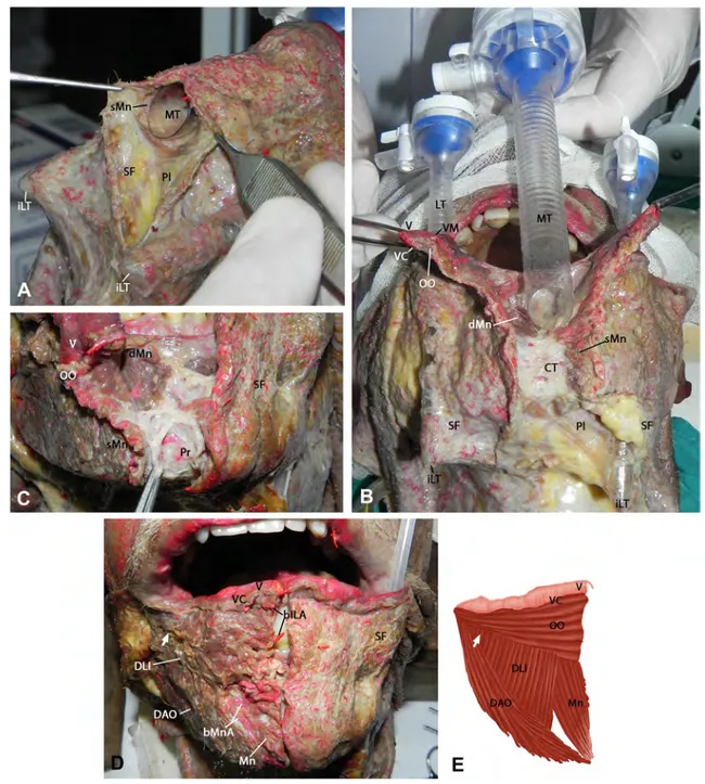
Fig. 4 Anatomical passage of median trocar. A Tip of median tro-car between epiplatysmal part of superficial fascia and platysma. B Removing the trocar and the connective tissue under it. C Deep dis-section after removing the median trocar presents superficial and deep fibres of the mentalis muscle, and below them the periosteum of the mandible. D Cadaveric muscles related to the TOETVA trocar tun-nels are shown. E Schematic diagram of muscles related to TOETVA trocars. bILA, branch of inferior labial artery; bMnA, branch of
men-tal artery; CT, connective tissue; DAO, depressor anguli oris muscle; DLI, depressor labii inferioris muscle; dMn, superficial laying fibres of mentalis muscle; iTP, inferior tip of lateral trocar; LT, lateral tro-car; Mn, mentalis muscle; MT, median trotro-car; OO, orbicularis oris muscle; Pl, platysma; Pr, periosteum; sMn, superficial laying fibres of mentalis muscle; SF, superficial fascia; V, vermilion; VC, vermilion cutaneous border; VM, vermilion mucosa border; Arrow, site of LT where it exits below OO muscle
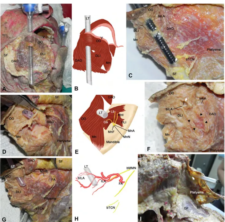
Fig. 5 Anatomical course of lateral trocar, and transmission to the subplatysmal plane. A After skin and superficial fascia remove the anatomical passage of LT is exposed. B Schematic diagram of A presents the muscles related to TOETVA trocars and the penetration site of LT. C The LT pierces depressor labii inferioris muscle and transmits to a subplatysmal plane through platysma fibres. D After superolateral deviation of the depressor labii inferioris muscle, men-tal neurovascular structures were shown under it. E Schematic dia-gram of D. F Perioral muscles and the effects of LT are presented after removing the LT. G Superior elevation of depressor anguli oris and platysma fibres to show neurovascular structures beneath them. H Schematic diagram of G. I Transmission of LT to the subplatys-mal plane without penetrating the platysma. The trocar penetrates
the superficial facia. bILA, branch of inferior labial artery; bMMB, branch of marginal mandibular branch of facial nerve; bMnA, branch of mental artery; bTCN, branch of transverse cervical nerve; DAO, depressor anguli oris muscle; DF, deep facia; DLI, depressor labii inferioris muscle; FA, facial artery; Gb, gingival branch of mental nerve; ILA, inferior labial artery; iTP, inferior tip of lateral trocar; Lb, labial branch of mental nerve; LT, lateral trocar; Mn, mentalis mus-cle; MnF, mental foramen; MnN, mental nerve; Mnt, mental branch of mental nerve; OO, orbicularis oris muscle; Pl, platysma; SF, super-ficial fascia; St, strap muscles; V, vermilion; Arrow, penetration site of LT (to DLI muscle or platysma); Black triangle, tear in depressor labii inferioris muscle; Asterisk (*), fibres of DAO muscle reach over LT LT bMMN
I
y
/
bTCNG
muscles. Then, it approximates to the lateral corner of the OO muscle and to LT (Fig. 5G, H).
Inferior labial artery (ILA): This arises from the facial artery beneath and near the lateral border of the DAO mus-cle. It goes superomedially towards the OO muscle and comes too close to LT (Fig. 5G, H). Close to the lateral edge of LT, the ILA and its branches enter the OO muscle and course medially among its lateral fibres that are for-wardly pushed by LT. These branches pass anterior to the LT. Below the level where LT exits from the DLI muscle, the medial courses of the remaining (2 or 3) branches of ILA are positioned posterior to the trocar and one of them is commonly larger (Fig. 5F–H). Here, branches are under the layer of the DLI, DAO and platysma fibres. Both side branches of ILA anastomose with each other on the median plane. The small anastomotic branches that are anterior to the OO muscle are in the superficial fascia of the lower lip. Also, the small anastomotic branches are located posterior to the OO muscle fibres, under the labial mucosa. These small branches are cauterised during MI (Fig. 1B). Large anasto-motic branches are between muscle fibres. The large anas-tomotic branches are between the fibres of the OO muscle and the small branches in the superficial fascia are pushed forward by MT (Fig. 4A, D).
Mental artery (MnA): It exits from the mental foramen located under the DLI and DAO muscles and just below the LT route (Fig. 5D, E). The MnA branches course medi-ally and superomedimedi-ally under the DLI muscle. The large branches run towards the median plane and pass between the DLI and Mn muscles fibres (Fig. 4D). The arterial anas-tomosis of large MnA branches is superficial to MT. The small MnA branches enter between the muscle fibres and lie between the periosteum and the median connective tissue structures (Fig. 4C, D).
The determined relations of the trocars with nerves are as follows:
Mental nerve (MnN): Beneath the DAO muscle and on the LT route, MnN exits from the mental foramen. Its branches accompany the mental artery branches and approach the median plane under the DLI muscle (Fig. 5D, E). The gingival MnN branch goes in a superomedial direc-tion, under the DLI muscle. The cutaneous and mental MnN branches lie transversely more medial to the chin, under the DLI muscle, just medial to the LT route (Fig. 5D, E). On the MT route, superficial to the OO and Mn muscles, gross nerve MnN branches were not observed during the anatomical dissections but they were observed histologically at zones M1–M3. This is described in detail below.
Facial nerve (FN): A superior MMB branch of the facial nerve is observed deep in the platysma. This branch crosses the facial artery above the inferior border of the mandible. More medially, just lateral to the LT direction, the superior MMB branch crosses deep to the DAO and DLI muscles (Fig. 5G,
H). This branch cannot be traced until it reaches the menton during anatomical dissections. Near the inferior border of the mandible, the inferior MMB branch is more lateral to the LT passage. FN branches were not observed macroscopically on the MT route during dissections.
Transverse cervical branch of the cervical plexus (TCN): Those branches exit from platysma more laterally to LT and lie on this muscle (in the epiplatysmal plane) away from LT (Fig. 5G, H).
Histological results and anatomical
correlations
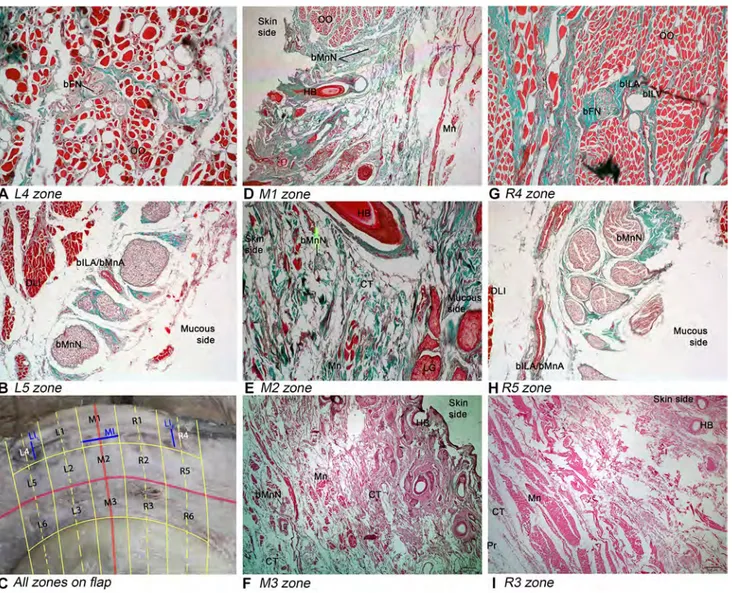
The zones M1–M3 are on the MT route. The zones on the right LT route are the R4–R6 zones and the zones on the left LT route are the L4–L6 zones. The remaining zones are positioned between the trocars.
Histologically, a great number of cutaneous MnN branches were observed near the inferior border of the OO muscle in front of the Mn muscle fibres in zone M1 (Fig. 6D). In zones M2 and M3, the sparse nerve fibres suitable for the terminal MnN branches were observed mostly between the Mn muscle and the dermis (Fig. 6E, G). The observations made during the anatomical dissections of the median connective tissue as an attachment point for the Mn muscle fibres were also proven histologically. The connective tissue occurs between the periosteum and the Mn muscle fibres in the M3 and R4 zones (Fig. 6G, K).
The R4 and L4 zones were symmetrical in histological appearance. The muscle fibres oriented in the circular direc-tion in these zones suit for the OO muscle. Nerve branches and accompanying vessels were observed between those mus-cle fibres. These large and lateral- to medial-directed nerve branches are congruent with the MMB branches of the facial nerve that extend to the innervate OO muscle (Fig. 6A, H). The accompanying vessels suit for ILA branches. The R4 and L4 zones took place at the LI projection and were pushed ante-riorly by LTs.
The R5 and L5 zones seem symmetrical. In these zones, the oblique oriented fibres suit for the DLI muscle. The large nerve fibres under DLI correspond to the proximal MnN branches, close to mental foramen. Accompanied large ves-sels to these nerve branches are considered to be either ILA or MnA branches as observed in cadaver dissections (Fig. 6B, I). Due to the thickness of the R6 and L6 zones that are under LT, trimming could not be performed effectively for histological examination.
Discussion
In this study, the surgical anatomy of the perioral, chin and submental regions for the initial TOETVA steps has been defined. The MI and LI locations and proportions, the detailed anatomical pathways, the relations of the trocars to the mimetic muscles and neurovascular structures and, finally, the histological correlations of surgical anatomy are presented.
The aim of the NOTES trials modifications, including the TOETVA surgery, is to avoid damaging the anatomical structures, such as the mental nerve, facial nerve branches and vessels, while carrying out a scarless surgery.
The onset incision for the trocar inlet at the oral ves-tibule affects the entire downwards anatomical passage. Before TOETVA, the trocars were applied through the oral vestibule and mouth floor in different combinations [2,
8, 9]. Richmond et al. placed a 1.5-cm LI for LT on the BG sulcus at the level of the first molar tooth in order to avoid MnN injury during transoral robotic-assisted thy-roidectomy [22]. It appears that the oral vestibular inci-sions, described by Anuwong for the TOETVA surgery, have been optimised [19, 23]. Our study presents further anatomical details for these optimised incisions that, to our knowledge, have not been reported previously.
Fig. 6 Histological findings. A and B present respectively upper and middle zone of the lower lip on the left LT route. D, E and F pre-sent respectively upper, middle and lower zone of the lower lip on the MT route. G, H and I present respectively upper middle and lower zones of the lower lip on the right LT route. All zones are presented for orientation in the left lower corner of the picture (C) and also see legends of Fig. 3. bFN, branch of facial nerve; bILA, branch of
infe-rior labial artery; bILV, branch of infeinfe-rior labial vein; bMnA, branch of mental artery; bMnN, branch of mental nerve; CT, connective tis-sue; DLI, depressor labii inferioris muscle; HB, hair bulb; LG, labial gland; LI, lateral incision; LIL, left lateral Incision; LIR, right lateral
incision; MI, median incision; Mn, mentalis muscle; OO, orbicularis oris muscle; Pr, periosteum; M1–M3, median zones on the flap; R1– R6, right zones of the flap; L1–L6 left zones on the flap
C All zones on flap
,/ Mucous side
ilJ
' {'.. .r F M3zone I R3zone Mucous sideThe transverse MI 1 cm in width is placed almost at the middle of the VM–BG length or, as described previously, at the midpoint of the lower lip internal surface [19]. The vertical LI 0.5 cm in width is placed opposite the second premolar tooth. The distance above LI towards VM is nearly one-third of the distance below LI towards the BG sulcus. The mean distance between the MI and LI marks, on both sides with the lower lip half-pulled, was measured at 2.3 cm. These previously unreported proportions may facilitate the practical surgical application and education of TOETVA. The usage of these incision locations has been increasing on a daily basis due to their reliability in lowering complication rates [7, 14, 19, 24, 25].
MT and LTs are in close anatomical relation with OO, M, DAO, DLI and platysma. The trocars are related to and may affect those muscles at the perioral, chin and submental regions as presented in Figs. 4, 5.
One of the candidates subjected to the trocar impacts is the OO muscle. Histologically, the OO muscle and the neu-rovascular content of its fibres were observed in M1, L4 and R4 zones that were positioned in front of all trocar (MT and LTs) passages. The OO fibres and neurovascular contents are pushed anteriorly by MT and LTs. A small group of lower OO fibres may remain behind LTs. To prevent injury of the OO muscle and FN branches, the lower OO edge should be observed with blunt dissection after performing MI and LI, before creating the tunnel with the tunnelling probe and inserting the trocars.
The MT passage through the Mn muscle was previously reported in NOTES techniques, including TOETVA [10, 19,
20, 26]. However, this passage has not been described well. In a cadaveric study about the NOTES technique, MT was passed through the Mn muscle. In that technique, the Mn muscle was dissected through a larger MI located at bottom the BG sulcus. This can potentially result in the ‘pucker-ing’ or sagging of the chin and to an aesthetic deformity of the mentum [10]. To prevent this complication during the Robotic Transoral Periosteal Thyroidectomy (TOPOT), carried out through a 2-cm MI at bottom BG sulcus, the Mn muscle was elevated with a periosteal flap using an elevator also without being seen by the surgeon. In this series, MT passed downwards under the periosteum. This technique was believed to be advantageous for the dissection of the subplat-ysmal flap [11, 27]. During the dissection of the Mn mus-cle with monopolar diathermy, a flap perforation over the chin, resulting from the transmitted heat, has been reported [28]. Understanding the anatomy of the trocar pathways is of utmost importance for preventing such complications. Our dissections show that a blindly replaced MT during TOE-TVA separates the Mn muscle fibres in both superficial and deep layers. Fibres that lie superficially pass the lateral side of the trocar and attach to the overlying skin. Deep fibres of the Mn muscle attach to the connective tissue structure
behind the MT passage. That connective tissue lies over the periosteum. It means that MT goes downwards to the sub-platysmal plane above the periosteum. Conversely, during TOPOT and the transoral robotic-assisted thyroidectomy (TRAT), the mandibular periosteum is elevated for the MT and LT placements subperiosteally to reach the subplatysmal plane [22, 27].
LT pierces the DLI muscle and laceration occurs in some fibres. This may result from the position of the muscle fibres. The DLI muscle fibres are slightly oblique according to the sagittal plane. The LT transition from the oral vestibule to the superficial DLI site is near the angle between the OO and DAO muscles. These findings have not been reported previ-ously. MT stretches only the most medial DLI fibres without laceration. In one report, the DLI fibres were shown only at the lateral edges of the retracted MI [29].
In the lower part of the mandible body, LT passes over the DAO muscle after piercing the DLI muscle (Fig. 5A, C). As shown in Fig. 5c, some of the upper medial DAO muscle fibres stretch over LT without laceration. There is only little tension in those fibres. The lowermost medial DAO fibres are below and not affected by LT (Fig. 5A–C). However, those fibres may be compressed between LT and the sharp edge of the mandible during surgical manoeuvres.
Terms like ‘premandibular space’, ‘submental pocket’ and ‘subplatysmal plane’ are common in the TOETVA and NOTES papers. ‘Premandibular space’ is a misnomer in our opinion because that area is not found in front of the mandi-ble but just below it. For this reason, the term ‘submental’ is advised to ensure an exact description. According to a paper that described the transoral video-assisted neck surgery (TOVANS), MT was placed in the subplatysmal tunnel just below the mandible [30]. In one of the NOTES cadaver stud-ies, MT was inserted through a deeply situated MI at the BG sulcus and its course to the submental space was between the two digastric muscles and the mandible. However, the position the platysma was not mentioned in that report [31]. Most reports about TOETVA describe that, during the ini-tial steps of the procedure, the working space (or submental pocket) was created beneath the platysma layer via a straight vascular tunnelling probe that was inserted through the MI to the so-called premandibular or submental space [16, 17, 19,
20]. Our study showed that MT did not penetrate the platy-sma at the submental region just below the mandible. Near the lower border of the mandible, the submental pocket or the so-called premandibular space is found above the platy-sma not under it (Fig. 7A). However, TOETVA was carried out in the subplatysmal plane at lower levels, where both sides of the platysma are further away from each other. In other words, MT is not totally in the subplatysmal plane, especially above the mandible. The platysma fibres decus-sate on different levels in the midline. In 43% to 75% of instances, the platysma fibres decussate 1–20 mm below
the mandibular border (Type 1). In 10% to 14%, there is no decussation (Type 3). The rest of platysma decussation is much below 20 mm (Type 2) [32]. Moreover, depending on the lack of platysma fibres in the midline, MT traverses the superficial fascia that is present there. In our study, the platy-smas were type 1 and MT passed to the subplatysmal plane through the superficial fascia between the non-decussated fibres. This indicates that most MTs may not penetrate the platysma during the initial TOETVA surgery phase and that the submental pocket may not be subplatysmal.
There are unreported findings regarding LT relations with the platysma. We found different ways of LT transmission to the subplatysmal plane. LT is epiplatysmal at the chin and the submental or submandibular levels, after it exits towards the superficial DLI muscle surface. Afterwards, LT reaches the subplatysmal plane by passing through the entire layer of the superficial neck fascia, piercing the platysma near its medial border.
The lower lip mimetic muscles related to the initial TOE-TVA steps are supplied mainly by the facial nerve MMB. The OO muscle is innervated by MMB (100%), buccal branch (88%) and zygomatic branch (40%), respectively [33]. MMB also innervates the DAO, DLI and Mn muscles [34, 35]. The medial course of MMB is under the platysma and parallel to the inferior mandible border [36]. In 74% of cases, MMB crosses the facial artery and lies above the
inferior mandible border as it does in our study also [35]. The anatomical LT passage is epiplatysmal in the perioral region. In our TOETVA application, the MMB course was beneath the platysma and LT passed over it. Such an LT course does not injure the nerve and proves that the inci-sions described by Anuwong are safe with respect to FN injury. Some studies advise a more lateral LT passage, run-ning under the platysma, to avoid MnN injury in one of the NOTES techniques [4, 37]. However, such approaches have the potential to injure MMB according to our findings.
We did not observe FN branches among the OO muscle fibres histologically in M1, M2 and M3 zones, unlike the R4 and L4 zones (Fig. 6). Care must be taken not to cut the OO fibres while making the MI and LIs and during the trocar placements at the beginning of the TOETVA surgery. This may reduce the facial branch injury risks among the OO fibres.
The platysma is innervated by the cervical FN branch [32,
35]. In the current study, we did not consider this nerve due to its posterior position. Nevertheless, we saw that the infe-rior MMB branch and the transverse cervical nerve branches lay lateral to LT, over the platysma (Fig. 5C).
The mental foramen, which transmits the mental nerve and vessels, is at the level of the premolar tooth. Classically, MnN is a branch of the inferior alveolar nerve that exits from the mental foramen. It lies under the DLI muscle and
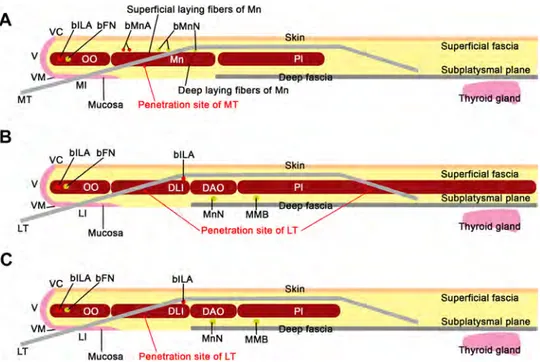
Fig. 7 Schematic diagram of the anatomical passage of the trocars used during TOETVA. A Anatomical passage and related structures of the median trocar. B, C Two different types of anatomical pas-sage and related structures of the lateral trocars. bFN, branch of facial nerve; bILA, branch of inferior labial artery; bMnA, branch of mental artery; bMnN, branch of mental nerve; DAO, depressor anguli oris
muscle; DLI, depressor labii inferioris muscle; LI, lateral incision; LT, lateral trocar; MI, median incision; MMB, marginal mandibular branch of facial nerve; Mn, mentalis muscle; MnF, mental foramen; MT, median trocar; OO, orbicularis oris muscle; Pl, platysma; V, ver-milion; VC, vermilion cutaneous border; VM, vermilion mucosa bor-der
A Superficial laying fibers of Mn
.
12::r
1
l""r
,.,
VM- • • \
µ.
,
..
,n:g=fib:e .. r•s-of• M'Ol!niep;aflnK!lt- - . a a . - - - • - - •Tliihiiiiyilliroiild•g.:la•n•d -MT Mucosa Penetration site of MTSuperficial fascia B
vc}'/N
V - VM-Superficial fasciaS
u
b
,at smal laneLI
LT Mucosa
I
Thyroid gla!)_dC VC blLA bFN
v
.p
Superficial fascia VM -LT LI MucosaI
MnN MMBcommonly gives off the gingival, mental and labial branches. MnN branches may cross the OO muscle as they spread to the centre. The mental nerve innervates the skin of the lower lip and chin, the mucosa and the gingiva of the inferior ves-tibule [38]. Many attempts have been made to determine the ultimate LI location in different NOTES techniques to avoid MnN injury. The LI for LT in endoscopic minimally inva-sive thyroidectomy (eMIT) was made at the bottom the BG sulcus, 1 cm lateral to the lower lip frenulum near the canine teeth. After this procedure, 75% of patients had paraesthe-sia due to various degrees of MnN injury [8]. In TOVANS cases, where an MI 2.5-cm deep in the BG sulcus was used, sensory disorders around the chin persisted in all patients for more than 6 months [30]. According to our anatomical find-ings, we suggest that an LT course at the level of the canine teeth, as well as wide and deep incisions at the BG sulcus, may result in MnN injuries.
The MnNs in our dissections showed a common pattern. This common pattern was previously reported by Alsaad et al. MnN crosses medially beneath the DLI, DAO and platysma muscles [38]. These muscles protect the nerve from LT’s pressure. As a result, appropriate trocar applica-tion sites during TOETVA are safe and prevent MnN injury. As shown in Fig. 3, the projection of mental foramina is in the R5 and L5 zones, where we also observed thick MnN fibres histologically (Fig. 6). The medial borders of these zones are the lines that pass from the lateral margins of the canine teeth. The R5 and L5 zones are on the LT routes. This anatomical fact can explain the high MnN injury risk during the bone close by or the subperiosteal downward LT passage. Also, this might explain the intact MnN in the TOETVA procedure [8, 13, 14].
Branches of MnN were not observed macroscopically during the MT passage. However, low numbers of small calibre branches were detected on histological samples from the M2 and M3 zones. These zones are located where MT passes downwards to the subplatysmal plane. Injury to these branches may cause the loss of sensation in the chin. The histology of the M1 zone includes a great number of MnN fibres but those are pushed forward by MT. This means that the risk of MnN injury by MT is low.
Wang et al. reported ecchymosis on the skin without nerve injury findings after ETOVA in two cases [6]. Karakas et al. reported haematomas in the chin and perioral region in a TOETVA case [25]. These complications seem to be due to the close relation of LTs with the ILA branches as shown in this study. The vessel injury risk, mostly of arter-ies, is higher with LTs than with MT because ILA and its branches cross just below the site of the LT passage through the DLI muscle. This relation is obvious in the histological samples (Fig. 6). When LT is directed towards the mandibu-lar base and grazes the bone, it is possible to injure ILA. Normally, this vessel is expected to be beneath LT during
optimal TOETVA surgery. The surgeon should be aware that LTs, which are more laterally placed to avoid MnN injury, may increase the risk of injury to ILA or the facial artery [4, 10, 22]. Although bigger in size, the risk of arterial inju-ries resulting from MT is lower than that from LT, because the anastomoses between the large ILA branches are among the OO muscle fibres and the small anastomotic branches are located in the superficial fascia. All these structures are superficial to MT and are pushed forward by the trocar (Fig. 4A, D). The arterial anastomoses between large MnA branches are also superficial to the MT. Histologically, small branches are seen in the M1 and M3 zones (Fig. 6).
We hope to facilitate practical TOETVA surgical instruc-tions and applicainstruc-tions with the outcomes of this study. Besides the mimetic muscles, it may also be possible to affect the neurovascular structures during the TOETVA pro-cedure. These findings highlight the importance of checking the function of every single mimetic muscle during post-operative care. The endocrine surgeons who are willing to deal with the TOETVA procedure should know the anatomy of the perioral region thoroughly. The patients should be informed before surgery that these muscles may be affected.
In conclusion, the surgical anatomy of the perioral, chin and submental regions for initial TOETVA-related steps were investigated and defined using cadavers. The MI and LI locations and proportions in the oral vestibule, the detailed anatomical pathways and the relations of the TOETVA tro-cars with the mimetic muscles and neurovascular structures were studied. The histological correlation of the surgical anatomy to describe possible complications for the purpose of safer surgery was presented. Since the TOETVA tech-nique is novel and its new, possible pitfalls were described; these should be well defined for patients during the informed consent procedure. An examination of the mimetic muscles in the postoperative period should be a part of patient care for those cases receiving the TOETVA surgery.
Compliance with ethical standards
Disclosure Drs. Servet Celik, Ozer Makay, Mustafa Deniz Yoruk, Ilke Bayzit Kocer, Murat Ozdemir, Kubilay Dogan Kilic, Canberk Tomruk, Okan Bilge, Yigit Uyanikgil and Gianlorenzo Dionigi declare that they have no conflicts of interest or financial ties to disclose.
References
1. Witzel K, von Rahden BH, Kaminski C, Stein HJ (2008) Tran-soral access for endoscopic thyroid resection. Surg Endosc 22:1871–1875
2. Benhidjeb T, Wilhelm T, Harlaar J, Kleinrensink GJ, Schneider TA, Stark M (2009) Natural orifice surgery on thyroid gland: totally transoral video-assisted thyroidectomy (TOVAT): report of
first experimental results of a new surgical method. Surg Endosc 23:1119–1120
3. Karakas E, Steinfeldt T, Gockel A, Schlosshauer T, Dietz C, Jager J, Westermann R, Sommer F, Richard HR, Exner C, Ses-terhenn AM, Bartsch DK (2011) Transoral thyroid and parathy-roid surgery—development of a new transoral technique. Surgery 150:108–115
4. Wilhelm T, Harlaar JJ, Kerver A, Kleinrensink GJ, Benhidjeb T (2010) Surgical anatomy of the floor of the oral cavity and the cervical spaces as a rationale for trans-oral, minimal-invasive endoscopic surgical procedures: results of anatomical studies. Eur Arch Otorhinolaryngol 267:1285–1290
5. Karakas E, Steinfeldt T, Gockel A, Westermann R, Kiefer A, Bar-tsch DK (2010) Transoral thyroid and parathyroid surgery. Surg Endosc 24:1261–1267
6. Wang C, Zhai H, Liu W, Li J, Yang J, Hu Y, Huang J, Yang W, Pan Y, Ding H (2014) Thyroidectomy: a novel endoscopic oral vestibular approach. Surgery 155:33–38
7. Makay O, Dionigi G, Celik S (2018) Scarless Thyroidectomy-Atlas of TOETVA (Transoral Endoscopic Thyroidectomy Vestibu-lar Approach) (Turkish: İzsiz Tiroidektomi Vestibüler Yaklaşim ile Transoral Endoskopik Tiroidektomi TOETVA Atlası), 1st edn. İstanbul Tıp Kitabevleri, İstanbul
8. Wilhelm T, Metzig A (2010) Video. Endoscopic minimally invasive thyroidectomy: first clinical experience. Surg Endosc 24:1757–1758
9. Wilhelm T, Metzig A (2011) Endoscopic minimally invasive thyroidectomy (eMIT): a prospective proof-of-concept study in humans. World J Surg 35:543–551
10. Richmon JD, Holsinger FC, Kandil E, Moore MW, Garcia JA, Tufano RP (2011) Transoral robotic-assisted thyroidectomy with central neck dissection: preclinical cadaver feasibility study and proposed surgical technique. J Robot Surg 5:279–282
11. Lee HY, Hwang SB, Ahn KM, Lee JB, Bae JW, Kim HY (2014) The safety of transoral periosteal thyroidectomy: results of Swine models. J Laparoendosc Adv Surg Tech A 24:312–317
12. Anuwong A (2016) Transoral Endoscopic Thyroidectomy Ves-tibular Approach: a series of the first 60 human cases. World J Surg 40:491–497
13. Anuwong A, Ketwong K, Jitpratoom P, Sasanakietkul T, Duh QY (2018) Safety and Outcomes of the Transoral Endoscopic Thy-roidectomy Vestibular Approach. JAMA Surg 153:21–27 14. Jitpratoom P, Ketwong K, Sasanakietkul T, Anuwong A (2016)
Transoral endoscopic thyroidectomy vestibular approach (TOE-TVA) for Graves’ disease: a comparison of surgical results with open thyroidectomy. Gland Surg 5:546–552
15. Yang J, Wang C, Li J, Yang W, Cao G, Wong HM, Zhai H, Liu W (2015) Complete endoscopic thyroidectomy via oral vestibular approach versus areola approach for treatment of thyroid diseases. J Laparoendosc Adv Surg Tech A 25:470–476
16. Dionigi G, Chai YJ, Tufano RP, Anuwong A, Kim HY (2018) Transoral endoscopic thyroidectomy via a vestibular approach: why and how? Endocrine 59:275–279
17. Dionigi G, Bacuzzi A, Lavazza M, Inversini D, Boni L, Rausei S, Kim HY, Anuwong A (2017) Transoral endoscopic thyroidec-tomy: preliminary experience in Italy. Updates Surg 69:225–234 18. Dionigi G, Lavazza M, Wu CW, Sun H, Liu X, Tufano RP, Kim
HY, Richmon JD, Anuwong A (2017) Transoral thyroidectomy: why is it needed? Gland Surg 6:272–276
19. Anuwong A, Sasanakietkul T, Jitpratoom P, Ketwong K, Kim HY, Dionigi G, Richmon JD (2018) Transoral endoscopic thyroidec-tomy vestibular approach (TOETVA): indications, techniques and results. Surg Endosc 32:456–465
20. Dionigi G, Lavazza M, Bacuzzi A, Inversini D, Pappalardo V, Tufano R, Kim HY, Anuwong A (2017) Transoral endoscopic
thyroidectomy vestibular approach (TOETVA): from A to Z. Surg Technol Int 30:103–112
21. Bilge O, Celik S (2017) Cadaver embalming fluid for surgical training courses: modified Larssen solution. Surg Radiol Anat 39:1263–1272
22. Richmon JD, Pattani KM, Benhidjeb T, Tufano RP (2011) Tran-soral robotic-assisted thyroidectomy: a preclinical feasibility study in 2 cadavers. Head Neck 33:330–333
23. Anuwong A, Kim HY, Dionigi G (2017) Transoral endoscopic thyroidectomy using vestibular approach: updates and evidences. Gland Surg 6:277–284
24. Inabnet WB 3rd, Fernandez-Ranvier G, Suh H (2018) Transoral endoscopic thyroidectomy—an emerging remote access technique for thyroid excision. JAMA Surg 153:376–377
25. Karakas E, Anuwong A, Ketwong K, Kounnamas A, Schopf S, Klein G (2018) Transoral thyroid and parathyroid surgery: imple-mentation and evaluation of the transoral endoscopic technique via the vestibular approach (TOETVA). Chirurg 89:537–544 26. Clark JH, Kim HY, Richmon JD (2015) Transoral robotic thyroid
surgery. Gland Surg 4:429–434
27. Lee HY, Richmon JD, Walvekar RR, Holsinger C, Kim HY (2015) Robotic transoral periosteal thyroidectomy (TOPOT): experience in two cadavers. J Laparoendosc Adv Surg Tech A 25:139–142 28. Bakkar S, Al Hyari M, Naghawi M, Corsini C, Miccoli P (2018)
Transoral thyroidectomy: a viable surgical option with unprec-edented complications—a case series. J Endocrinol Invest 41:809–813
29. Zhang M, Xie Q, Wang Y, Wang P (2017) Surgical anatomy for transoral endoscopic thyroid surgery through vestibular approach (TOETVA). Ann Thyr 2:1–7
30. Nakajo A, Arima H, Hirata M, Mizoguchi T, Kijima Y, Mori S, Ishigami S, Ueno S, Yoshinaka H, Natsugoe S (2013) Trans-oral video-assisted neck surgery (TOVANS). A new transoral tech-nique of endoscopic thyroidectomy with gasless premandible approach. Surg Endosc 27:1105–1110
31. Park JO, Kim MR, Kim DH, Lee DK (2016) Transoral endo-scopic thyroidectomy via the trivestibular route. Ann Surg Treat Res 91:269–272
32. Kim HJ, Hu KS, Kang MK, Hwang K, Chung IH (2001) Decus-sation patterns of the platysma in Koreans. Br J Plast Surg 54:400–402
33. Hwang K, Jin S, Hwang SH, Chung IH (2006) Innervation of upper orbicularis oris muscle. J Craniofac Surg 17:1116–1117 34. Mitsukawa N, Moriyama H, Shiozawa K, Satoh K (2014) Study
on distribution of terminal branches of the facial nerve in mimetic muscles (orbicularis oculi muscle and orbicularis oris muscle). Ann Plast Surg 72:71–74
35. Saylam C, Ucerler H, Orhan M, Uckan A, Ozek C (2007) Locali-zation of the marginal mandibular branch of the facial nerve. J Craniofac Surg 18:137–142
36. Batra AP, Mahajan A, Gupta K (2010) Marginal mandibular branch of the facial nerve: an anatomical study. Indian J Plast Surg 43:60–64
37. Cai C, Huang Y, Zhang T, Chai L, Wang G, Shi L, Wiegand S, Güldner C, Günzel T, Wilhelm T (2015) Anatomical study of surgical approaches for minimally invasive transoral thyroid-ectomy: eMIT and TOPP. Minim Invasive Ther Allied Technol 24:340–344
38. Alsaad K, Lee TC, McCartan B (2003) An anatomical study of the cutaneous branches of the mental nerve. Int J Oral Maxillofac Surg 32:325–333
Publisher’s Note Springer Nature remains neutral with regard to jurisdictional claims in published maps and institutional affiliations.
Affiliations
Servet Celik1 · Ozer Makay2 · Mustafa Deniz Yoruk3 · Ilke Bayzit Kocer1 · Murat Ozdemir2 · Kubilay Dogan Kilic4 ·
Canberk Tomruk4 · Okan Bilge1 · Yigit Uyanikgil4,5 · Gianlorenzo Dionigi6
Ozer Makay
[email protected] Mustafa Deniz Yoruk [email protected] Ilke Bayzit Kocer [email protected] Murat Ozdemir
[email protected] Kubilay Dogan Kilic [email protected] Canberk Tomruk [email protected] Okan Bilge [email protected] Yigit Uyanikgil [email protected] Gianlorenzo Dionigi [email protected]
1 Faculty of Medicine Department of Anatomy, Ege
University, 35100 Bornova, Izmir, Turkey
2 Faculty of Medicine Department of General Surgery, Ege
University, 35100 Bornova, Izmir, Turkey
3 Faculty of Medicine, Department of Anatomy, Mugla
Sitki Kocman University, Kotekli Quarter, Marmaris Road Boulevard, No: 50, 48000 Mentese, Mugla, Turkey
4 Faculty of Medicine Department of Histology
and Embryology, Ege University, 35100 Bornova, Izmir, Turkey
5 Cord Blood, Cell and Tissue Research and Application
Center, Ege University, 35100 Bornova, Izmir, Turkey
6 Division of Endocrine and Minimally Invasive Surgery,
“G. Martino” University Hospital, Messina University, Sicily, Italy