A Giant Bladder Stone In A Middle-Aged Female Patient Without
Any Predisposing Factor: Case Report
Orta Yaș Bayan Hastada Predispozan Faktör Olmaksızın Olușan Dev Mesane Tașı: Olgu Sunumu
Mirze Bayındır
1,Berkan Reșorlu
1,Selçuk Sarıkaya
1,Erhan Șahin
1,Ali Ünsal
11 Keçiören Education and Research Hospital, Department of
Urology Mesane tașları tüm idrar yolu tașı hastalıklarının %5'ini olușturmaktadır. Genellikle prostat
büyümesi, nörojenik ișeme bozukluğu gibi infravezikal obstrüksiyondan dolayı yașlı erkeklerde görülür. Enfeksiyon, yabancı cisim, kadınlarda nadiren gebelik ve anti inkontinans cerrahisi de mesane tașı için predispozan faktör olabilir. Nadiren herhangi bir predispozan faktörleri olmayan çocuk ve genç sağlıklı hastalarda görülebilir ve bu duruma “endemik mesane tașı “ denir. Biz de endemik dev mesane tașı olan ve perkütan sistolitotomi gerçekleștirilen 43 yașındaki bayan hastayı sunduk. Taș boyutu 61x56 mm olarak ölçüldü. Taș analizi kalsiyum-oksalat olarak raporlandı. Hastamızda tüm araștırmalara rağmen etyolojiye yönelik bir neden yoktu ancak hasta Çin'den gelen bitkisel bir hap kullanmıștı. Bu olgu sunumunun amacı, mesane tașının herhangi bir predispozan faktörü olmayan orta yașlı hastalarda gelișebileceği gerçeğini vurgulamaktır.
Anahtar Sözcükler: Dev mesane tașı, predispozan faktör, tedavi yaklașımı
Bladder stones account for 5% of all urinary tract’s stone diseases. They are usually seen in older men because of infravesical obstructions such as prostate hyperplasia and neurogenic voiding dysfunction. Infection, foreign bodies, anti-incontinence surgery in woman can also be predisposing factors for bladder calculus. They can rarely be seen in children and young, healthy patients without any predisposing factors and are then called “endemic bladder calculi”. In this case report we present a 43 year old woman who has had an endemic giant bladder calculus and on whom we performed percutaneous cystolithotomy. The stone measured 61x56 mm in size. It was composed of calcium oxalate. In our patient, despite all the researches there was no etiological cause but the patient, had used herbal pills that came from China. The aim of this case report is to emphasize the fact that bladder calculi can develop in middle-aged patients without any predisposing factors.
Key Words: Giant bladder Stone, predisposing factor, treatment strategy
Bladder stones compose %5 of the urinary tract stones and they generally develop secondary to the outflow tract obstruction of bladder, voiding dysfunction, foreign body or recurrent urinary tract infection (1, 2). It is more often seen in the developing nations than the developed. Also it is more frequently seen in males older than 50 years and in females after urinary incontinence surgeries (3).
Bladder stones generally develop secondary to the outflow tract obstructions of the bladder and recurrent urinary tract infection, hematuria, urine retansion are
accompanied. Bilateral ureterohydronephrosis, and sometimes chronic renal failure also can be seen in these patients. Therapy must be planned after the etiology is identified. Otherwise the therapy will be insufficient. Endemic bladder stones are frequently
seen in Turkey and Iran, but also seen in Thailand, Indonesia, and
Papua New Guinea. Endemic
bladder stones are seen more often in males and it peaks at the age of five. They are rarely composed in kidney and fall into the bladder. These stones are generally uric acid or calcium oxalate stones (4). The
Ankara Üniversitesi Tıp Fakültesi Mecmuası 2012, 65 (3)
DOI: 10.1501/Tıpfak_000000832 CERRAHİ TIP BİLİMLERİ/SURGICAL SCIENCES
Olgu Sunumu / Case Reports
Received: 10.04.2012 Accepted: 19.06.2013 Corresponding author
Dr. Mirze Bayındır Phone: 356 90 00/1072 E-Mail: [email protected]
Keçiören Education and Research Hospital, Department of Urology
Ankara Üniversitesi Tıp Fakültesi Mecmuası2012, 65 (3)
A Giant Bladder Stone In A Middle-Aged Female Patient Without Any Predisposing Factor: Case Report
180
etiology is not identified exactly but the dietary factors are accused. In this case report, a giant bladder
stone of a mid-aged woman without any predisposing factor is presented with the literature related to it.
CASE REPORT
A 43 year-old female patient was admitted with complaints of hematuria and dysuria. Urinalysis
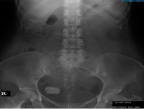
result revealed; 43 erythrocytes, 14 leukocytes/ml. Abdominal plain film showed a 60 mm diameter opacity compatible with the stone at the right side of the bladder wall (Figure 1). Urinary system ultrasonography showed a 61x56 mm stone at the base part of the bladder with a posterior acoustic shadow (Figure 2). The upper urinary tract was normal and there was no dilatation of the collecting system. Postvoiding residual volume was 20 cc. There was no
other features about the patient’s history except hematuria and dysuria. The patient had a C/S history for two times and didn’t have any other urinary or pelvic surgery history.
At first we used cefuroxime axetil, a second–generation cephalosporin, for the urinary tract infection of the patient. After the urine was sterile; we performed a filling cystometry and a pressure flow study. The capacity of the bladder was 510 cc and there wasn’t any over-active contractions and voiding dysfunction and the results of the uroflowmetry was; Qmax: 27 Qaverage: 11.
On cystoscopic examination; the urethra was normal but mucosa of the bladder was edematous and hyperemic. There was not any obstruction findings such as increased trabeculation or diverticules in the bladder.
Afterwards percutaneus cystolit hotomy was performed and the stone was fragmented with the ultrasonic lithotriptor, a sample was taken for the stone analysis. Both a cystostomy catheter and an urethral foley catheter was placed. On postoperative first day the cystostomy catheter and on the second day the foley catheter were removed and the patient was discharged. The result of the stone analysis was reported as a calcium oxalate stone.
In the first control at postoperatively second month, 24-hour urine parameters and blood parathormone levels were analyzed and all were found to be normal. The follow-up period was 18 months and patient was seen on every 6th month. At each visit, urinanalysis, serum creatinine analysis, plain film and abdominal ultrasonography were performed. No urinary tract infection or new stone formation was seen in this period.
Figure 1 : A giant stone formation in bladder.
Journal of Ankara University Faculty of Medicine 2012, 65 (3)
Mirze Bayındır, Berkan Reșorlu, Selçuk Sarıkaya, Erhan Șahin, Ali Ünsal 181
DISCUSSION
The first bladder stone was determined in 4800 BC at a male child patient (5). Endemic bladder stones are usually seen in child patients however it is rare in developed countries as a result of healthy eating and antimicrobial therapies. It is more often seen in the developing countries.
Bladder stones can be determined when researching the symptoms of a patient with lower urinary tract sypmtoms. The common symptoms of primary bladder stones are intermittant or continuous hematuria and suprapubic pain. The most common symptom is dysuria. In our patient the predominant symptoms were hematuria and dysuria.
Although urinary system stone diseases are frequently seen in the community, the bladder stone disease is rare. In male patients, underlying causes are usually infravesical obstructions, urinary
tract infections and foreign bodies. In our patient despite all the researches no etiological cause was identified, only that the patient had used herbal pills that came from China. As we were unable to contact with other patients using these pills the place of this drug in the etiology of this disease remains undetermined.
Extracorporeal shock wave
lithothripsy (ESWL), cystolithotomy, endoscopic cystolithotomy (transurethral or percutaneous) can be used for the treatment of bladder stones. The success rate of ESWL ranges from %72 to %100 and it is a suitable method for patients with severe comorbidities(6). However, ESWL has a low success rate in large bladder stones.
The success rate of open surgery is %100 (7), the main limitation of this procedure is its invasiveness. Both cystoscopy and endoscopic cystolithotomy are the most suitable treatment options for
these stones, as the bladder underlying obstructive pathologies can be checked by this method. When the stone diameter is over 5 cm, percutaneous cystolithotomy and open surgery are more effective. In this patient, we dilated to 24 F under cystoscopic vision then we used a 22 F nephroscope. The stone was fragmented with the ultrasonic lithotriptor. The treatment option is decided depending on the patient’s age, sex, stone size and the presence of infravesical obstruction.
Although the bladder stones are mostly seen in patients with
infravesical obstruction, neurogenic bladder, bladder diverticulum, chronic bacteriuria, upper urinary system stones and foreign bodies, it can also be seen in patients without an etiologic factor. Treatment of the bladder stone is not only extracting the stone. The etiological cause must also be cleared and treated. Otherwise recurrent bladder stones would be seen.
REFERENCES
1. Hammad FT, Kaya M, Kazim E.
Bladder calculi: Did the clinical picture change? Urology. 2006;67:1154-1158
2. Rahman M, Uddin A, Das GC, et al. A
giant vesical calculus. Mymensingh Med J. 2007;16(2 Suppl):57-59.
3. Razvi HA, Song TY, Denstedt JD:
Management of vesical calculi: Comparison of lithotripsy devices. J Endourol 1996; 10:559-563
4. Andersen DA. The nutritional
significance of bladder stones. Br J Urol 1962;34:160-177.
5. Papatsoris AG, Varkarakis I, Dellis A,
Deliveliotis C. Bladder lithiasis: From open surgery to lithotripsy. Urol Res 2006;34:163-167.
6. Al-Ansari A, Shamsodini A, Younis N,
et al. Extracorporeal shock wave lithotripsy
monotherapy for treatment of patients with urethral and bladder stones presenting with acute urinary retention. Urology 2005;66:1169-1171.
7. Takasaki E, Suzuki T, Honda M, et al.
Chemical compositions of 300 lower urinary tract calculi and associated disorders in the urinary tract. Urol Int 1995;54:89-94.
Ankara Üniversitesi Tıp Fakültesi Mecmuası2012, 65 (3)
A Giant Bladder Stone In A Middle-Aged Female Patient Without Any Predisposing Factor: Case Report