www.bjorl.org
Brazilian
Journal
of
OTORHINOLARYNGOLOGY
ORIGINAL
ARTICLE
The
role
of
nasopharyngeal
examination
and
biopsy
in
the
diagnosis
of
malignant
diseases
夽
Necmi
Arslan
a,
Arzu
Tuzuner
a,
Alper
Koycu
b,∗,
Songul
Dursun
a,
Sema
Hucumenoglu
caUniversityofHealthSciences,AnkaraTrainingandResearchHospital,DepartmentofOtolaryngology,HeadandNeckSurgery, Ankara,Turkey
bBaskentUniversityHospital,DepartmentofOtolaryngology,HeadandNeckSurgery,Ankara,Turkey
cUniversityofHealthSciences,AnkaraTrainingandResearchHospital,DepartmentofPathology,Ankara,Turkey
Received10January2018;accepted6April2018 Availableonline15May2018
KEYWORDS Nasopharynx carcinoma; Nasopharynxbiopsy; Endoscopic nasopharyngoscopy Abstract
Introduction:Indirectproportiontotheincreasingrateofnasopharynxexaminationsapplied, the earlydiagnosisand treatmentoflesions inthisregionispossible. Attimes theclinical findingsandthebiopsyresultsarenotconsistent,sobiopsiesmayhavetoberepeated.
Objectives: Theaimofthisstudy wasto evaluatethedistributionofpathologytestresults obtained from cases of nasopharynx biopsy, to determine with which methods determina-tion mostoften was made, andto investigate which kinds ofcases required the biopsy to berepeated.
Methods:Thestudyincludedatotalof1074patients(500female,574male)whounderwent
nasopharyngeal biopsyinourclinicbetween June2011andJune2017.Data wereobtained frompatientrecordsofage,gender,clinicalfindings,imagingfindingsifavailableand patho-logical diagnosis. The pathologicaldiagnoseswere separated into 3maingroups as chronic nasopharyngitis,benigncytologyandmalignantcytology.
Results:The examinations resultedin 996cases reportedas chronicnasopharyngitis, 47 as benigncytologyand31asmalignantcytology.Ofthe31malignantlesions,diagnosiswasmade in15patients(48.4%)withasinglebiopsy,andin16patients(51.6%),asaresultofthepathology reportwhen2ormorebiopsiesweretaken.Inthecomparisonofthebenignandmalignant lesions inrespect oftheneedfor repeatedbiopsies,thecasesdeterminedwithmalignancy werefoundtohaveastatisticallysignificantlyhigherrateofrepeatedbiopsy(p<0.001).
夽 Pleasecitethisarticleas:ArslanN,TuzunerA,KoycuA,DursunS,HucumenogluS.Theroleofnasopharyngealexaminationandbiopsy inthediagnosisofmalignantdiseases.BrazJOtorhinolaryngol.2019;85:481---5.
∗Correspondingauthor.
E-mail:[email protected](A.Koycu).
PeerReviewundertheresponsibilityofAssociac¸ãoBrasileiradeOtorrinolaringologiaeCirurgiaCérvico-Facial.
https://doi.org/10.1016/j.bjorl.2018.04.006
1808-8694/©2018Associac¸˜aoBrasileiradeOtorrinolaringologiaeCirurgiaC´ervico-Facial.PublishedbyElsevierEditoraLtda.Thisisanopen accessarticleundertheCCBYlicense(http://creativecommons.org/licenses/by/4.0/).
Conclusion:Incomparisonwithcasesofbenigntumor,astatisticallysignificantlygreater num-berofrepeatedbiopsieswererequiredincasesdiagnosedasmalignanttumorstoconfirmthe pathologicaldiagnosisorwhentherewascontinuedsuspicionofmalignancy.Therefore,when thereisclinicalsuspicion,eveniftherearenofindingsofmalignancyonthefirstbiopsy,the biopsyshouldberepeatedexpeditiously.
© 2018 Associac¸˜ao Brasileira de Otorrinolaringologia e Cirurgia C´ervico-Facial. Published by Elsevier Editora Ltda. This is an open access article under the CC BY license (http:// creativecommons.org/licenses/by/4.0/). PALAVRAS-CHAVE Carcinomade nasofaringe; Biópsiade nasofaringe; Nasofaringoscopia endoscópica
Opapeldoexameclínicoedabiópsiadenasofaringenodiagnósticodedoenc¸as malignas
Resumo
Introduc¸ão:Emproporc¸ãodiretaàtaxacrescentedeexamesdenasofaringequesãofeitos,o diagnósticoprecoceeotratamentodelesõesnessaregiãotêmsidopossíveis.Nemsempreos achadosclínicoseosresultadosdaprimeirabiópsiasãoconsistentes,levandoànecessidadede biópsiasrepetidas.
Objetivos: Avaliaradistribuic¸ãodosresultadosdostesteshistopatológicosobtidospelabiópsia denasofaringe,determinarquaismétodosforammaisfrequentementeusadosnaidentificac¸ão einvestigaroscasosnosquaisabiópsiaprecisouserrepetida.
Método: Oestudoincluiu1.074pacientes(500mulheres,574homens)submetidosabiópsia
denasofaringeemnossaclínicaentrejunhode2011ejunhode2017.Osdadosforamobtidos dosprontuáriosdospacienteseincluíramidade,sexo,achadosclínicos,achadosdeimageme diagnósticohistopatológico.Osdiagnósticoshistopatológicosforamseparadosemtrêsgrupos principaiscomonasofaringitecrônica,citologiabenignaecitologiamaligna.
Resultados: Osexamesresultaramem996casoslaudadoscomonasofaringitecrônica,47como citologiabenignae31comocitologiamaligna.Das31lesõesmalignas,odiagnósticofoifeito em15(48,4%)comumaúnica biópsiaeem 16(51,6%),quandoduasoumaisbiópsiasforam realizadas.Nacomparac¸ãodaslesõesbenignasemalignasemrelac¸ãoànecessidadedebiópsias repetidas,oscasosdeterminadoscomosendomalignosmostraramumataxaestatisticamente maiordebiópsiarepetida(p<0,001).
Conclusão:Emcomparac¸ãocomoscasosdetumoresbenignos,um númeroestatisticamente
maiordebiópsiasrepetidas foinecessárioem casosdiagnosticadoscomo tumoresmalignos, para confirmac¸ãodo diagnósticohistopatológico ounasuspeitacontinuada demalignidade. Portanto,quandohásuspeitaclínica,mesmoquenãohajaachadosdemalignidadenaprimeira biópsia,eladeveserrepetidatãologosejapossível.
© 2018 Associac¸˜ao Brasileira de Otorrinolaringologia e Cirurgia C´ervico-Facial. Publicado por Elsevier Editora Ltda. Este ´e um artigo Open Access sob uma licenc¸a CC BY (http:// creativecommons.org/licenses/by/4.0/).
Introduction
Severalmalignantandbenigntumorsareencounteredinthe nasopharynx,andeven thoughfindings suchasotitis with effusionoramassintheneckmaybepresent,lesionsmay have an asymptomatic course until an advanced stage.1---3
Endoscopicnasopharyngoscopyappliedintheear,noseand throat examination can increase the rates of diagnosing nasopharynxcanceratanearlystage.4,5
The most frequently encountered findings in symp-tomatic cases are nasal obstruction, otitis media with effusionandepistaxis;endoscopicnasopharyngoscopyisthe gold standard inclinical examination.6 In thepresence of
findingssuchaschronicmiddleeareffusion,amassonthe neckorasuspiciousappearancedeterminedonendoscopic
nasopharyngeal examination, investigation of malignancy withabiopsycanbenecessary,evenifnospecificfocusis seen.Althoughmanydifferentscanningandspecific exami-nationmethodshavebeendescribedinthedeterminationof nasopharyngealmalignancies,biopsyunderendoscopy guid-ance remains indispensable for both adefinitivediagnosis andpathologicalclassification.7Whentheclinical
examina-tion findings and the biopsy results are notconsistent, it maybenecessary torepeatthebiopsywithoutanylossof time.
Theaimofthisstudywastoevaluatethedistributionof pathologytest resultsobtainedfromcasesofnasopharynx biopsy, to determine with which methods determination mostoftenwasmade,andtoinvestigatewhichkindsofcases requiredrepeatbiopsy.
Methods
This retrospective study included the records of 1074 patients (574 males, 500 females) who underwent endo-scopic transnasal nasopharynx biopsy between June 2011 and June 2017 in the Ear, Nose and Throat (ENT) Clinic of Ankara Training and Research Hospital. Permission for thestudywasgrantedbytheEducation Planning Commis-sion of Ankara Training and Research Hospital (Ref n◦ 0670/5618-04.01.2017). In the presence of findings such as chronic middle ear effusion, a mass in the neck or a suspicious appearance determined on endoscopic naso-pharynx examination, transnasal endoscopic nasopharynx biopsywasperformed. Patientsinwhomgeneral anesthe-siaconstitutedahighriskand/orthosewhoacceptedlocal anesthesia underwent endoscopic transnasal nasopharynx biopsies under local anesthesia in the operating theatre. Inother cases, thepatients wereoperated onunder gen-eral anesthesia. The patient records were examined and data were recorded including age, gender, clinical find-ings (nasal obstruction, otitis media with effusion, mass in the neck), imaging findings if available and patholog-ical diagnosis. Immunohistochemical analysis was applied to all patients to define the tumor type. The pathologi-caldiagnoses wereseparatedinto 3main groups: chronic nasopharyngitis,benigncytologyandmalignantcytology.In cases wherethe material wasinsufficient for a pathology report,thosepatientswere excludedfromthe study.Two ormorecoreandtransitionaldeepbiopsieswereobtained fromall patients. As the tumor localization wasnot fully specifiedintherecordsofallpatients,noevaluationcould bemadeofthenasopharyngealtumorsite.
Statistical analyses of the data were made using IBM SPSSfor Windows Version 22.0software.Numerical varia-bles were stated as mean±standard deviation (SD) and minimum---maximum values, and categorical variables as number (n) and percentage (%). Differencesbetween the malignantand benigngroups wereexamined withtheChi squaretest.Avalueofp<0.05wasacceptedasstatistically significant.
Results
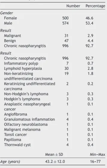
Atotal of 904,609patients were examinedbetween June 2011 and June 2017 in the Ear, Nose and Throat (ENT) Clinic of Ankara Training and Research Hospital. Endo-scopic transnasal nasopharynx biopsy was performed on 1074 patients (ratio of all examined patients: 0.118%). Allpatientsunderwentendoscopictransnasal nasopharynx biopsiesundergeneralorlocalanesthesiaintheoperating theatreandnomajorcomplications wererecorded,other thanminorbleedingthatwascontrolledwithbipolar cau-terisation. The patients comprised574 (53.4%) males and 500(46.6%) femaleswithamean ageof43.2±12.0years (range16---77years).Nodifferencewasdeterminedbetween thegroupsinrespectofgender.Theexaminationsresulted in 996 (92.7%) cases reportedas chronic nasopharyngitis, 47 (4.4%) as benign cytology and 31 (2.9%) as malignant cytology.The numbersandpercentages ofthebenign and malignantdiagnosesareshowninTable1.
Table1 Demographicdata.
Number Percentage Gender Female 500 46.6 Male 574 53.4 Result Malignant 31 2.9 Benign 47 4.4 Chronicnasopharyngitis 996 92.7 Result Chronicnasopharyngitis 996 92.7 Inflammatorypolyp 7 0.7 Lymphoidhyperplasia 30 2.8 Non-keratinizing undifferentiatedcarcinoma 19 1.8 Keratinizingundifferentiated carcinoma 2 0.2 Non-Hodgkin’slymphoma 3 0.3 Hodgkin’slymphoma 3 0.3 Anaplasticnasopharyngeal cancer 1 0.1 Angiofibroma 1 0.1 Granulomatousinflammation 4 0.4 Olfactoryneuroblastoma 1 0.1 Malignantmelanoma 1 0.1 Tonsilcancer 1 0.1 Papilloma 1 0.1 Thornwaldcyst 4 0.4 Mean±SD Min---Max Age(years) 43.2±12.0 16---77
Table2 Distributionofmalignantlesions.
Number Percentage Non-keratinizing undifferentiated carcinoma 19 61.3 Keratinizing undifferentiated carcinoma 2 6.5 Non-Hodgkin’slymphoma 3 9.7 Hodgkin’slymphoma 3 9.7 Anaplastic nasopharyngealcancer 1 3.2 Olfactoryneuroblastoma 1 3.2 Malignantmelanoma 1 3.2 Tonsilcancer 1 3.2
Thepathologyreportsofmalignantdiagnosisin31cases includednon-keratinizingundifferentiatedcarcinomain19 (61.3%), anaplastic nasopharynx carcinoma in 1 (3.2%), keratinizing undifferentiated carcinoma in 2 (6.5%), non-Hodgkin’s lymphoma in 3 (9.7%), Hodgkin’s lymphoma in 3 (9.7%), olfactory neuroblastoma in 1 (3.2%), malignant melanoma metastasis in 1 (3.2%) and metastasis of tonsil carcinomain1(3.2%)(Table2).
Table3 Statisticalanalysisofthefindingsofthephysical examination,numberofbiopsiesandimagingmethodsused.
Malignant lesions Benign lesions p Imaging MRI --- 8 NA CT 2 4 Findings Effusion 2 --- NA
Massintheneck 6 3 Nasalobstruction 4 7 Preseptalcellulitis 1
---Numberofbiopsies
1 15(48.4%) 46(97.9%) <0.001 >1 16(51.6%) 1(2.1%)
At the time of diagnosis of malignant lesions, com-putedtomography(CT)wasrequestedin only2casesand in the remainder; the diagnosis was made as a result of existing symptoms and findings causing suspicion in the clinical examination. In the cases of benign lesions, a biopsywastakenafterthelesionwasdeterminedon mag-netic resonance imaging (MRI) in 8 cases and on CT in 4 (Table3).
Ifamassintheneckconformingtomalignancycriteria was discovered during the examination and neck imag-ing,or ifpersistentmiddleeareffusionwithan ipsilateral orcontralateral nasopharygealmass wasdetected,biopsy wasrepeated without any loss of time. Of the 31 malig-nant lesions, diagnosis was made in 15 (48.4%) with a single biopsy, and in 16 (51.6%) the pathological diagno-sis was reported as malignant when more than 1 biopsy wastaken at different times. The diagnosis of malignant lesionsin16 cases withrecurrentbiopsy (2or 3 separate biopsies)werenon-keratinizingundifferentiatedcarcinoma in12 (75%)cases,Hodgkin’slymphoma in3 (18.75%),and non-Hodgkin’s lymphoma in 1 (6.25%). Of the 5 patients from whom 3 separate biopsies at different times were taken, the definitive diagnosis was malignant in 3 cases as follows; non-keratinizing undifferentiated carcinoma, Hodgkin’slymphoma,andnon-Hodgkin’slymphoma.Ofthe benigncases,diagnosiswasmadefromasecond biopsyin only 1 case. In the comparison of the benign and malig-nantlesionsin respectofthe needfor repeatedbiopsies, thecasesdeterminedwithmalignancywerefoundtohave a statistically significantly higherrate of repeatedbiopsy (p<0.001)(Table3).
Discussion
Nasopharynx carcinoma has differentcharacteristics from other head and neck epithelial tumors in respect of epi-demiology,histopathology,findings,diagnosisandtreatment modalities.8 As nasopharynx carcinoma is a tumor which
growssilentlyandhasadeeplocationextending fromthe region,it isanadvancedstagetumor atthetimeof diag-nosisofseveraldiseases.9,10 Severaldifferentscanningand
specificexaminationmethodshavebeendescribedsuchas
theserologicalexaminationofEpstein---Barrvirusantibodies andsmearsamplinginadditiontotheuseofMRIandCTas imagingmethods.However,biopsyunderendoscopic guid-anceremainsindispensablebothfordefinitivediagnosisand forpathologicalclassification.7---10
Thesensitivityofendoscopicimaginginthediagnosisof nasopharynxcarcinomahasbeenshowntobeover90%.6In
thecurrentstudy,ofthe1074patientswhounderwent naso-pharynxbiopsyunderendoscopyguidance,imagingmethods wereonlyappliedto14,asCTin6casesandMRIin 8.In themajority,diagnosiswasmadefrombiopsywhena suspi-ciouslesionwasobservedonendoscopicnasopharyngoscopy or fromblind nasopharynx biopsy when there was persis-tentunilateral chronicotitismediaintheexamination,or thepresenceofamalignantmasswhichwasobviouslynot primary. However,in the literaturetherearereports that advocatethe necessity forimaging beforebiopsyin cases of clinical suspicion.6---11 According to some publications,
imagingis necessary tobeabletodiscountmalignancy in cases of suspicious lesions such as those discovered with mild swellingor asymmetrydeterminedin theendoscopic examinationandthusthepatientcanbesparedan unnec-essaryinvasiveinterventionandalossofworkforcecanbe avoided.11Kingetal.6reportedanaccuracyrateof95%for
nasopharynxMRIinprimarynasopharynxcancers.
InastudybyBercinetal.11of983nasopharynxbiopsies,
45(4.6%)werereportedasmalignant.Inthecurrentstudy, therateofmalignancywasfoundtobe2.9%.Althoughthe rateofmalignancyinthecurrentstudywaslower,theresults aresimilar.Thereasonforthedifferencecanbeexplained by the fact that the first preference of Bercin et al. for suspicious nasopharynx lesions (mild swellingor asymme-try)wastoapplyMRIexaminationsratherthanbiopsyand ifcontrastinvolvement,invasionofsofttissueorerasureof bordersweredeterminedontheMRIreport,thenabiopsy wasperformed.
AccordingtoLiuetal.,12whilemalignantlymphomasare
the second mostcommon headand necktumor, the most common location of their extra-nodal involvement is the Waldeyer lymphatic ring. In the current study, lymphoma (n=6)wasdeterminedtobethesecondmostcommon malig-nanttumor,whichwasconsistentwithliterature.
Of the 31 malignant lesions in the current study, the pathological diagnosis of 16 (51.6%) could only be con-firmedwithrepeatedseparatebiopsiesatdifferenttimes. The diagnosis ofmalignantlesionsof 16caseswith recur-rentbiopsy(2or3separatebiopsies)werenon-keratinizing undifferentiatedcarcinomain12(75%)cases,Hodgkin’s lym-phoma in 3 (18.75%), and non-Hodgkin’s lymphoma in 1 (6.25%).Ahigherriskforrecurrentbiopsywasdetermined fornon-keratinizingundifferentiatedcarcinoma.Inthe com-parison ofthe benign and malignantlesions in respectto theneedforrepeatedbiopsies,thecasesdeterminedwith malignancywerefound tohavea statisticallysignificantly higherrateofrepeatedbiopsy(p<0.001).Ifthephysician hassuspicionofmalignancybecauseof eitherthe appear-anceofthelesiononendoscopicnasopharyngoscopyorother clinical findings, when the suspicion persists even if the results of the first biopsy are benign or there is chronic nasopharyngitis,thendespiteanincreaseinpatient morbid-ityandcost-effectiveness,itcanbeconsideredappropriate torepeatthebiopsywithoutanylossoftime.Itisclearthat
early diagnosisand treatmentof thepatientwillincrease survivalrates.
Bercinetal.11reportedthesensitivityofMRItobe88.2%
in nasopharynx malignancies and the sensitivity of endo-scopic biopsy to be 84.4%. This can be attributed to not applying imaging first in cases where problems could be experiencedin determiningthe lesionlocation,especially intumorswithadeeplocationandthattherecouldbe neg-ativebiopsyresults.Thereasonthattherewasaneedfor suchahighrateofrepeatedbiopsiestobeabletoreacha diagnosisofmalignancyinthecurrentstudycanbe consid-eredtobethatthelesionlocationwasnotdeterminedwith preoperativeimagingorthatthebiopsywasnottakenfrom asufficientdepth.
Alimitationofthecurrent studywasthatpreoperative imagingwasnotappliedroutinelytoallpatientswitha sus-picious lesion or to those whowere being considered for biopsy.Iftherehadbeenasufficientnumberof preopera-tiveimages,thesefindingscouldhavebeencomparedwith theendoscopicbiopsyresults.Undercurrent conditionsin Turkey, asthere is a lengthywaiting timefor an imaging appointment at our hospital,it is preferred toperform a biopsyinashorttimetoreducethespreadofthedisease andprovidediagnosisandtreatmentquickly,and,when sus-picionofmalignancypersists,totakerepeatedbiopsies.
Conclusion
Inthevastmajorityofthepatientsinthisstudydiagnosed withmalignantandbenignlesions,thedeterminationofthe lesiononthepre-diagnosticendoscopicnasopharyngoscopy imagewasseen tobethefirstfindingwithoutradiological examination and findings of disease outside the nasopha-rynx. Routineendoscopic examination of all ENTpatients canbeconsiderednecessary.
In comparison with cases of benign tumor, a statisti-callysignificantlygreaternumberofrepeatedbiopsieswere takenincasesdiagnosedasmalignanttumorstoconfirmthe pathologicaldiagnosis.Therefore,whenthereisclinical sus-picion, eveniftherearenofindingsof malignancyonthe firstbiopsy,itcanberecommendedthatthebiopsyshould berepeatedwithoutanylossoftime.
Funding
PermissionforthestudywasgrantedbytheEducation Plan-ningCommissionof AnkaraTrainingand ResearchHospital (Refn◦0670/5618-04.01.2017).
Conflicts
of
interest
Theauthorsdeclarenoconflictsofinterest.
References
1.GentileMS,YipD,LiebschNJ,AdamsJA,BussePM,ChanAW. Definitiveproton beamtherapy foradenoid cysticcarcinoma of the nasopharynx involvingthe base of skull. OralOncol. 2017;65:38---44.
2.MakhasanaJA,KulkarniMA,VazeS,ShroffAS.Juvenile nasopha-ryngealangiofibroma.JOralMaxillofacPathol.2016;20:330.
3.Luo M, Peng G, Shi L, Ming X, Li Z, Fei S, et al. Intensity-modulatedradiotherapyforlocalizednasopharyngeal amyloidosis: case report and literature review. Strahlenther Onkol.2016;192:944---50.
4.Agrawal S, Jayant K. Breast cancer with metastasis to the nasopharynx and paranasal sinuses. Breast J. 2016;22: 476---7.
5.NakaoY,ShibataR,MuroharaT,TanigawaT.Primary nasopha-ryngeal tuberculosis:acasereport.BMCInfect Dis.2016;16: 121.
6.KingAD,VlantisAC,BhatiaKS,ZeeBC,WooJK,TseGM,etal. Primarynasopharyngealcarcinoma:diagnosticaccuracyofMR imagingversusthatofendoscopyandendoscopicbiopsy. Radi-ology.2011;258:531---7.
7.NiXG, ZhangQQ,Wang GQ.Classificationofnasopharyngeal microvesselsdetectedbynarrowbandimagingendoscopyand its role in the diagnosis of nasopharyngeal carcinoma. Acta Otolaryngol.2016;14:1---8.
8.Wei WI, Sham JST. Nasopharyngeal carcinoma. Lancet. 2005;365:2041---54.
9.Lee AW, Ng WT, Chan YH, Sze H, Chan C, Lam TH. The battle against nasopharyngeal cancer. Radiother Oncol. 2012;104:272---8.
10.Ng RH, Ngan R, Wei WI, Gullane PJ, Phillips J. Trans-oral brushbiopsiesandquantitativePCRforEBVDNAdetectionand screeningofnasopharyngealcarcinoma.OtolaryngolHeadNeck Surg.2014;150:602---9.
11.BercinS,YalcinerG,MuderrisT,GulF,DegerHM,KirisM. Patho-logic evaluation ofroutine nasopharynxpunch biopsy inthe adultpopulation:isitreallynecessary?ClinExp Otorhinolaryn-gol.2017;10:283---7.
12.LiuXW,XieCM,MoYX,ZhangR,Li H,HuangZL,etal. Mag-neticresonanceimagingfeaturesofnasopharyngealcarcinoma andnasopharyngealnon-Hodgkin’slymphoma:arethere differ-ences?EurJRadiol.2012;81:1146---54.