INTRODUCTION
Nonsurgical endodontic treatment is a highly predictable treatment option in most cases. However, periapical surgery is indicated for teeth with persistent periradicular pathosis unresponsive to nonsurgical approaches or when nonsurgical retreatment is impossible1,2). Resection and retrograde preparation of the root canal is followed by placement of a retrograde filling material to seal the apical canal anatomy2). An ideal root-end cavity preparation can be best described as a Class I cavity of at least 3 mm depth with parallel walls3). For root-end cavity preparations, a microhandpiece with a rotating bur has been the traditional tool of choice for many years. With the advent of ultrasonic tips, they have since become the standard tool of choice. This technique offers various benefits: minimal or no bevel after root-end resection, smaller cavities with more preservation of dental material, deeper and more consistent root-end cavity preparations, and better preparation of root canals with difficult anatomic structures (such as an isthmus)4).
In endodontic therapy, root dentin permeability is an important consideration. In endodontic treatment, lasers have been used as an alternative for root-end cavity preparations. To increase dentin permeability and improve apical sealing after canal obturation, different types of lasers have been suggested for smear layer removal and melting and solidification of root canal dentin walls5-8).
During the surgical procedure, a root-end filling is key to providing an apical seal that prevents the penetration of bacteria or diffusion of bacterial products from the leaking root canal system into the periapical tissues9,10). Mineral trioxide aggregate (ProRoot MTA,
Dentsply Tulsa Dental Specialties, Tulsa, OK, USA) has shown promising results as a root-end filling material because of its sealing properties11,12), bioactivity13), and cementogenesis potential14). It has rapidly become the ‘gold’ standard for root-end filling materials15). However, poor handling characteristics, initial looseness, and slow setting time make MTA difficult to use16).
Recently, a calcium silicate-containing bioceramic cement (iRoot BP, Innovative BioCeramix Inc., Vancouver, BC, Canada) is introduced to the market. It is available either in a premixed moldable putty form (iRoot BP Plus) or packaged in a preloaded syringe (iRoot BP) and supplied with disposable tips. These hydraulic materials are recommended for perforation and root resorption repair, root-end filling, apexification, and pulp capping. The manufacturer claimed that these novel materials possess physical and mechanical properties comparable to those of MTA, but with improved handling characteristics and shorter setting times.
Apical leakage remains a top-priority consideration when evaluating new root-end filling materials17-19). Thus, the objective of this study was to evaluate the in vitro apical sealing abilities of root-end cavities filled with MTA and iRoot BP cements at 4 weeks after treated with either 17% EDTA solution or Er,Cr:YSGG laser irradiation. The null hypotheses tested were that Er,Cr:YSGG laser treatment would not influence the sealing ability of the filling materials compared with EDTA application and that the sealing ability of iRoot BP would not be superior to that of MTA.
MATERIALS AND METHODS
This study was approved by the Baskent University Institutional Review Board (Project No. D-DA11/04). A
Effect of Er,Cr:YSGG laser irradiation on apical sealing ability of calcium
silicate-containing endodontic materials in root-end cavities
Emel Olga ONAY1, Christos GOGOS2, Mete UNGOR1, Nikolaos ECONOMIDES2, Vasileios LYSSARIS2, Ersin OGUS3 and Theodoros LAMBRIANIDIS2
1 Department of Endodontics, Baskent University, School of Dentistry, 82. sok. No. 26, Bahcelievler 06490, Ankara, Turkey 2 Department of Endodontology, Aristotle University, School of Dentistry, University Campus, Thessaloniki 54006, Greece 3 Department of Biostatistics, Baskent University, School of Medicine, University Campus, Baglica 06810, Ankara, Turkey
Corresponding author, Emel Olga ONAY; E-mail: [email protected]
The aim of this research was to evaluate the apical sealing abilities of 60 root-end cavities filled with mineral trioxide aggregate (MTA) and iRoot BP cements after treated with either 17% EDTA solution or Er,Cr:YSGG laser irradiation. After the filling procedure, apical leakage quantity was measured at 4 weeks using a fluid filtration method. One root from each group was processed for scanning electron microscopy and energy dispersive X-ray spectroscopy analyses. Both EDTA/MTA and laser irradiation/MTA combinations showed significantly lower microleakage than EDTA/iRoot BP and laser irradiation/iRoot BP combinations (p<0.05). Between groups of the same filling material, there were no significant differences among specimens treated with EDTA or laser (p>0.05). Both MTA and iRoot-BP demonstrated tag-like structures within the dentinal tubules when used in conjunction with EDTA.
Keywords: Calcium silicate-containing biomaterials, Er,Cr:YSGG laser, Fluid filtration method, Mineral trioxide aggregate, Root-end filling
Received Mar 13, 2014: Accepted May 29, 2014
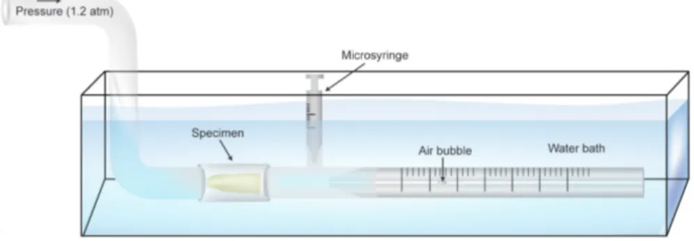
Fig. 1 Schematic illustration of the modified fluid filtration model used in this study.
total of 70 extracted, single-rooted, human teeth were used. Each tooth was placed in sodium hypochlorite (NaOCl) for 2 h for surface disinfection, and then stored in distilled water until its use. Standard access cavities were prepared, and the canal orifices located and apical patency confirmed with a size 15 K-file (Maillefer, Ballaigues, Switzerland). Working length was established at 1 mm from the apex. Middle and coronal thirds were prepared using Gates Glidden drills (Produits Dentaires S.A., Vevey, Switzerland) of ISO sizes 1-4. All teeth were instrumented using a crown-down technique with a set of ProTaper rotary instruments (Dentsply Maillefer, Ballaigues, Switzerland) as follows. S1 hand file (0.17 mm tip diameter; variable taper) was taken into the canal just short of the depth at which the hand file was taken previously. The other files were used in the following sequence, and all were advanced to working length: S2 (0.20 mm tip diameter; variable taper), F1 (0.20 mm tip diameter; 7% taper), F2 (0.25 mm tip diameter; 8% taper), F3 (0.30 mm tip diameter; 9% taper), and F4 (0.40 mm tip diameter; 6% taper) to achieve apical preparation of size #40. Two milliliters of 2.5% NaOCl was used for irrigation between each instrument change. Smear layer was removed during instrumentation with 2 mL of 17% EDTA (pH 7.4). Finally, root canals were successively flushed with 5 mL of 2.5% NaOCl, 5 mL of 17% EDTA, and 3 mL of distilled water. Crowns were sectioned using a water-cooled diamond disk to standardize the root length at 14 mm.
Root-end resection and preparation
The apical 3 mm of each instrumented root was resected 90° to the longitudinal axis of the root. To provide an intracanal matrix, a prefitted gutta-percha cone (Dentsply Maillefer, Ballaigues, Switzerland) was inserted into the root canal leaving a root-end void of 3 mm, which served as a barrier for root-end filling material placement. A provisional restoration (IRM, Caulk, Dentsply, Milford, DE, USA) was placed in the coronal access cavity to stabilize the gutta-percha cone. Root-end cavities were prepared to a depth of 3 mm using ultrasonic tips (S12/90°D, Satelec, Merignac Cedex, France) powered by an ultrasonic unit (Suprasson P5
Booster; Satelec, F-33708, Merignac Cedex, France). The roots were randomly divided into six groups: four experimental groups of 15 roots each and two control groups of five roots each. In Groups 1 and 2, the resected and ultrasonically prepared root-end cavities were rinsed with 5 mL of 17% EDTA for 3 min. In Groups 3 and 4, root-end cavities were treated with 40 s of Er,Cr:YSGG laser (Biolase Technology, San Clemente, CA, USA) using a 400-µm endodontic fiber tip (MZ4, Biolase Technology, San Clemente, CA, USA ) with these operating parameters: 1.25 W, 20 pulses per s, 25% water, and 35% air. Finally, all root-end cavities were irrigated with 2 mL of distilled water and obturated with one of the filling materials:
Group 1 (without irradiation): Retrofilled with MTA Group 2 (without irradiation): Retrofilled with iRoot BP
Group 3 (with irradiation): Retrofilled with MTA Group 4 (with irradiation): Retrofilled with iRoot BP
In the negative control group (Group 5), roots were filled a single gutta-percha cone without a root canal sealer and root-ends were neither filled nor covered. In the positive control group (Group 6), two layers of nail varnish were applied over the root surfaces of teeth with intact crowns.
All retrofilling materials were prepared according to manufacturers’ instructions and condensed using a microplugger (Obtura Spartan, Fenton, MO, USA). Adequacy of root-end fillings was verified using both buccolingual and mesiodistal radiographs. All specimens were stored at 37°C with 100% humidity for 4 weeks. However, after the initial set (48 h) of the tested filling materials, the gutta-percha cone was removed from each root canal except for root canals in the negative control group (Group 5).
Microleakage evaluation
Microleakage of the retrofilled roots was evaluated by using a modified fluid filtration method (Fig. 1), as reported by Pashley and Depew20). In the experimental groups (Groups 1 to 4), external root surfaces were
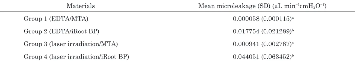
Table 1 Mean microleakage (µL/cmH2O/min−11.2 atm) values and standard deviations (SD) for experimental groups.
Groups identified by the same superscript letters are not significantly different (p>0.05). Different superscript letters identify significantly different groups (p<0.05).
Materials Mean microleakage (SD) (µL min−1cmH2O−1)
Group 1 (EDTA/MTA) 0.000058 (0.000115)a
Group 2 (EDTA/iRoot BP) 0.017754 (0.021289)b
Group 3 (laser irradiation/MTA) 0.000941 (0.002787)a
Group 4 (laser irradiation/iRoot BP) 0.044051 (0.063452)b
coated with two layers of nail varnish up to 3 mm below each resected root end. Each root section specimen was connected to a plastic tube, which was filled with distilled water from either side of the specimen. A 20-µL glass capillary tube was connected to the plastic tube on the outlet side of the specimen. Using a syringe, water was retracted approximately 2 mm into the open end of the glass capillary tube. The whole set-up was placed in a water bath (20°C), and the syringe was used to adjust the air bubble to a suitable position within the capillary tube. A pressure of 120 kPa (1.2 atm) was applied at the apical side. A 5-min pressurization preload of the system was completed before readings were taken. Fluid movement was measured at 2-min intervals for a total of 8 min, and then the measurements were averaged. Microleakage quantity was expressed as µL/cm H2O per min.
Scanning electron microscope (SEM) evaluation
At the end of week 1, one root from each experimental group was selected for SEM analysis. Each root was cross-sectioned into 4-mm-thick segments. Specimen preparation for SEM observation followed a routine procedure: specimens were fixed in 10% buffered formaldehyde overnight, dehydrated through ascending concentrations of alcohol up to 100% alcohol, and finally critical-point dried (Bio-Rad E3000, Bio-Rad Microscience Ltd., Hertfordshire, UK). Specimens were mounted in stubs, sputter-coated with gold, and then observed under SEM (JSM-840A, Jeol Co., Akishima, Tokyo, Japan) to assess the contact and adaptation between root canal walls and filling materials and the presence of intratubular mineralization. In some specimens, elemental compositions of the interfacial layer and intratubular mineralization were investigated using energy dispersive X-ray spectroscopy (EDS) with SEM (JSM-840A, Jeol Co., Akishima, Tokyo, Japan). Statistical analysis
For statistical analysis, normality was tested using the Shapiro-Wilk test. Levene’s test was performed to test for homogeneity of variances. Variables were found to be heterogeneous, and data were analyzed using non-parametric statistical analysis, Mann-Whitney U test. All analyses were performed using SPSS statistical package for Windows, version 17.0 (September 2012;
license number: 1093910, Baskent University). All levels of significance were set at α=0.05.
RESULTS
Table 1 shows the means and standard deviations of the microleakage values of tested root-end filling materials. EDTA/MTA combination exhibited the least microleakage, and laser irradiation/MTA combination ranked second in this regard. Both combinations showed significantly lower microleakage values than EDTA/ iRoot BP and laser irradiation/iRoot BP combinations (p<0.05). Between groups of the same filling material, there were no significant differences among specimens treated with EDTA or laser (p>0.05).
The negative controls showed extreme apical leakage with values exceeding the capillary tube length. The positive controls registered no detectable bubble movement at 1.2 atm.
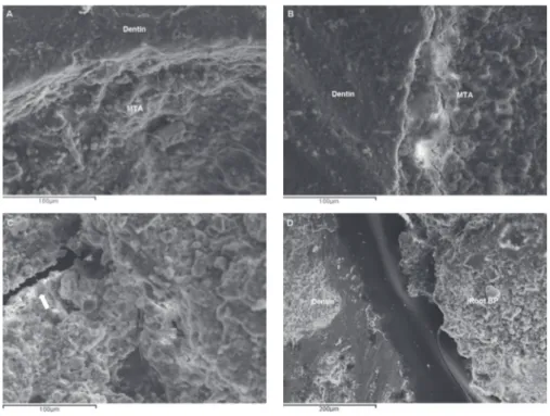
SEM observations agreed with the fluid filtration analysis. Root-end cavities treated with EDTA/MTA (Group 1) and laser irradiation/MTA (Group 3) showed best adaptation to the apical root canal walls. In Group 2 (EDTA/iRoot BP) and Group 4 (laser irradiation/iRoot BP), lack of continuity between the filling material and root canal walls increased in direct proportion to their higher microleakage values (Fig. 2).
Representative SEM images of Group 1 (EDTA/ MTA) and Group 2 (EDTA/iRoot BP) also showed intratubular mineralization (tag-like structures) within the dentinal tubules just beneath the dentin-filling material interface. EDS analysis of the elemental compositions of these tags revealed mainly the presence of calcium, phosphorus, and oxygen for Group 1 (EDTA/ MTA), and calcium, silicon, zirconium, tantalum, and oxygen for Group 2 (EDTA/iRoot BP) (Fig. 3).
DISCUSSION
In light of the current findings, the null hypotheses of this study were accepted: Er,Cr:YSGG laser treatment would not influence the sealing ability of the filling materials when compared with EDTA application and that the sealing ability of iRoot BP would not be superior to that of MTA.
Fig. 2 Representative SEM micrographs of Group 1 (EDTA/MTA) (A) and Group 3 (laser irradiation/MTA) (B) reveal good adaptation of MTA filling to root dentin.
Representative SEM micrograph of Group 2 (EDTA/iRoot BP) shows a gap (white arrowhead) inside the filling material (C). Representative micrograph of Group 4 (laser irradiation/iRoot BP) (D) shows an area of evident interfacial separation between the filling material and root dentin.
Fig. 3 Representative SEM micrographs of Group 1 (EDTA/MTA) (A) and Group 2 (EDTA/ iRoot BP) (C) show intratubular mineralization with tag-like structures.
EDS analyses (B and D) reveal the elemental compositions of intratubular mineralization seen in (A) and (C) respectively (area marked with *).
erbium-type lasers on microleakage. Controversy remains as to whether or not smear layer removal with laser application is necessary before root-end filling procedure to promote sealing ability. Karlovic et al.7) and Kocak et al.21) showed that MTA-filled root-end cavities prepared by Er:YAG laser had significantly lower microleakage than those prepared using ultrasonic devices. However, in the absence of smear layers, there seemed to be no apparent advantages in the use of Er,Cr:YSGG laser over conventional root-end cavity preparation methods to address apical microleakage of root-end fillings22,23). In agreement with the results of these studies, Er,Cr:YSGG laser treatment did not influence the sealing ability of the root-end filling materials tested in this study when compared with EDTA application.
Recent studies on calcium silicate-containing biomaterials showed that they yielded comparable results to MTA, and the authors also concluded that these biomaterials offered no apparent advantages over MTA19,24). In the present study, comparison of groups with the same smear layer removal method yielded the same results. Group 1 (EDTA/MTA) had higher sealing ability than Group 2 (EDTA/iRoot BP), and Group 3 (laser irradiation/MTA) had higher sealing ability than Group 4 (laser irradiation/iRoot BP). Consistent with the findings of previous studies19,24), our results based on fluid filtration method showed that iRoot BP did not provide a better seal than MTA.
The mechanism by which MTA provides superior sealing ability is not completely understood. Sarkar et al.13) analyzed the interactions of MTA with a synthetic tissue fluid and root canal dentin, and suggested that MTA initially produced a mechanical seal. MTA then dissolved, leading to the formation of hydroxyapatite crystals which reacted with dentin to create a chemical adhesion19).
Representative SEM images of Group 1 (EDTA/ MTA) and Group 2 (EDTA/iRoot BP) showed tag-like structures within the dentinal tubules just beneath the dentin-filling material interface. These tags were previously reported when root-end cavities were filled with MTA25) or a calcium silicate-based coronal restorative material (Biodentine, Septodont, Saint Maur des Fosses, France)26,27). Following hydration, the flowable consistency of these filling materials aided their penetration through opened dentinal tubules to crystallize in situ over time28).
However, the SEM images of iRoot BP specimens also showed gap-containing segments along the dentin-filling material interface and within the material itself. A problem encountered in this study was the inability to achieve complete setting of iRoot BP filling material. These gaps might be associated with its long setting time, which then led to material washout during setting. In pilot studies, it was observed that this material started to set only when completely covered with water29). A recent study reported that EndoSequence Root Repair Material (marketed as iRoot BP in Canada; Brasseler USA, Savannah, GA,
USA) incubated at 37°C in 100% humidity was unable to withstand a 500-g load until after a duration of 168 h (7 days)30). This has important clinical implications, especially where apical surgeries are involved, in that any unset material may be washed out by tissue fluids and blood in the surgical field and consequently lead to microleakage and treatment failure.
Our results based on fluid filtration method and SEM evaluation suggested that the root canal adaptation and sealing ability of MTA were superior to that of iRoot BP cement when used as a root-end filling material in vitro. Er,Cr:YSGG laser treatment did not enhance the sealing ability of the filling materials when compared with EDTA application.
Good long-term sealing capability is a pivotal quality of endodontic materials. Although a clear correlation between in vitro and in vivo biocompatibility was already described for MTA, further investigations for iRoot BP cement on its biocompatibility, dimensional stability, and bone formation induction need to be performed to establish consensus on the clinical performance of this material.
ACKNOWLEDGMENTS
This study was supported by the Baskent University Research Fund (Project No. D-DA11/04) and was presented at the 6th International Association of Dental Research Pan-European Region (IADR/PEF) Meeting in Helsinki, Finland, on September 2012.
The authors would like to thank Mr. Enver Atali and Mr. Aslan Atali for their extensive help in technical support and for their generous material support to this work.
REFERENCES
1) Gutmann JL. Surgical endodontics: quo vadis? Endod Topics 2005; 11: 1-3.
2) Kim S, Kratchman S. Modern endodontic surgery concepts and practice: a review. J Endod 2006; 32: 601-623.
3) Johnson BR, Fayad MI, Witherspoon DE. In: Hargreaves KM, Cohen S, Berman LH, editors. Cohen’s pathways of the pulp. 10th ed. St Louis: Mosby Elsevier; 2011. pp. 720-776. 4) de Lange J, Putters T, Baas EM, van Ingen JM. Ultrasonic
root-end preparation in apical surgery: a prospective randomized study. Oral Surg Oral Med Oral Pathol Oral Radiol Endod 2007; 104: 841-845.
5) Bader G, Lejeune S. Prospective study of two retrograde endodontic apical preparations with and without the use of CO2 laser. Endod Dent Traumatol 1998; 14: 75-78.
6) Aranha AC, Domingues FB, Franco VO, Gutknecht N, Eduardo Cde P. Effects of Er:YAG and Nd:YAG lasers on dentin permeability in root surfaces: a preliminary in vitro study. Photomed Laser Surg 2005; 23: 504-508.
7) Karlovic Z, Pezelj-Ribaric S, Miletic I, Jukic S, Grgurevic J, Anic I. Erbium:YAG laser versus ultrasonic in preparation of root-end cavities. J Endod 2005; 31: 821-823.
8) de Souza EB, de Amorim CV, Marques JL. Effect of diode laser irradiation on the apical sealing of MTA retrofillings. Braz Oral Res 2006; 20: 231-234.
9) Gutmann JL, Pitt Ford TR. Management of the resected root end: a clinical review. Int Endod J 1993; 26: 273-283. 10) Rubinstein RA, Kim S. Long-term follow-up of cases
considered healed one year after apical microsurgery. J Endod 2002; 28: 378-383.
11) Torabinejad M, Watson TF, Pitt Ford TR. Sealing ability of a mineral trioxide aggregate when used as a root end filling material. J Endod 1993; 19: 591-595.
12) Fischer EJ, Arens DE, Miller CH. Bacterial leakage of mineral trioxide aggregate as compared with zinc-free amalgam, intermediate restorative material, and Super-EBA as a root-end filling material. J Endod 1998; 24: 176-179.
13) Sarkar NK, Caicedo R, Ritwik P, Moiseyeva R, Kawashima I. Physicochemical basis of the biologic properties of mineral trioxide aggregate. J Endod 2005; 31: 97-100.
14) Baek SH, Plenk H Jr, Kim S. Periapical tissue responses and cementum regeneration with amalgam, SuperEBA, and MTA as root-end filling materials. J Endod 2005; 31: 444-449. 15) Bodrumlu E. Biocompatibility of retrograde root filling
materials: a review. Aust Endod J 2008; 34: 30-35.
16) Ber BS, Hatton JF, Stewart GP. Chemical modification of ProRoot MTA to improve handling characteristics and decrease setting time. J Endod 2007; 33: 1231-1234. 17) Maltezos C, Glickman GN, Ezzo P, He J. Comparison of the
sealing of Resilon, Pro Root MTA, and Super-EBA as root-end filling materials: a bacterial leakage study. J Endod 2006; 32: 324-327.
18) Gandolfi MG, Sauro S, Mannocci F, Watson TF, Zanna S, Capoferri M, Prati C, Mongiorgi R. New tetrasilicate cements as retrograde filling material: an in vitro study on fluid penetration. J Endod 2007; 33: 742-745.
19) Leal F, De-Deus G, Brandao C, Luna AS, Fidel SR, Souza EM. Comparison of the root-end seal provided by bioceramic repair cements and White MTA. Int Endod J 2011; 44: 662-668.
20) Pashley DH, Depew DD. Effects of the smear layer, Copalite and oxalate on microleakage. Oper Dent 1986; 11: 95-102. 21) Kocak MM, Kocak S, Aktuna S, Görücü J, Yaman SD. Sealing
ability of retrofilling materials following various root-end
cavity preparation techniques. Lasers Med Sci 2011; 26: 427-431.
22) Winik R, Araki AT, Negrao JAA, Bello-Silva MS, Lage-Marques JL. Sealer penetration and marginal permeability after apicoectomy varying retrocavity preparation and retrofilling material. Braz Dent J 2006; 17: 323-327. 23) Çalışkan MK, Parlar NK, Oruçoğlu H, Aydın B. Apical
microleakage of root-end cavities prepared by Er,Cr:YSGG laser. Lasers Med Sci 2010; 25: 145-150.
24) Nair U, Ghattas S, Saber M, Natera M, Walker C, Pileggi R. A comparative evaluation of the sealing ability of 2 root-end filling materials: an in vitro leakage study using Enterococcus
faecalis. Oral Surg Oral Med Oral Pathol Oral Radiol Endod 2011; 112: e74-e77.
25) Reyes-Carmona JF, Felippe MS, Felippe WT. Biomineralization ability and interaction of mineral trioxide aggregate and white Portland cement with dentin in a phosphate-containing fluid. J Endod 2009; 35: 731-736.
26) Han L, Okiji T. Uptake of calcium and silicon released from calcium silicate-based endodontic materials into root canal dentine. Int Endod J 2011; 44: 1081-1087.
27) Atmeh AR, Chong EZ, Richard G, Festy F, Watson TF. Dentin-cement interfacial interaction: calcium silicates and polyalkenoates. J Dent Res 2012; 91: 454-459.
28) Weller RN, Tay KC, Garrett LV, Mai S, Primus CM, Gutmann JL, Pashley DH, Tay FR. Microscopic appearance and apical seal of root canals filled with gutta-percha and ProRoot Endo Sealer after immersion in a phosphate-containing fluid. Int Endod J 2008; 41: 977-986.
29) Lovato KF, Sedgley CM. Antibacterial activity of EndoSequence root repair material and ProRoot MTA against clinical isolates of Enterococcus faecalis. J Endod 2011; 37: 1542-1546.
30) Damas BA, Wheater MA, Bringas JS, Hoen MM. Cytotoxicity comparison of mineral trioxide aggregates and EndoSequence bioceramic root repair materials. J Endod 2011; 37: 372-375.