© 2019 by the Serbian Biological Society How to cite this article: Korkmaz ÖA, Sadi G, Kocabas A, Yildirim OG, Sumlu E, 265 Koca HB, Nalbantoglu B, Pektaş MB, Akar F. Lactobacillus helveticus and Lactobacillus plantarum modulate renal antioxidant status in a rat model of fructose-induced metabolic syndrome. Arch Biol Sci. 2019;71(2):265-73.
Lactobacillus helveticus and Lactobacillus plantarum modulate renal antioxidant status
in a rat model of fructose-induced metabolic syndrome
Ömer Adil Korkmaz1, Gökhan Sadi2, Aytaç Kocabaş2, Onur Gökhan Yildirim3, Esra Sumlu4, Halit Buğra
Koca5, Barbaros Nalbantoglu1, Mehmet Bilgehan Pektaş6,* and Fatma Akar4
1 Department of Chemistry, Faculty of Science, Yildiz Technical University, Istanbul, 34220 Turkey
2 Department of Biology, K.Ö. Science Faculty, Karamanoglu Mehmetbey University, 70100, Karaman, Turkey
3 Department of Pharmacy Service, Health Services Vocational School, Artvin Coruh University, Artvin, 08100 Turkey
4 Department of Pharmacology, Faculty of Pharmacy, Gazi University, Ankara, 06330 Turkey
5 Department of Medical Biochemistry, Faculty of Medicine, Afyonkarahisar Health Sciences University, Afyonkarahisar,
03200 Turkey
6 Department of Medical Pharmacology, Faculty of Medicine, Afyonkarahisar Health Sciences University, Afyonkarahisar,
03200 Turkey
*Corresponding author: [email protected]
Received: January 23, 2019; Revised: February 20, 2019; Accepted: February 21, 2019; Published online: February 26, 2019 Abstract: High dietary fructose intake causes a metabolic disorder and augments the risk of chronic kidney disease most
likely due to oxidative stress. Probiotics could have antioxidant, antiinflammatory and immunoregulatory properties. The present study examined the influence of Lactobacillus helveticus and Lactobacillus plantarum supplementation on dietary fructose-induced metabolic changes and renal antioxidant/oxidant status of rats. Male Wistar rats were divided into four groups as follows: control; fructose; fructose plus L. helveticus; fructose plus L. plantarum. Fructose was given to the rats as a 20% solution in drinking water for 15 weeks. The probiotic supplementation was applied by gastric gavage once a day for six weeks. Several metabolic parameters in the plasma, gene and protein expressions of the main antioxidant enzymes in renal tissues of rats were measured. Dietary fructose-induced elevations in plasma insulin, triglyceride, VLDL, creatinine as well as renal urea levels were alleviated after treatment with L. helveticus and L. plantarum. Moreover, L. helveticus and
L. plantarum supplementation recovered the changes in renal protein expression level of SOD1, SOD2 and CAT. In
conclu-sion, supplementation with L. helveticus and L. plantarum has an improving effect on specific metabolic parameters and renal antioxidative enzymes in a fructose-induced metabolic disorder.
Keywords: dietary fructose; renal antioxidant status; antioxidant enzymes; Lactobacillus helveticus; Lactobacillus plantarum. Abbreviations: CAT (catalase); Fruc (fructose); FAS (fatty acid synthase); GAPDH (glyceraldehyde 3-phosphate
dehydro-genase); GPx (glutathione peroxidase); HDL (high density lipoprotein); IL-1β; 6 (interleukin-1β; 6); iNOS (inducible nitric oxide synthase); MDA (malondialdehyde); NF-κB (nuclear factor-kappa B); PAGE (polyacrylamide gel electrophoresis); PVDF (polyvinylidene floride); SOD (superoxide dismutase); qRT-PCR (quantitative real-time PCR); SDS (sodium dodecyl sulfate); SREBP1 (sterol regulatory element-binding transcription factor 1); TBARS (thiobarbituric acid reactive substances); TNF-α (tumor necrosis factor-α); VLDL (very low-density lipoprotein).
INTRODUCTION
Excess sugar intake, especially of fructose, has been shown to induce metabolic syndrome and kidney diseases via several molecular mechanisms, including activation of inflammatory processes and generation of oxidative stress [1,2]. We previously described the upregulation of the inflammatory pathway and induction of the superoxide-producing system in various tissues with high dietary fructose intake [3-9]. Development of a proinflammatory condition, as manifested by nuclear factor-kappa B (NF-κB) activation, high expression of tumor necrosis factor-α (TNF-α), inducible nitric oxide synthase (iNOS) and interleukin-6 (IL-6), was demonstrated in the renal tissue of high-fructose-fed rats [10]. Furthermore, oxidative stress, as evidenced by decreased antioxi-dant enzyme activities and glutathione levels and increased malondialdehyde (MDA) contents in kidney tissues was shown in high-fructose-fed rats [11-14]. Fructose-induced uric acid generation is proposed to lead to increased mitochondrial oxidative stress, endothelial dysfunction and maladaptive immune and inflammatory responses [15,16].
Probiotics are currently defined as nonpathogenic microorganisms and have been reported to improve metabolic diseases such as obesity and diabetes through modulation of intestinal microorganisms [17,18]. Their beneficial health effects could be attributed to their antiinflammatory and immunoregulatory properties [19]. Some probiotic strains were shown to induce significant antioxidant activity through ac-tion on superoxide dismutase (SOD), catalase (CAT) and glutathione peroxidase (GPx) [20]. Lactic acid bacterial strains, which are the principal representa-tives of probiotics in both food and pharmaceuti-cal forms, have been shown to ameliorate metabolic diseases by increasing insulin sensitivity, reducing inflammatory reactions and oxidative stress [18,21]. Supplementation with Lactobacillus plantarum along with a high-fat fructose diet in rats was shown to improve metabolic dysfunction by modulating blood proinflammatory cytokines, IL-1β, IL-6 and TNF-α, as well as the antioxidant enzymes, SOD, CAT and GPx [22]. The combination of L. curvatus and L. plantarum in high-fructose-fed rats lowered plasma glucose, insulin, triglyceride and oxidative stress, and also suppressed hepatic lipogenesis via downregulation of
sterol regulatory element-binding transcription factor 1 (SREBP1) and fatty acid synthase (FAS) mRNA levels [23]. Knowledge of the influence of probiotic treat-ment on high-fructose-induced metabolic syndrome is limited. Therefore, in the present study, an attempt was made to investigate whether supplementation with
L. helveticus and L. plantarum improves metabolic
parameters and the renal antioxidant/oxidant status of fructose-fed rats.
MATERIALS AND METHODS Chemicals
Chemicals were purchased from the Sigma Chemical Co. (St. Louis, MO) unless otherwise stated. Fructose was obtained from Danisco Sweeteners OY (Finland).
L. helveticus and L. plantarum strains were obtained
from Chr. Hansen (Denmark; ATCC: 15009 and ATCC: 14917 respectively) and grown in our laboratory.
Animals and diets
All protocols for animal usage were approved by the Ethical Animal Research Committee of Afyon Kocatepe University (Akuhadyek-49533702). Three-week-old male Wistar rats were housed in temperature- and humidity-controlled rooms (at 20-22°C and 40-60% humidity), with a 12-h light-dark cycle. The animals were fed with a standard rodent chow diet composed of 62% starch, 23% protein, 4% fat, 7% cellulose, stan-dard vitamins and salt. At the end of the acclimation for one week, the rats were randomly divided into four groups, designated as Control; Fructose (Fruc); Fructose+L. helveticus (Fruc+LH) and Fructose+L.
plantarum (Fruc+LP). Fructose was given to the rats
as a 20% solution (w/v) in drinking water for 15 weeks.
L. helveticus and L. plantarum (1x10⁹ CFU per 100 g of
body weight of animal) in 2 mL of saline were given by gastric gavage once a day during the final six weeks. The control and fructose groups were administered the same volume of saline by gavage for the same period. Body weights, food, and fluid intake were recorded weekly during the follow-up period. At the end of the follow-up period, the rats were anesthetized with a mixture of ketamine-xylazine (100 and 10 mg/ kg, respectively, i.p.) and blood samples were rapidly
collected via cardiac puncture. The kidneys of rats were blotted dry, weighed, frozen in liquid nitrogen and stored at -85°C.
Preparation of L. helveticus and L. plantarum
L. helveticus and L. plantarum were cultured in de Man,
Rogosa and Sharpe broth (MRS; Oxoid; Unipath Ltd., Basingstoke, Hampshire, England) at 30°C in a rotary shaker at 150 rpm. Stock cultures were stored at -80°C in MRS broth containing 20% (v/v) glycerol. Erlenmeyer flasks containing 20 mL of MRS were inoculated with 1.5 mL of glycerol stock culture and the cultures were incubated at 35°C±1°C in a rotary shaker at 150 rpm and grown to an optical density of 1.0 at 600 nm (cell density corresponding to 1x10⁸ CFU/mL). The culture
was divided into 10 mL tubes (1x10⁹ CFU) and the
cells were harvested at 5000 x g for 5 min at 4°C. The cell pellets were washed with isotonic saline solution and lyophilized under a freeze drier.
Measurement of metabolic parameters in plasma and renal tissue
Cardiac blood samples of non-fasted rats were imme-diately centrifuged at 10000 x g and 4°C for 30 min. Kidney samples were homogenized in 0.1 M phosphate buffer 1:10 (w/v), pH 7.4, and 24000 cycles/min (Ultra Turrax, IKA Works Inc., USA), and ultrasonicated at 20000 cycles/s for 1 min (Dr. Hielscher, Germany). Homogenates were centrifuged at 10000 x g and 4°C for 15 min, and the supernatants were collected. All the samples were stored at -85°C until analysis. Plasma triglyceride, very low-density lipoprotein (VLDL), high-density lipoprotein (HDL) and total cholesterol levels were determined by standard enzymatic techniques. Insulin (Elabscience, USA), creatinine, urea, uric acid, albumin and total protein levels (Biolabo, France) were assessed using enzyme-linked immunosorbent assay (ELISA) kits according to the manufacturers’ instruc-tions. Glucose levels were measured using a glucometer (Roche Diagnostics, USA) in blood collected from the tail veins of rats. MDA levels were measured with a TBARS assay kit (Cayman Chemical, USA).
Determination of sod1, sod2, cat, and gpx gene expressions by real-time polymerase chain reaction (qRT-PCR)
Total RNAs were isolated from kidney tissue using RNeasy total RNA isolation kit (Qiagen, Venlo, Neth-erlands) as described in the manufacturer’s protocol. After isolation, the amount and the quality of the total RNA was determined by spectrophotometry and agarose gel electrophoresis. One µg of total RNA was reverse-transcribed to cDNA using a commercial first strand cDNA synthesis kit (Thermo Scientific, USA). Expression levels of sod1, sod2, cat, and gpx were determined by qRT-PCR (LightCycler480 II, Roche, Basel, Switzerland). One μL of cDNA, 5 μL 2X SYBR Green Master mix (Roche FastStart Universal SYBR Green Master Mix) and 2 μL primer pairs (sod1-F: GCAGAAGGCAAGCGGTGAAC; sod1-R: TAGCAG-GACAGCAGATGAGT; sod2-F: GCACATTAACGCG-CAGATCA; sod2-R: AGCCTCCAGCAACTCTCCTT;
cat-F: GCGAATGGAGAGGCAGTGTAC; cat-R:
GAGTGACGTTGTCTTCATTAGCACTG; gpx-F: CTCTCCGCGGTGGCACAGT; gpx-R: CCACCAC-CGGGTCGGACATAC; gapdh-F: TGATGACAT-CAAGAAGGTGGTGAAG; gapdh-R: TCCTTGGAG-GCCATGTGGGCCAT) were mixed and qRT-PCR was performed as follows; initial denaturation at 95°C for 10 min, denaturation at 95°C for 10 s, annealing at 58°C for 15 s, extension at 72°C for 15 s, with 40 repeated thermal cycles measuring the green fluores-cence at the end of each extension step. All reactions were performed in triplicate and the specificity of PCR products was confirmed by melt analysis. The relative expression of genes to internal control glyceraldehyde 3-phosphate dehydrogenase (gapdh) was calculated with the quantification tool provided by LightCycler® 480 SW 1.5.1 software.
Immunoblot analysis of antioxidant enzymes, SOD1, SOD2, CAT and GPx
For determination of SOD1, SOD2, CAT and GPx protein contents, kidney tissue was homogenized in 2-fold volumes of homogenization medium (50 mM Tris, 150 mM NaCl, 5 mM EDTA, 1 % (w/w) Triton X-100, 0.26% (w/v) sodium deoxycholate, 50 mM sodium fluoride, 0.1 mM sodium orthovanadate and 0.2 mM phenylmethylsulfonyl fluoride (PMSF)) with a
Tissue RuptorTM (Qiagen, Netherlands) homogenizer.
The homogenates were centrifuged at 1500 x g for 10 min at 4ºC. After the removal of the supernatants, the protein concentrations were determined by the Lowry method [24]. Ten to 50 µg of total proteins were separated by SDS-PAGE and transferred on to PVDF membranes using a semi-dry electroblotting apparatus (TransBlot Turbo, BioRad, Germany). Blot-ted membranes were blocked with 5% (w/v) nonfat dried milk and incubated with primary antibodies for SOD1 (Anti-SOD1 Sheep IgG, Calbiochem-574597, 1:5,000), SOD2 (Anti-SOD2 Rabbit IgG, Santa Cruz; sc:30080, 1:100), CAT (Anti-CAT Rabbit IgG, Abcam, ab:6731,1:6,000), GPx (Anti-GPx Rabbit IgG, Santa Cruz, sc:30147,1:100) for 2 h at room temperature or overnight at 4ºC. As an internal control, GAPDH proteins were also labeled with anti-GAPDH Rab-bit IgG (Santa Cruz, sc:25778, 1:2,000). Horseradish peroxidase-conjugated secondary antibody (Santa Cruz, sc:2030 or sc:2770, 1:10,000) was incubated for 1 h, and the blots were incubated in ClarityTM Western
ECL (Bio-Rad Laboratories, Hercules CA, USA) sub-strate solution. Images of the blots were obtained using the ChemiDocTM MP Chemiluminescence detection
system (Bio-Rad Laboratories, Hercules CA, USA) equipped with a CCD camera. The relative expression of proteins with respect to GAPDH was calculated using ImageLab5.2 software.
Statistical analysis
All data are presented as the mean±standard error of the mean; n is the number of rats. Statistical compari-sons were performed using unpaired Student’s t-test
or one-way ANOVA followed by the Tukey post hoc test. P values smaller than 0.05 were considered as statistically significant.
RESULTS
The effects of dietary fructose and supplementation with L. helveticus and L. plantarum on body weight, caloric intake and kidney weight
As shown in Table 1, the high-dietary-fructose inter-vention did not change the final body weights of rats despite the increase in total caloric intake. There was a trend toward an increase in final body weight in Fruc+LH group when compared to the fructose group, but the differences were not significant. Besides, no significant change was found in the final body weights of rats between Fruc and Fruc+LP groups. The total caloric intakes were not changed after supplementation with L. helveticus and L. plantarum when compared to the fructose group. Moreover, the right and left kidney absolute weights, as well as the ratio of right or left kidney weights to the body weight were not significantly altered between the groups (Table 1).
The effects of dietary fructose and supplementation with L. helveticus and L.
plantarum on metabolic and biochemical
parameters in plasma and kidney
As shown in Table 2, high dietary fructose increased the plasma glucose and insulin levels as compared to control group. The probiotic supplementation did not
Table 1. The effects of dietary fructose, L. helveticus and L. plantarum supplementation on body weight, kidney weight, food, liquid and
caloric intake.
Groups Control Fruc Fruc+LH Fruc+LP
Initial body weight (g) 90.7±5.2 92.1±3.7 93.1±3.4 92.3±1.8
Final body weight (g) 354.6±5.2 359.3±12.7 373.4±17.7 354.2±19.2
Food intake (g/day) 21.4±0.2 14.7±0.4* 15.6±1.6 16.9±1.5
Liquid intake (mL/day) 48.3±2 40.1±2.8* 37.5±1.2 38.1±0.7
Total caloric intake (kcal) 74.9±3.1 84.3±2.1* 85.4±2.1 90.4±4.2
Left kidney absolute weight (g) 1.5±0.1 1.4±0.05 1.5±0.1 1.4±0.1
Ratio of left kidney weight to body weight (%) 0.4±0.01 0.4±0.02 0.4±0.01 0.4±0.01
Right kidney absolute weight (g) 1.5±0.1 1.4±0.05 1.5±0.1 1.4±0.1
Ratio of right kidney weight to body weight (%) 0.4±0.01 0.4±0.02 0.4±0.01 0.4±0.01
alter the plasma glucose levels. However, L. plantarum supplementation significantly reduced the plasma insulin levels. The dietary fructose-induced increase in plasma triglyceride and VLDL levels was markedly reduced with L. helveticus and L. plantarum. Fructose feeding and L. helveticus supplementation did not affect the HDL and total cholesterol levels, but L.
plantarum supplementation significantly enhanced
plasma HDL contents.
Plasma MDA levels, but not the renal concentration, increased in rats that were fed with fructose. Moreover, supplementation with L. helveticus and L. plantarum did not modulate the MDA levels. Dietary fructose also increased the plasma creatinine levels, and conversely,
L. helveticus and L. plantarum supplementation reduced
the creatinine levels to the control value. Significant changes in plasma urea, uric acid, albumin and total protein contents were determined between groups: plasma uric acid levels in the L. helveticus-treated group were decreased. High-fructose intake increased renal urea concentration, which was decreased by L. helveticus
and L. plantarum supplementation. Additionally, L.
plantarum treatment significantly decreased the renal
uric acid levels as compared to the fructose group.
The effects of dietary fructose and supplementation with L. helveticus and L.
plantarum on SOD1, SOD2, CAT and GPx gene
and protein expression
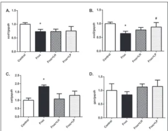
Relative changes in antioxidant enzyme gene and pro-tein expression levels in kidney tissue were measured by qRT-PCR and Western blot analysis, respectively. The results show the suppression of sod1 and sod2 mRNA expression in fructose-fed rats. However, treatments with L. helveticus and L. plantarum did not alter sod1 expression, but L. plantarum significantly increased
sod2 mRNA levels (Fig. 1A and 1B). Moreover,
high-fructose intake significantly increased cat mRNA expressions but not gpx levels. Supplementation of L.
helveticus and L. plantarum did not modulate the cat
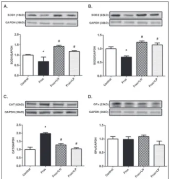
and gpx gene expressions significantly (Fig. 1C and D). Fig. 2 summarizes the changes in renal protein expression of the main antioxidant enzymes, SOD1, SOD2, CAT and GPx, by dietary fructose and probiotic bacteria. As shown in Fig. 2A and B, SOD1 and SOD2 protein expression was decreased in fructose-fed rats. Supplementation with L. helveticus and L. plantarum
Table 2.The effects of dietary fructose, L. helveticus and L.
plan-tarum supplementation on metabolic parameters in the plasma
and kidney tissue.
Groups Control Fruc Fruc+LH Fruc+LP
Glucose (mmol/L) 6.1±0.3 8.5±0.3* 8.7±0.9 8.2±0.5 Insulin (pmol/L) 2.77±0.7 5.9±1.4* 5.5±1.6 3.62±0.5# Triglyceride (mmol/L) 1.39±0.1 3.75±0.1* 2.11±0.1# 1.73±0.1# VLDL (mmol/L) 0.58±0.05 1.63±0.04* 0.97±0.03 # 0.79±0.02 # HDL (mmol/L) 0.61±0.03 0.61±0.05 0.66±0.04 0.76±0.03 # Total Cholesterol (mmol/L) 1.14±0.1 1.42±0.1 1.29±0.1 1.53±0.1 MDA (µmol/L) 21.5±1.1 27.1±2* 29.1±3.2 23.6±0.9 Creatinine (µmol/L) 15±1.8 21.2±2.7* 14.1±2.6# 15±2.7# Urea (mmol/L) 11.6±0.9 12.6±1 10.9±0.4 11.5±0.7 Uric Acid (µmol/L) 142.7±11.9 127.9±11.8 107.1±5.9# 136.8±11.9
Albumin (g/L) 44±3 44±3 44±4 42±4 Total Protein (g/L) 74±1 79±1 79±2 81±1 Renal MDA (µmol/g protein) 1.47±0.1 1.6±0.2 1.32±0.2 1.27±0.2 Renal Urea (mg/g protein) 14.7±1.2 21.6±1.3* 16.3±2.1# 14.1±1.2# Renal Uric Acid
(mg/g protein) 8.6±0.6 10.2±0.9 8.8±0.6 7.2±0.8#
Values are expressed as the mean±SEM, n = 6-8; *P<0.05, significantly different from the control; #P<0.05, significantly different from the
fructose-treated rats.
Fig. 1. Changes in expression levels of sod1 (A), sod2 (B), cat (C)
and gpx (D) mRNAs in kidney tissues of Control, Fruc, Fruc+LH, and Fruc+LP groups. Data were normalized using gapdh. Each bar represents the means from at least six rats.*P<0.05, significantly different from the control; #P<0.05, significantly different from
increased both SOD1 and SOD2 protein expression (Fig. 2A and B). Parallel with the gene expression pattern, CAT protein expression was increased in the fructose group (Fig. 2C) but significantly decreased after treatments with L. helveticus and L. plantarum. No change was observed in GPx levels among all examined groups (Fig. 2D).
DISCUSSION
Fructose is commonly used as an industrial sweetener and is excessively consumed in the regular human diet. Overconsumption of fructose has been proposed as a risk factor for the metabolic syndrome, manifesting in dysfunctional adipose, liver, kidney, intestine and cardiovascular tissues [25]. One of the leading driv-ing forces for the fructose-induced disturbances is the oxidative stress arising from an imbalance between the prooxidant and antioxidant systems [26]. This study demonstrated some favorable influences of probiotic
supplementation with L. helveticus and L. plantarum on the metabolic parameters and renal antioxidant/ oxidant status of rats maintained on a high-fructose diet. Probiotics have emerged as a therapeutic potential for metabolic syndrome, especially in the high-fat diet model [27-29]. Data describing the influence of probiotic treatment on high-fructose-induced metabolic syndrome is limited [22,30]. Therefore, we evaluated the effects of the supplementation of two commonly used probiotic microorganisms, L.
helveticus and L. plantarum, on metabolic, plasma
and renal antioxidant/oxidant markers. The findings presented herein demonstrated hyperinsulinemia and hypertriglyceridemia with high-fructose intake, which is in accordance with our previous studies [3,4,6,9]. There were no significant alteration in the body weights and the ratio of renal weights to body weights in rats fed with high-fructose diet. L.
hel-veticus and L. plantarum supplementation
dimin-ished the increase in plasma triglyceride. Moreover,
L. plantarum treatment restored the elevated plasma
insulin levels. The plasma glucose- and lipid-lowering effects of L. plantarum were previously reported in high-fat- or high-fructose-induced metabolic disor-ders [22,27,28,30]. Moreover, the consumption of a combination of L. curvatus and L. plantarum probiotic strains was shown to reduce plasma triglyceride levels in hypertriglyceridemic subjects [31]. Administration of the probiotic L. kefiri to mice fed with a fructose-rich diet prevented the increase in plasma triglycer-ides and leptin [32]. An early sign of nephropathy is an increase in serum creatinine, which is associated with the progression of renal damage [33]. Serum urea and uric acid levels are also essential parameters of renal functions. Fructose-induced hyperglycemia may impair renal functions and increase plasma cre-atinine, urea and uric acid levels as well as oxidative stress [15,34]. In this study, a significant increase in the levels of plasma creatinine and renal urea could be taken as a reflection of a fructose-induced renal disorder. The correction of these abnormalities by L.
helveticus and L. plantarum supplementation could
be valuable in prevention of the disease. Previously, it has been demonstrated that probiotic therapy with L.
plantarum strains improves urinary oxalate, calcium,
uric acid, creatinine and serum uric acid levels in rats with increased renal calcium oxalate deposition [35].
Fig. 2. Changes in protein expression of the antioxidant enzymes
in kidney tissues of Control, Fruc, Fruc+LH, and Fruc+LP groups. SOD1 (A), SOD2 (B), CAT (C) and GPx (D) protein levels were quantified using densitometry and normalized with GAPDH. Representative Western blot images are included above the cor-responding figures. Each bar represents at least six rats. *P<0.05, significantly different from control; #P<0.05, significantly different
A diet rich in fructose can lead to the induction of marked oxidative stress and mitochondrial dysfunction [36]. Oxidative stress arising from a high-fructose diet has been shown to suppress antioxidant enzymes in different tissues [37-41]. Renal SOD, CAT and GPx levels were reduced in rats with fructose-induced metabolic syndrome [12,42,43]. However, contradic-tory results have demonstrated the upregulation of catalase in hepatic tissues [44]. Besides, the enzymes in the liver or kidney tissues were also reported to be unchanged in similar animal models [45]. Recently, we demonstrated significant suppression of both CAT, GPx and SOD gene and protein expression in the re-nal tissues of diabetic rats [46]. The suppression was attributed to a moderate increase in tissue oxidative biomarkers. In this study, we demonstrated alterations of oxidative stress markers in plasma and reduction in renal SOD1 and SOD2 gene and protein expressions. These results indicate the presence of oxidative stress in fructose-fed rats. On the contrary, both CAT gene and protein expression were upregulated in renal tissue of fructose-fed rats. Such discrepancy might result from differences in intracellular locations since most of the CAT is present in peroxisomes which could prevent it from participating in primary oxidation reactions arising from excessive mitochondrial input through the electron transport systems [47]. Upregulation of CAT could be seen as an adaptation process to the moderate increase in oxidative stress biomarkers in peroxisomes.
Recent studies proposed that probiotics can stimu-late the antioxidant systems of the host and augment the activities of antioxidant enzymes [20]. Supplementation with probiotics and vitamin C increased the activity of antioxidant enzymes in the serum, brain and kid-neys in type 1 diabetic rats [48]. Administration of L.
casei with inulin to healthy volunteers resulted in a
significant increase in plasma CAT activity [49] and in a significant decrease in plasma MDA and oxidized glutathione concentrations and increased free thiol and glutathione contents [50]. Moreover, probiotics pro-vided significant protection against mercury-induced toxicity by preventing alterations in the levels of GPx and SOD [51]. In another study, upregulation of serum SOD, GPx and hepatic CAT activities with dietary L.
fermentum was demonstrated [52]. Moreover, yeast
probiotic supplementation increased the body weights
and serum antioxidant enzyme activities in chicks [53]. In line with these findings, a study conducted with humans demonstrated increased SOD and GPx activities in erythrocytes of diabetic patients supple-mented with L. acidophilus and Bifidobacterium lactis [54]. In our study, treatment with L. helveticus and
L. plantarum strains did not affect the gene expression
patterns of sod1 and cat, however, L. plantarum upregu-lated sod2. In contrast to gene expression levels, the changes in protein expression levels of these enzymes were normalized with both probiotics. These results suggested that L. helveticus and L. plantarum might have a positive influence on renal antioxidant capacity in the dietary fructose-induced metabolic disorder.
In conclusion, supplementation with L. helveticus and L. plantarum has an improving effect on specific metabolic parameters and renal antioxidative enzymes in a fructose-induced metabolic disorder.
Acknowledgments: This study was supported by grants from the
Karamanoglu Mehmetbey University Research Fund (29-M-15) and Yildiz Technical University Research Fund (FDK-2018-3392).
Author contributions: OAK, OGY, ES, and HBK performed the
research. AK grew the bacteria. GS helped during the experimen-tal work, statistical analysis and in writing the manuscript. MBP drafted the manuscript. BN and FA conceived and designed the study and critically revised the manuscript.
Conflict of interest disclosure: The authors declare no conflict
of interest. REFERENCES
1. Prasad K, Dhar I. Oxidative stress as a mechanism of added sugar-induced cardiovascular disease. Int J Angiol. 2014;23:217-26.
2. Hannou SA, Haslam DE, McKeown NM, Herman MA. Fructose metabolism and metabolic disease. J Clin Invest. 2018;128:545-55.
3. Akar F, Uludağ O, Aydın A, Aytekin YA, Elbeg S, Tuzcu M, Sahin K. High-fructose corn syrup causes vascular dysfunc-tion associated with metabolic disturbance in rats: protective effect of resveratrol. Food Chem Toxicol. 2012;50:2135-41. 4. Babacanoglu C, Yildirim N, Sadi G, Pektas MB, Akar F. Res-veratrol prevents high-fructose corn syrup-induced vascular insulin resistance and dysfunction in rats. Food Chem Toxi-col. 2013;60:160-7.
5. Sadi G, Ergin V, Yilmaz G, Pektas MB, Yildirim OG, Menevse A, Akar F. High-fructose corn syrup-induced hepatic dysfunction in rats: improving effect of resveratrol. Eur J Nutr. 2015;54(6):895-904.
6. Pektas MB, Yücel G, Koca H, Sadi G, Yıldırım O, Öztürk G, Akar F. Dietary fructose-induced hepatic injury in male and female rats: influence of resveratrol. Drug Res. (Stuttg). 2017;67:103-10.
7. Pektas MB, Koca HB, Sadi G, Akar F. Dietary fructose activates insulin signaling and inflammation in adipose tissue: modulatory role of resveratrol. Biomed Res Int. 2016;2016:8014252.
8. Pektas MB, Sadi G, Akar F.Long-term dietary fructose causes gender-different metabolic and vascular dysfunction in rats: modulatory effects of resveratrol. Cell Physiol Biochem. 2015;37:1407-20.
9. Yildirim OG, Sumlu E, Aslan E, Koca HB, Pektas MB, Sadi G, Akar F. High-fructose in drinking water initi-ates activation of inflammatory cytokines and testicular degeneration in rat. Toxicol Mech Methods. 2018;DOI: 10.1080/15376516.2018.1543745.
10. Prince PD, Lanzi CR, Toblli JE, Elesgaray R, Oteiza PI, Fraga CG, Galleano M. Dietary (–)-epicatechin mitigates oxida-tive stress, NO metabolism alterations, and inflammation in renal cortex from fructose-fed rats. Free Radic Biol Med. 2016;90:35-46.
11. Palanisamy N, Viswanathan P, Anuradha CV. Effect of Genistein, a soy isoflavone, on whole body insulin sensitiv-ity and renal damage induced by a high-fructose diet. Ren Fail. 2008;30:645-54.
12. Nasri R, Abdelhedi O, Jemil I, Daoued I, Hamden K, Kallel C, Elfeki A, Lamri-Senhadji M, Boualga A, Nasri M, Karra-Châabouni M. Ameliorating effects of goby fish protein hydrolysates on high-fat-high-fructose diet-induced hyper-glycemia, oxidative stress and deterioration of kidney func-tion in rats. Chem Biol Interact. 2015;242:71-80.
13. Wang W, Ding X-Q, Gu T-T, Song L, Li J-M, Xue Q-C, Kong L-D. Pterostilbene and allopurinol reduce fructose-induced podocyte oxidative stress and inflammation via microRNA-377. Free Radic Biol Med. 2015;83:214-26. 14. Qiao Y, Xu L, Tao X, Yin L, QiY, Xu Y, Han X, Tang Z, Ma X,
Liu K, Peng J. Protective effects of dioscin against fructose-induced renal damage via adjusting Sirt3-mediated oxidative stress, fibrosis, lipid metabolism and inflammation. Toxicol Lett. 2018;284:37-45.
15. Chaudhary K, Malhotra K, Sowers J, Aroor A. Uric Acid - key ingredient in the recipe for cardiorenal metabolic syn-drome. Cardiorenal Med. 2013;3:208-20.
16. Johnson RJ, Nakagawa T, Sanchez-Lozada LG, Shafiu M, Sundaram S, Le M, Ishimoto T, Sautin YY, Lanaspa MA. Sugar, uric acid, and the etiology of diabetes and obesity. Diabetes. 2013;62:3307-15.
17. Wang J, Tang H, Zhang C, Zhao Y, Derrien M, Rocher E, van-Hylckama Vlieg JE, Strissel K, Zhao L, Obin M, Shen J. Modulation of gut microbiota during probiotic-mediated attenuation of metabolic syndrome in high fat diet-fed mice. ISME J. 2015;9:1-15.
18. Rad AH, Abbasalizadeh S, Vazifekhah S, Abbasalizadeh F, Hassanalilou T, Bastani P, Ejtahed H-S, Soroush A-R, Javadi M, Mortazavian AM, Khalili L. The future of diabetes man-agement by healthy probiotic microorganisms. Curr Diabe-tes Rev. 2017;13:582-9.
19. Plaza-Díaz J, Ruiz-Ojeda FJ, Vilchez-Padial LM, Gil A. Evidence of the anti-inflammatory effects of probiotics and synbiotics in intestinal chronic diseases. Nutrients. 2017;9(6):555.
20. Wang Y, Wu Y, Wang Y, XuH, Mei X, Yu D, Wang Y, Li W. Antioxidant properties of probiotic bacteria. Nutrients. 2017;9(5):521.
21. Zielińska D, Kolożyn-Krajewska D. Food-origin lactic acid bacteria may exhibit probiotic properties: review. Biomed Res Int. 2018;2018:5063185.
22. Huang H-Y, Korivi M, Tsai C-H, Yang J-H, Tsai Y-C. Supple-mentation of Lactobacillus plantarum K68 and fruit-vege-table ferment along with high fat-fructose diet attenuates metabolic syndrome in rats with insulin resistance. Evid Based Complement Alternat Med. 2013;2013:943020. 23. Park D-Y, Ahn Y-T, Huh C-S, McGregor RA, Choi M-S.
Dual probiotic strains suppress high fructose-induced meta-bolic syndrome. World J Gastroenterol. 2013;19(2):274-83. 24. Lowry OH, Rosebrough NJ, Farr AL, Randall RJ. Protein measurement with the Folin phenol reagent. J Biol Chem. 1951;193:265-75.
25. Zhang D-M, Jiao R-Q, Kong L-D. High dietary fructose: direct or indirect dangerous factors disturbing tissue and organ functions. Nutrients. 2017;9(4):335.
26. Della Corte KW, Perrar I, Penczynski KJ, Schwingshackl L, Herder C, Buyken AE. Effect of dietary sugar intake on biomarkers of subclinical inflammation: a systematic review and meta-analysis of intervention studies. Nutrients. 2018;10(5):606.
27. Andersson U, Bränning C, Ahrné S, Molin G, Alenfall J, Önning G, Nyman M, Holm C. Probiotics lower plasma glu-cose in the high-fat fed C57BL/6J mouse. Benef Microbes. 2010;1:189-96.
28. Choi I-D, Kim S-H, Jeong J-W, Lee DE, Huh C-S, Hong SS, Sim J-H, Ahn Y-T. Triglyceride-lowering effects of two pro-biotics, Lactobacillus plantarum KY1032 and Lactobacillus curvatus HY7601, in a rat model of high-fat diet-induced hypertriglyceridemia. J Microbiol Biotechnol. 2016;26:483-7. 29. Martinic A, Barouei J, Bendiks Z, Mishchuk D, Heeney DD, Martin R, Marco ML, Slupsky CM. Supplementation of Lactobacillus plantarum improves markers of metabolic dysfunction induced by a high fat diet. J Proteome Res. 2018;17:2790-802.
30. Yakovlieva M, Tacheva T, Mihaylova S, Tropcheva R, Tri-fonova K, Toleкova A, Danova S, Vlaykova T. Influence of Lactobacillus brevis 15 and Lactobacillus plantarum 13 on blood glucose and body weight in rats after high-fructose diet. Benef Microbes. 2015;6:505-12.
31. Ahn HY, Kim M, Chae JS, Ahn Y-T, Sim J-H, Choi I-D, Lee S-H, Lee JH. Supplementation with two probiotic strains, Lactobacillus curvatus HY7601 and Lactobacillus plantarum KY1032, reduces fasting triglycerides and enhances apolipo-protein A-V levels in non-diabetic subjects with hypertri-glyceridemia. Atherosclerosis. 2015;241:649-56.
32. Zubiría MG, Gambaro SE, Rey MA, Carasi P, Serradell M de L.Á, Giovambattista A. Deleterious metabolic effects of high fructose intake: the preventive effect of Lactobacillus kefiri administration. Nutrients. 2017;9:470.
33. Dabla PK. Renal function in diabetic nephropathy. World J Diabetes. 2010;1:48-56.
34. Aoyama M, Isshiki K, Kume S, Chin-Kanasaki M, Araki H, Araki S-I, Koya D, Haneda M, Kashiwagi A, Maegawa H, Uzu T. Fructose induces tubulointerstitial injury in the kid-ney of mice. Biochem Biophy Res Commun. 2012;419:244-9. 35. Sasikumar P, Gomathi S, Anbazhagan K, Abhishek A, Paul
E, Vasudevan V, Sasikumar S, Selvam GS. Recombinant Lac-tobacillus plantarum expressing and secreting heterologous oxalate decarboxylase prevents renal calcium oxalate stone deposition in experimental rats. J Biomed Sci. 2014;21:86. 36. Cioffi F, Senese R, Lasala P, Ziello A, Mazzoli A, Crescenzo
R, Liverini G, Lanni A, Goglia F, Iossa S. Fructose-rich diet affects mitochondrial DNA damage and repair in rats. Nutri-ents. 2017;9(4)323.
37. Girard A, Madani S, Boukortt F, Cherkaoui-Malki M, Bel-leville J, Prost J. Fructose-enriched diet modifies antioxidant status and lipid metabolism in spontaneously hypertensive rats. Nutrition. 2006;22:758-66.
38. Francini F, Castro MC, Schinella G, García ME, Maiztegui B, Raschia MA, Gagliardino JJ, Massa ML. Changes induced by a fructose-rich diet on hepatic metabolism and the anti-oxidant system. Life Sci. 2010;86:965-71.
39. Taleb-Dida N, Krouf D, Bouchenak M. Globularia alypum aqueous extract decreases hypertriglyceridemia and amelio-rates oxidative status of the muscle, kidney, and heart in rats fed a high-fructose diet. Nutr Res. 2011;31:488-95. 40. Ali Hussei S, M Abd El-O, S Hemdan H. Protective effect
of L-carnitine on metabolic disorders, oxidative stress, anti-oxidant status and inflammation in a rat model of insulin resistance. Int J Biol Chem. 2014;8:21-36.
41. Abdel-Kawi SH, Hassanin KMA, Hashem KS. The effect of high dietary fructose on the kidney of adult albino rats and the role of curcumin supplementation: A biochemi-cal and histologibiochemi-cal study. Beni-Suef Univ J Basic Appl Sci. 2016;5:52-60.
42. Rajasekar P, Viswanathan P, Anuradha CV. Renoprotective action of L-carnitine in fructose-induced metabolic syn-drome. Diabetes Obes Metab. 2008;10:171-80.
43. Sivakumar AS, Viswanathan P, Anuradha CV. Dose-dependent effect of galangin on fructose-mediated insulin resistance and oxidative events in rat kidney. Redox Rep. 2010;15:224-32.
44. Nogales F, Ojeda ML, del Valle PM, Serrano A, Murillo ML, Carreras Sánchez, O. Metabolic syndrome and selenium during gestation and lactation. Eur J Nutr. 2017;56:819-30. 45. Demirtas CY, Pasaoglu OT, Bircan FS, Kantar S, Turkozkan
N. The investigation of melatonin effect on liver antioxidant and oxidant levels in fructose-mediated metabolic syndrome model. Eur Rev Med Pharmacol Sci. 2015;19:1915-21. 46. Sadi G, Şahin G, Bostancı A. Modulation of renal insulin
signaling pathway and antioxidant enzymes with strepto-zotocin-induced diabetes: effects of resveratrol. Medicina (B. Aires). 2019;55(1):3.
47. Sadi G, Sadi Ö.Antioxidants and regulation of antioxi-dant enzymes by cellular redox status. Turkish J Sci Rev. 2010;3:95-107.
48. Aluwong T, Ayo J, Kpukple A, Oladipo O. Amelioration of hyperglycaemia, oxidative stress and dyslipidaemia in alloxan-induced diabetic wistar rats treated with probiotic and vitamin C. Nutrients. 2016;8(5):151.
49. Kleniewska P, Hoffmann A, Pniewska E, Pawliczak R. The influence of probiotic Lactobacillus casei in combination with prebiotic inulin on the antioxidant capacity of human plasma. Oxid Med Cell Longev. 2016;2016:1340903. 50. Kleniewska P, Pawliczak R. Influence of synbiotics on
selected oxidative stress parameters. Oxid Med Cell Longev. 2017;2017:9315375.
51. Majlesi M, Shekarforoush SS, Ghaisari HR, Nazifi S, Sajedi-anfard J, Eskandari MH. Effect of probiotic Bacillus coagu-lans and Lactobacillus plantarum on alleviation of mercury toxicity in rat. Probiotics Antimicrob Proteins. 2017;9:300-9. 52. Wang AN, Yi XW, Yu HF, Dong B, Qiao SY. Free radical
scavenging activity of Lactobacillus fermentum in vitro and its antioxidative effect on growing-finishing pigs. J Appl Microbiol. 2009;107:1140-8.
53. Aluwong T, Kawu M, Raji M, Dzenda T, Govwang F, Sinkalu V, Ayo J. Effect of yeast probiotic on growth, antioxidant enzyme activities and malondialdehyde concentration of broiler chickens. Antioxidants (Basel). 2013;2(4):326-39. 54. Ejtahed HS, Mohtadi-Nia J, Homayouni-Rad A, Niafar M,
Asghari-Jafarabadi M, Mofid V. Probiotic yogurt improves antioxidant status in type 2 diabetic patients. Nutrition. 2012;28(5):539-43.