Funda Sibel Pala and
Hakan Gürkan*
Trakya University, Faculty of Medicine, Medical Biology Dept. 22030, Edirne - Turkey
Abstract
Free radicals can be defined as atoms or molecules containing one or more unpaired electrons in their orbitals. Their formation occurs continuously in the cells as a consequence of both enzymatic and non-enzymatic reactions. It has been estimated that the average person has around 10000–20000 free radicals attacking each body cell each day. Some free radicals are good in that they enable your body to fight inflammation, kill bacteria, and control the tone of smooth muscles, which regulate the working of internal organs and blood vessels. On the other hand increased or uncontrolled free radical activity might combine with other factors to cause some diseases such as neurodegenerative diseases, heart disease, cancers etc. The balance between the production of free radicals and the antioxidant defences in the body has important health implications. Under the normal conditions the antioxidant defense system within the body can easily handle free radicals that are produced. If there are too many free radicals produced and too few antioxidants, this may cause chronic damage. The aim of this study is review the data on diseases which may be linked to free radicals in order to clarify the role of them in ethiopathogenesis of these diseases.
Key Words: Free radicals, biological effects, pathological effects.
Introduction
Free radicals can be defined as molecules or molecular fragments containing one or more unpaired electrons in atomic or molecular orbitals (Halliwell and Gutteridge, 1999). This unpaired electron(s) usually gives a considerable degree of reactivity to the free radical. Radicals derived from oxygen represent the most important class of radical species generated in living systems (Miller et al., 1990). The harmful effect of free radicals causing potential biological damage is termed oxidative stress and nitrosative stress (Kovacic et al., 2001; Ridnour et al., 2005; Valko et al., 2001).
Oxygen-free radicals (OFR), or more generally, reactive oxygen species (ROS), as well as reactive nitrogen species (RNS) are products of normal cellular metabolism. ROS and RNS are well recognised for playing a dual role as both deleterious and beneficial species, since they can be either harmful or beneficial to living systems. It has been estimated that the average person has around 10000–20000 free radicals attacking each body cell each day (Valko et al., 2006). Despite the cell’s antioxidant defence system to counteract oxidative damage from ORF, radical-related damage of DNA and proteins have been proposed to play a key role in the development of degenerative processes including amyotrophic lateral sclerosis, ischemic heart disease, Alzheimer disease, Parkinson disease, cancer, arthritis and aging. ROS are generated by mitochondria as the toxic by-products of oxidative phosphorylation, their energy generating pathway (Ridnour et al., 2005).
Oxidative stress has been implicated in various pathological conditions involving cardiovascular disease, cancer, neurological disorders, diabetes, ischemia/reperfusion, other diseases and ageing (Dalle-Donne et al., 2006; Dhalla et al., 2000; Jenner, 2003; Sayre et al., 2001). These diseases fall into two groups: (i) the first group involves diseases characterised by pro-oxidants shifting the thiol/disulphide redox state and impairing glucose tolerance the so-called “mitochondrial oxidative stress” conditions (cancer and diabetes mellitus); (ii) the second group involves disease characterised by * Correspondence Author:
Trakya University, Faculty of Medicine,
Medical Biology Department 22030 Edirne - Turkey E-mail: [email protected]
Received: July 10, 2007; Accepted: November 10, 2007.
Review
“inflammatory oxidative conditions” and enhanced activity of either NAD(P)H oxidase (leading to atherosclerosis and chronic inflammation) or xanthine oxidase-induced formation of ROS (implicated in ischemia and reperfusion injury). The process of ageing is to a large extent due to the damaging consequence of free radical action (lipid peroxidation, DNA damage, protein oxidation) (Harman, 1956).
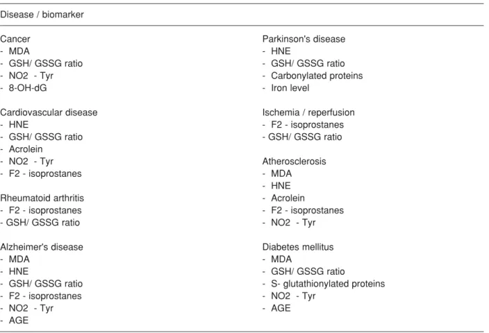
Convincing evidence for the association of oxidative/ nitrosative stress and acute and chronic diseases lies on validated biomarkers of oxidative stress. Such biomarkers have to be objectively measured and evaluated on healthy and ill subjects for long periods. Table 1 summarises most representative biomarkers of oxidative damage associated with human diseases discussed below (Dalle-Donne et al., 2006).
Cancer
Oxidative stress induces a cellular redox imbalance which has been found to be present in various cancer cells compared with normal cells; the redox imbalance thus may be related to oncogenic stimulation. Permanent modification of genetic material resulting from “oxidative damage” incidents represents the first step involved in mutagenesis, carcinogenesis, and ageing. DNA mutation is a critical step in carcinogenesis and elevated levels of oxidative DNA lesions have been noted in various tumours, strongly implicating such damage in the etiology of cancer.
ROS-induced DNA damage involves single- or double-stranded DNA breaks, purine, pyrimidine, or deoxyribose modifications, and DNAcross-links. DNA damage can result in either arrest or induction of transcription, induction of signal transduction
Disease / biomarker
Cancer Parkinson's disease
- MDA - HNE
- GSH/ GSSG ratio - GSH/ GSSG ratio
- NO2 - Tyr - Carbonylated proteins
- 8-OH-dG - Iron level
Cardiovascular disease Ischemia / reperfusion
- HNE - F2 - isoprostanes
- GSH/ GSSG ratio - GSH/ GSSG ratio
- Acrolein
- NO2 - Tyr Atherosclerosis
- F2 - isoprostanes - MDA
- HNE
Rheumatoid arthritis - Acrolein
- F2 - isoprostanes - F2 - isoprostanes
- GSH/ GSSG ratio - NO2 - Tyr
Alzheimer's disease Diabetes mellitus
- MDA - MDA
- HNE - GSH/ GSSG ratio
- GSH/ GSSG ratio - S- glutathionylated proteins
- F2 - isoprostanes - NO2 - Tyr
- NO2 - Tyr - AGE
- AGE
Abbreviations: MDA, malondialdehyde; HNE, 4-hydroxy-2-nonenal; AGE, advanced glycation end products; 8-OH-dG, 8-hydroxy-20- deoxyguanosine; GSH, reduced glutathione; GSSG, oxidised glutathione; NO2-Tyr, 3-nitro-tyrosine
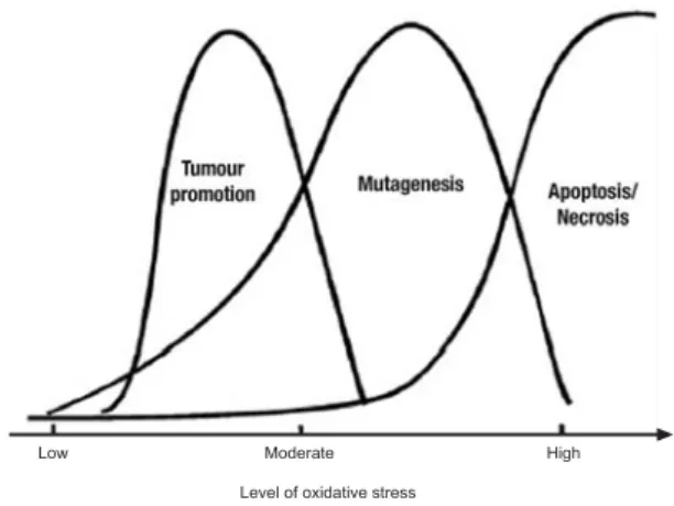
pathways, replication errors, and genomic instability, all of which are associated with carcinogenesis (Valko et al., 2006; Martnett, 2000). DNA damage, mutations, and altered gene expression are thus all key players in the process of carcinogenesis. The involvement of oxidants appears to be the common denominator to all these events (Valko et al., 2001; Valko et al., 2004; Valko et al., 2006). The role of oxidative stress at various stages of carcinogenic process and the process of apoptosis are outlined in the Figure 1.
There are many different sources-induced free radicals which have been linked with different types of cancers:
Hexavalent chromium is considered a potential lung carcinogen; Cr (VI)-induced cytotoxicity is associated with mitochondrial/lysosomal toxicity substantiated by the enhanced formation of free radicals (Pourahmad and O’Brien, 2001). Iron-induced oxidative stress is considered to be a principal determinant of human colorectal cancer (Valko et al., 2001). Occupational exposure to asbestos containing about 30% (weight) of iron is related to increased risk of asbestosis -the second most important cause of lung cancer (Stayner et al., 1996). Occupational exposure to cadmium has been associated with occurence of increased oxidative stress and cancer (Santos et al., 2005). Cadmium itself is unable to generate free radials directly, however, via indirect mechanisms, it can cause free radical-induced
damage to the gene expression. It has been reported that cadmium can cause activation of cellular protein kinases (protein kinase C), which result in enhanced phosphorylation of transcription factors and consequently lead to the transcriptional activation of target gene expression (Valko et al., 2005). It has been suggested that cadmium might also be implicated in the pathogenesis of human pancreatic cancer and renal carcinoma. Arsenic compounds are well-established human carcinogens, capable of binding to –SH groups and thus inhibiting various enzymes, including glutathione reductase (Roy and Saha, 2002). Studies support the hypothesis that arsenic may act as a co-carcinogen -not by causing cancer directly, but by allowing other factors, such as cigarette smoke or UV radiation, to cause DNA mutations more effectively (Waalkes et al., 2004). It tobacco smoke, a well known carcinogenic source of ROS, increased the oxidative DNA damage rate by 35–50%, as estimated from the urinary excretion of 8-OH-G, or by 20–50%, estimated from the level of 8-OH-G in leukocytes (Loft and Poulsen, 1996). In addition to the extensive studies devoted to the role of oxidative nuclear DNA damage in neoplasia, there exists evidence about the involvement of mitochondrial oxidative DNA damage in the carcinogenesis process. Mutations and altered expression in mitochondrial genes encoding for complexes I, III, IV and V, and in the hypervariable regions of mitochondrial DNA, have been identified in various human cancers. Hydrogen peroxide and other reactive oxygen species have been implicated in the activation of nuclear genes that are involved in mitochondrial biogenesis, transcription, and replication of the mitochondrial genome. Although the region of tumour cells that possess mutated mitochondrial DNA and the extent to which mitochondrial DNA alterations participate in the cancer process have not been satisfactorily established, a significant amount of information supporting the involvement of the mitochondria in carcinogenesis exists (Valko et al., 2006). This connection supports the fact that fragments of mitochondrial DNA have been found to be inserted into nuclear DNA, suggesting a possible mechanism for activation of oncogenes.
Apart from DNA damage, the lipid peroxidation process has been implicated in the mechanism of carcinogenesis. Once formed, lipoperoxyl radicals (ROO•) can be rearranged via a cyclisation reaction to endoperoxides with the final product of the peroxidation process being malondialdehyde (MDA) (Valko et al.,
Low Moderate High
Level of oxidative stress
Figure 1. The dose-dependent effect of relationship between level of oxidative stress and the tumour promotion process, process of mutagenesis and the process of apoptosis/necrosis (Valko et al., 2007).
2005). MDA is mutagenic in bacterial and mammalian cells and carcinogenic in rats. 4-hydroxy -2-nonenal (HNE) is weakly mutagenic but appears to be the major toxic product of lipid peroxidation (Martnett, 1999). There are also other exocyclic DNA adducts that arise from lipid peroxidation. For example etheno-dA, etheno-dC and etheno-dG have been detected by both (Hare and Stamler, 2005) P-post-labelling and GC–MS (Fedtke et al., 1990).
ROS, antioxidant status and
cancer
Many of the biological effects of antioxidants appear to be related to their ability not only to neutralize deleterious free radicals but also modulate cell-signalling pathways (Mates et al., 1999). Thus the modulation of cell signalling pathways by antioxidants could help prevent cancer by (i) preserving normal cell cycle regulation; (ii) inhibiting proliferation and inducing apoptosis; (iii) inhibiting tumour invasion and angiogenesis; (iv) suppressing inflammation; (v) stimulating phase II detoxification enzyme activity and other effects. It has been demonstrated that activation of NFΚB by nearly all stimuli can be blocked by antioxidants, including l-cysteine, N-acetyl cysteine (NAC), thiols, green tea polyphenols, and Vitamin E (Valko et al., 2007).
A large number of studies have established an association between cancer incidence and various disorders of GSH-related enzyme functions, alterations of glutathione S transferases (GSTs) being most frequently reported (Pastore et al., 2003).
GSTs are a family of enzymes that utilize glutathione in reactions contributing to the transformation of a wide range of compounds, including carcinogens, therapeutic drugs, and products of oxidative stress. The GSH/GSSG ratio measured in the blood of patients with colon and breast cancer has been found to be significantly decreased compared to the control (Pastore et al., 2003). There exists significant experimental and clinical evidence connecting thioredoxin to cancer (Baker et al.,1997): (i) elevated levels of TRX have been reported in a wide range of human cancers including cervical carcinoma, hepatoma, gastric tumours, lung, and colorectal carcinomas; (ii) many cancer cells have been shown to secrete TRX; (iii) TRX is able to stimulate the growth of a wide variety of human leukemia and solid tumour cell lines; (iv) overexpression of TRX protected cells from
oxidative-stress induced apoptosis and provided a survival as well as a growth advantage to tumours; (v) the elevated levels of thioredoxin in human tumours may cause resistance to chemotherapy (e.g. doxorubicin, cis-platin and others). As it is well known, low-molecular weight antioxidants are involved directly in the conversion of ROS to less reactive species. However, antioxidant protection therapy in cancer patients should be used only with caution since its effects depend on the stage at which it is introduced (Valko et al., 2004; Dreher and Juno, 1996).
Matrix metalloproteinases,
angiogenesis and cancer
Angiogenesis is a multi-step process, involving degradation of the endothelial cell basement membrane, endothelial cell migration to the perivascular stroma and capillary sprouting. Previously, the tumour suppressor p53 was understood to regulate the process of angiogenesis through the activation of genes that inhibit neovascularization and the repression of genes that promote vessel growth. With the identification of p63 and p73, p53 family regulation of angiogenesis has broadened and become more complex (Cameliet and Jain, 2000). The cancer cell invasion is a critical point for cancer metastasis. It is generally accepted that remodeling of the extracellular matrix (ECM) is a required process for cancer cell invasion (Westermark and Kahari, 1999). Angiogenesis involves multiple interactions between endothelial cells, surrounding pericytes, and smooth muscle cells, ECM, and angiogenic cytokines/growth factors.
Cardivascular disease
The ROS-induced oxidative stress in cardiac and vascular myocytes has been linked with cardiovascular tissue injury (Dhalla et al., 2000). Regardless of the direct evidence for a link between oxidative stress and cardiovascular disease, ROS-induced oxidative stres plays a role in various cardiovascular diseases such as atherosclerosis, ischemic heart disease, hypertension, cardiomyopathies, cardiac hypertrophy and congestive heart failure (Kukreja and Hess, 1992). The major sources of oxidative stress in cardiovascular system involve: (i) the enzymes xanthine oxidoreductase (XOR), (ii) NAD(P)H oxidase (multisubunit membrane complexes) and (iii) NOS as well as (iv) the mitochondrial cytochromes and (v) hemoglobin (Berry and Hare, 2004; Hare and Stamler, 2005). Oxidative
stress is associated with increased formation of ROS that modifies phospholipids and proteins leading to peroxidation and oxidation of thiol groups (Molavi and Mehta, 2004). The assaults by ROS lead to changes in membrane permeability, membrane lipid bilayer disruption and functional modification of various cellular proteins. In addition to cellular protein and lipid damage, abnormalities in myocyte function due to increased oxidative stress are considered to be associated with the effects of ROS on subcellular organelles. Mitochondrial creatine kinase activity of rat heart was reported to decrease upon exposure to xanthine plus xanthine oxidase or hydrogen peroxide (Hayashi et al., 1998). Cardiac mitochondria treated with ROS exhibited decreased Ca2+ membrane
transport; cardiac mitochondria exposed to 4-hydroxy-2- nonenal cause a rapid decrease in NAD(P)H state 3 and uncoupled respiration. In view of these results, it may be concluded that oxidative stress may alter the activities of different subcellular structures, proteins, and lipids and thus changing myocyte function (Valko et al., 2007).
Deficiency in ATP synthesis in the ischemic heart may also impair Ca2+ handling mechanisms in the
sarcolemmal and sarcoplasmic reticular membranes and thus induce Ca2+ overload. Reperfusion of the
ischemic heart may also increase the uptake of extracellular Ca2+ into the myocardium and thus be
another factor for Ca2+ overload. Intracellular Ca2+
overload seems to be a common denominator for stimulation of neointimal hyperplasia and thus the occurrence of atherosclerosis, vasoconstriction for the development of hypertension, myocardial cell damage observed in ischemia-reperfusion, and cardiac hypertrophy in heart failure (Valko et al., 2007).
Since increased amounts of superoxide radical and hydrogen peroxide have been reported in hypertensive patients, the etiology of ROS-induced oxidative stres in the pathogenesis of hypertension is well established (Romero and Reckelhoff, 1999). Superoxide promotes cell proliferation whereas hydrogen peroxide induces apoptosis and activates protein kinase C, suggesting a role for protein kinase C in ROS-mediated vascular disease. ROS-induced oxidative stress in hypertensive patients is accompanied by decreased levels of antioxidants such as Vitamin E, GSH, and SOD, all good scavengers of free radicals (Li and Forsterman, 1999).
Ischemic preconditioning
Preconditioning ischemia triggers endogenous protective mechanisms in heart muscle, and is the most effective means for myocardial protection. The protection induced by short preconditioning ischemia periods disappears within 1–2 h, but reappears after 24–72 h (the so called “second window protection”).
The process involves such components as a paradoxical protective role of oxygen free radicals, adenosine, adenosine receptors, heat shock proteins (HSP), nitric oxide, the epsilon isoform of protein kinase C (PKC), mitogen-activated protein kinases, the mitochondrial ATP-dependent potassium (K+(ATP))
channels (Kalikiri and Sachan, 2004; Zhao et al., 2001; Skyschally et al., 1999).
Rheumatoid arthritis
Rheumatoid arthritis is an autoimmune disease that causes chronic inflammation of the joints and tissue around the joints with infiltration of macrophages and activated T cells (Bauerova and Bezek, 1999). The pathogenesis of this disease is linked predominantly with the formation of free radicals at the site of inflammation. Oxidative injury and inflammatory status in various rheumatic diseases was confirmed by increased levels of isoprostanes and prostaglandins in serum and synovial fluid compare to controls. Oxidative conditions in synovial tissue are also associated with a higher incidence of p53 mutations (Firestein et al., 1997). T cells isolated from the synovial fluid of patients with rheumatoid arthritis show signs of decreased intracellular GSH level, impaired phosphorylation of the adaptor protein linker for T-cell activation (LAT) and the “primed” CD45RO phenotype (Maurice et al., 1997). The migration of monocytes and lymphocytes into the rheumatoid arthritis synovium is mediated by the abnormal expression of several adhesion molecules (ELAM-1, VCAM-1, ICAM-1, ICAM-2) (Cunnane et al., 2001) ; this can be explained by the abnormal induction of redox-sensitive signalling pathways.
Diabetes
A relatively small amount (10%) of patients suffering from diabetes mellitus has type 1, or insulin dependent diabetes (Brownlee and Cerami, 1981; Niedowicz and Daleke, 2005). However, the majority of diabetes patients are non-insulin-dependent and capable at least initially of producing insulin, but are deficient in
their cellular response. This type of diabetes is called as the type 2 diabetes mellitus. Decreased uptake of glucose into muscle and adipose tissue leads to chronic extracellular hyperglycemia resulting in tissue damage and pathophysiological complications, involving heart disease, atherosclerosis, cataract formation, peripheral nerve damage, retinopathy and others (Brownlee and Cerami, 1981). Increased oxidative stress has been proposed to be one of the major causes of the hyperglycemia-induced trigger of diabetic complications. Hyperglycemia in an organism stimulates ROS formation from a variety of sources. These sources include oxidative phosphorylation, glucose autooxidation, NAD(P)H oxidase, lipooxygenase, cytochrome P450 monooxygenases,
and nitric oxide synthase (NOS) (Valko et al., 2007).
Neurological disorders
The brain is particularly vulnerable to oxidative damage because of its high oxygen utilisation, its high content of oxidisable polyunsaturated fatty acids, and the presence of redox-active metals (Cu, Fe). Oxidative stress increases with age and therefore it can be considered as an important causative factor in several neurodegenerative diseases, typical for older individuals (Valko et al., 2007).
Alzheimer’s disease
The brains of patients with Alzheimer’s disease (AD) show a significant extent of oxidative damage associated with a marked accumulation of amyloid- α peptide (Aα), the main constituent of senile plaques in brain, as well as deposition of neurofibrillary tangles and neurophil threads (Butterfield et al., 2002).
The direct evidence supporting increased oxidative stres in AD brain include (i) increased Cu, Fe, Al, and Hg content; (ii) increased lipid peroxidation and decreased polyunsaturated fatty acid content, and an increase in 4-hydroxynonenal, an aldehyde product of lipid peroxidation in AD ventricular fluid; (iii) increased protein and DNA oxidation; (iv) diminished energy metabolism and decreased cytochrome c oxidase content; (v) advanced glycation end products (AGE), malondialdehyde, carbonyls, peroxynitrite, heme oxygenase- 1, and SOD-1 in neurofibrillary tangles, (vi) the presence in activated microglia surrounding most senile plaques of nitrotyrosine, formed from peroxynitrite (ONOO•α). As mentioned above, elevated production of Aα, as a preventive antioxidant for brain lipoproteins under the action of increased oxidative
stress and neurotoxicity in ageing, is postulated to represent a major event in the development of Alzheimer’s disease (Butterfield et al., 2002).
Parkinson’s disease
Parkinson’s disease (PD) involves a selective loss of neurons in an area of the midbrain called the substantia nigra (Sayre et al., 2001). The cells of the substantia nigra use dopamine (a neurotransmitter-chemical messenger between brain and nerve cells) to communicate with the cells in another region of the brain called the stratium. Thus, a reduction in nigral dopamine levels results in a decrease in stratial dopamine that is believed to cause PD symptoms (Jenner, 2003). Neuronal loss and Lewy bodies, the pathological hallmarks of PD, have been fond in cerebral cortex, anterior thalamus, hypothalamus, amygdala and basal forebrain (Sayre et al., 2001). The major component of intracytoplasmic Lewy bodies are filaments consisting of α-synuclein. Two recently identified point mutations in α-synuclein are the genetic causes of PD (Jin and Yang, 2006). A majority of studies explored the effect of oxidative stress that contributes to the cascade of events leading to dopamine cell degeneration in PD (Tretter et al., 2004). The occurrence of oxidative stres in PD is supported by both postmortem studies and by studies demonstrating the capacity of oxidative stress to induce nigral cell degeneration. There is evidence that there are high levels of basal oxidative stress in the substantia nigra pars compacta (SNc) in the normal brain, but that this increases in PD patients. However, other factors involving inflammation, excitotoxic mechanisms, toxic action of nitric oxide, and mitochondrial dysfunction play roles in the etiology of PD (Andersen, 2004).
Although free radicals seems to be the main factor of many diseases, the human body has several mechanisms to counteract damage by free radicals and other reactive oxygen species. One important part of defence is enzyme systems, including superoxide dismutases, glutathione peroxidases and catalase, which decrease concentrations of the most harmful oxidants in the tissues. Several essential minerals including selenium, copper, manganese and zinc are necessary for the formation or activity of these enzymes.
The second part of defence against free radical damage is the antioxidants which can safely interact with free radicals and terminate the chain reaction before vital molecules are damaged. Some such
antioxidants, including glutathione, ubiquinol and uric acid, are produced during normal metabolism in the body. Other lighter antioxidants are found in the diet (Valko et al., 2006).
Recent years many antioxidants have been identified. The best known are vitamin E, vitamin C and the carotenoids. Many other non-nutrient food substances, generally phenolic or polyphenolic compounds and flavinoids show antioxidant properties and, thus, may be important for health. Some It should be noted, however, that while most flavonoids and phenolic compounds possess antioxidant properties and are considered safe, there have been reports of toxic flavonoid-drug interactions, as well as liver failure, contact dermatitis, hemolytic anemia and oestrogenic-related concerns, including male reproductive health and breast cancer associated with dietary flavonoid intake (Valko et al., 2006).
There is no doubt that antioxidants are necessary components for our health but we do not forget that the antioxidants and free radicals production should be in balance. Taking more synthetic antioxidants could be deleterius in order to useful. Reducing externally free radical sources such as smoking cigarettes, environmental pollutants, pesticides etc., in our life may be the better choise for eliminating hazardous effects of free radicals.
References
Andersen JK. Oxidative stress in neurodegeneration: Cause or consequence? Nat Rev Neurosci. 5:18-25, 2004.
Baker A, Payne CM, Briehl MM and Powis G. Thioredoxin, a gene found overexpressed in human cancer, inhibits apoptosis in vitro and in vivo. Cancer Res. 57:5162-5167, 1997.
Bauerova K and Bezek S. Role of reactive oxygen and nitrogen species in etiopathogenesis of rheumatoid arthritis. Gen Physiol Biophys.18:15-20, 1999. Berry CE and Hare JM. Xanthine oxidoreductase in the
cardiovascular system: Molecular mechanisms and pathophysiologic implications. J Physiol. 555:589-606, 2004.
Brownlee M and Cerami A. The biochemistry of the complications of diabetes-mellitus. Ann RevBiochem . 50:385-432, 1981.
Butterfield DA, Castegna A, Lauderback CM and Drake
J. Evidence that amyloid beta-peptide-induced lipid peroxidation and its sequelae in Alzheimer’s disease brain contribute to neuronal death. Neurobiol Aging. 23:655-664, 2002.
Carmeliet P and Jain RK. Angiogenesis in cancer and other diseases. Nature, 407: 249-57, 2000. Cunnane G, Fitzgerald O, Beeton C, Cawston TE and
Bresnihan B. Early joint erosions and serum levels of matrix metalloproteinase 1, matrix metalloproteinase 3, and tissue inhibitor of metalloproteinases 1 in rheumatoid arthritis. Arth Rheumat. 44:2263-2274, 2001.
Dreher D and Junod AF. Role of oxygen free radicals in cancer development. Eur J Cancer, 32A:30-38, 1996.
Dalle-Donne I, Rossi R, Colombo R, Giustarini D and Milzani A. Biomarkers of oxidative damage in human disease. Clin Chem. 52:601-623, 2006. Dhalla NS, Temsah RM and Netticadan T. Role of
oxidative stress in cardiovascular diseases. J Hypertens. 18:655-673, 2000.
Fedtke N, Boucheron JA, Walker VE and Swenberg JA. Vinyl chloride-induced DNA adducts. 2. Formation and persistence of 7-2-oxoethylguanine and n2,3-ethenoguanine in rat-tissue DNA. Carcinogenesis.11:1287-92, 1990.
Firestein GS, Echeverri F, Yeo M, Zvaifler NJ and Green DR. Somatic mutations in the p53 tumor suppressor gene in rheumatoid arthritis synovium. Proc Natl Acad Sci USA. 94:10895-10900, 1997. Halliwell B and Gutteridge JMC. Free Radicals in
Biology and Medicine (3rd ed.). Oxford University Press. 1999.
Hare JM and Stamler JS. NO/redox disequilibrium in the failing heart and cardiovascular system. J Clin Invest. 115: 509-17, 2005.
Harman D. Aging- A theory based on free-radical and radiation-chemistry. J Gerontol. 11: 298-300, 1956. Hayashi H, Iimuro M, Matsumoto Y and Kaneko M. Effects of gamma-glutamylcysteine ethyl ester on heart mitochondrial creatine kinase activity: Involvement of sulfhydryl groups. Eur J Pharmacol. 349:133-136, 1998.
Jenner P. Oxidative stress in Parkinson’s disease. Ann Neurol. 53:26–36. 2003.
Jin L and Yang H. Alpha-Synuclein aggregation and Parkinson’s disease: Factors affecting the aggregation of alpha-synuclein. Prog Biochem Biophys. 33:321-8, 2006.
Kalikiri PC and Sachan RS. Ischemic and anesthetic preconditioning of the heart: An insight into the concepts and mechanisms. Internet J Anesthesiol. 8:2, 2004.
Kaneko M, Elimban V and Dhalla NS. Mechanism for depression of heart sarcolemmal Ca2+ pump by oxygen freeradicals. Am J Physiol. 257:804-811, 1989.
Kovacic P and Jacintho JD. Mechanisms of carcinogenesis: Focus on oxidative stress and electron transfer. Curr Med Chem. 8:773-96, 2001. Kukreja RC and Hess ML. The oxygen free-radical
system-From equations through membrane–protein interactions to cardiovascular injury and protection. Cardiovasc Res. 26:641-655, 1992.
Li HG and Forstermann U. Nitric oxide in the pathogenesis of vascular disease. J Pathol.190:244-254, 2000.
Loft S and Poulsen HE. Cancer risk and oxidative DNA damage in man. J Mol Med. 74:297-312, 1996. Marnett LJ. Lipid peroxidation—DNA damage by
malondialdehyde. Mut Res-Fund Mol Mech Mutagen. 424: 83-95, 1999.
Marnett LJ. Oxyradicals and DNA damage. Carcinogenesis. 21:361-70, 2000.
Mates JM, Perez-Gomez C and De Castro IN. Antioxidant enzymes and human diseases. Clin Biochem. 32: 595-603, 1999.
Maurice MM, Nakamura H, Van der Voort EAM, Van Vliet AI, Staal FJT and Tak PP. Evidence for the role of an altered redox state in hyporesponsiveness of synovial T cells in rheumatoid arthritis. J Immunol. 158:1458-65, 1997.
Molavi B and Mehta, JL. Oxidative stress in cardiovascular disease: Molecular basis of its deleterious effects, its detection, and therapeutic considerations. Curr Opin Cardiol. 19:488-93, 2004.
Miller DM, Buettner GR and Aust SD. Transition metals’s catalysts of “autoxidation” reactions. Free Radic Biol Med. 8: 95-108, 1990.
Niedowicz DM and Daleke DL. The role of oxidative stres in diabetic complications. Cell Biochem Biophys. 43:289-330, 2005.
Pastore A, Federici G, Bertini E and Piemonte F. Analysis of glutathione: Implication in redox and detoxification. Clin Chim Acta. 333:19-39, 2003. Pourahmad J and O’Brien PJ. Biological reactive
intermediates that mediate chromium VI toxicity. Biol React Intermed. VI: Adv Exp Med Biol. 500:203-207, 2001.
Romero JC and Reckelhoff JF. Role of angiotensin and oxidative stress in essential hypertension. Hypertension. 34:943-949, 1999.
Roy P and Saha A. Metabolism and toxicity of arsenic: A human carcinogen. Curr Sci. 82:38-45, 2002. Ridnour LA, Isenberg JS, Espey MG, Thomas DD,
Roberts DD and Wink DA. Nitric oxide regulates angiogenesis through a functional switch involving thrombospondin-1. Proc Natl Acad Sci. 102:13147-52, 2005.
Santos FW, Zeni G, Rocha JB, Weis SN, Fachinetto JM and Favero AM. Diphenyl diselenide reverses cadmium-induced oxidative damage on mice tissues. Chem Biol Interact. 151:159-165, 2005. Sayre LM, Smith MA and Perry G. Chemistry and
biochemistry of oxidative stress in neurodegenerative disease. Curr Med Chem. 8:721-38, 2001.
Stayner LT, Dankovic DA and Lemen RA. Occupational exposure to chrysotile asbestos and cancer risk: A review of the amphibole hypothesis. Am J Public Health. 86;179-186, 1996.
Skyschally A, Schulz R, Gres P,Korth HG and Heusch G. Attenuation of ischemic preconditioning in pigs by scavenging of free oxyradicals with ascorbic acid. Am J Physiol Heart Circ Physiol. 284:698-703, 2003.
Tretter L, Sipos I and Adam-Vizi V. Initiation of neuronal damage by complex I deficiency and oxidative stress in Parkinson’s disease. Neurochem Res. 29:569-77, 2004.
Valko M, Morris H, Mazur M, Rapta P and Bilton RF. Oxygen free radical generating mechanisms in the colon: Do the semiquinones of Vitamin K play a role in the aetiology of colon cancer? Biochim Biophys Acta. 1527:161-166, 2001.
Valko M, Izakovic M, Mazur M, Rhodes CJ and Telser J. Role of oxygen radicals in DNA damage and cancer incidence. Mol Cell Biochem. 266: 37-56, 2004.
Valko M, Morris H and Cronin M TD. Metals, toxicity and oxidative stress. Curr Med Chem. 12:1161-208, 2005.
Valko M, Rhodes CJ, Moncol J, Izakovic M and Mazur M. Free radicals, metals and antioxidants in oxidative stress-induced cancer. Chem Biol Interact. 160:1-40, 2006.
Valko M, Leibfritz D, Moncola J, Cronin M, Mazura M and Telser I. Free radicals and antioxidants in normal physiological functions and human disease. Int J Biochem Cell Biol. 39(1):44-84, 2007. Waalkes MP, Liu J, Ward JM, Diwan LA. Mechanisms
underlying arsenic carcinogenesis: Hypersensitivity of mice exposed to inorganic arsenic during gestation. Toxicology. 198:31-38, 2004.
Westermarck J and Kahari VM. Regulation of matrix metalloproteinase expression in turner invasion. FASEB J. 13:781-792, 1999.
Zhao TC, Hines DS and Kukreja RC. Adenosine-induced late preconditioning in mouse hearts: Role of p38 MAP kinase and mitochondrial K (ATP) channels. Am J Physiol Heart Circ Physiol. 280:H1278-H1285, 2001.