531
Bull Vet Inst Pulawy 51, 531-534, 2007
GENERALISED TUBERCULOSIS IN A CAT
MEHMET HALIGUR, SEVIL ATALAY VURAL1, MEHMET SAHAL2,KEREM URAL2, AND SENAY BERKIN1
Department of Pathology, Faculty of Veterinary Medicine, University of Mehmet Akif Ersoy, 15100, Burdur, Turkey
1Department of Pathology, 2 Department of Internal Medicine, Faculty of Veterinary Medicine, University of Ankara, 06110, Ankara, Turkey
[email protected] [email protected]
Received for publication May 08, 2007
Abstract
Clinical, pathomorphological and immunohisto-logical findings of generalised tuberculosis diagnosed in a 1-year-old male tabby cat were evaluated. Macroscopic examination revealed greyish-white nodules, exceeding the surface of the organ, in the meninges and kidneys. The meninges nodules were of the size of a pinhead and those in the kidneys were 0.2-0.5 cm in diameter. The mediastinal lymph nodes were observed to be enlarged, and their cut surfaces displayed nodules of a similar appearance and size. Microscopic examination revealed typical tubercles in the cerebrum, cerebellum, kidneys, and mediastinal lymph nodes. The presence of Mycobacterium tuberculosis either free or located within the cytoplasm of macrophages was demonstrated by means of the avidin-biotin complex peroxidase method.
Key words: cat, tuberculosis, pathology.
Tuberculosis, which is a chronic zoonotic infection, is frequently encountered in all races of cats (7, 8, 19). However, Siamese cats have been reported to be more susceptible in comparison to other feline breeds. The infection is thought to be caused, generally by Mycobacterium bovis and to a less extent, by
Mycobacterium tuberculosis. Some cases of the
infection with Mycobacterium avium, Mycobacterium
microti, and Mycobacterium lepraemurium have also
been reported (16, 21).
Macroscopic findings of the infection have been observed in the lymph nodes, lungs, liver, kidneys, spleen, and adrenal glands (1, 2, 5, 11, 16). In certain cases, characterised with clinical observation of nervous symptoms, microscopic examination revealed lesions specific to tuberculosis, despite the absence of macroscopic findings (17).
The aim of this study was to investigate the clinical, pathomorphological and immunohistological findings in generalised tuberculosis in a cat.
Material and Methods
The material of this study was a one-year-old male tabby cat. After the necropsy of the cat, samples of the kidneys, mediastinal lymph nodes, cerebrum, and cerebellum, in which macroscopic changes were observed, were taken. Following fixation in 10% buffered formalin solution; the samples were trimmed, dehydrated, and blocked in paraffin. Five-six micron sections prepared from the blocks were stained with haematoxylin-eosin (HE) and Ziehl-Neelsen (ZN) methods for histopathological examination. In order to identify the causative agent, sections were stained in accordance with the immunoperoxidase staining method.
The Avidin-Biotin Complex Peroxidase technique (ABC-P) was employed for the detection of the agent using rabbit-anti M. tuberculosis serum (Dako). AEC (3 amino-9 ethylcarbazole) was used as a chromogen. Accordingly, following the deparaffinisation and dehydration of the sections in xylol and alcohol series, the sections were incubated in 3% solution of hydrogen peroxide in absolute methanol for 20 minutes in order to block endogenous peroxide activity. Subsequently, sections were subjected to pronase at 40°C for 10 min and treated with normal goat serum for 20 min at 40°C. Then, rabbit anti-M.
tuberculosis serum diluted 1:500 was poured onto the
sections and they were incubated for 1 h at 40°C. The next stage involved incubation of the sections in biotinised goat anti-rabbit IgG and streptoavidin peroxidase reagent (Cadenza Tags, Shandon) for 20 min and staining with AEC for 2 min. In all stages, excluding treatment with normal goat serum, sections were washed with PBS. Mayer’s haematoxylin was used for counterstaining the sections thatwere then examined under a light microscope.
532
Results
Clinical findings. Medical history of the cat
suggested respiratory distress, dyspnoea, tachypnoea, lethargy, and anorexia with body weight loss, ongoing for 3 months. According to the notification of the owner of the animal, the cat was kept in a single-roomed humid house under inadequate hygienic and nutritional conditions, together with 30 cats, and from time to time, was fed homemade food. Furthermore, it appeared that the owner of the animal has been infected with M.
tuberculosis for 24 years and was a carrier of the disease
having received treatment for a number of times in previous years.
Physical examinations revealed poor body condition and underweight (1.9 kg). of the cat. Moreover, generalised muscular weakness, pyrexia (39.9°C) and generalised lymphadenopathy was observed. Laboratory examination revealed only neutrophilia (14.7 x 10⁹/l, reference value 2.5-12.5 x 10⁹/l) and lymphopenia (0.76 x 10⁹/l) in the blood, and hypoalbuminaemia (17 g/l; reference value 25-35 g/l) in the serum. Numerous (generalised) foci of 12 mm in size were observed in the lungs by means of thoracic radiography.
Pulmonary infection was suspected in the patient, according to clinical findings, and therefore antibiotics (Reptopen-S 40 ml. flk., 0.1 ml/kg s.c., benzylpenicillin procain 200000 IU + dihydrostreptomycin 200 mg), vitamins B1 and B6 (Neurogriseovit), and lactated Ringer solution for supportive fluid therapy (200 ml i.v.) were administered as the initial treatment. No improvement was seen in the general condition of the patient on the 2nd d of treatment, and the animal eventually died.
Necropsy findings. Pathological changes were
observed in the kidneys, mediastinal lymph nodes, cerebellum, and cerebrum. Amongst the 3 subcapsularly and cranially located greyish-white rough nodules protruding the surface in the kidneys, one was located in the right kidney (diameter 0.2 cm), and the remaining two (diameters 0.4 and 0.5 cm) in the left kidney. The mediastinal lymph nodes were quite enlarged and the presences of numerous yellowish foci with borders of ambiguous distinction were observed in cross sections. Pinhead sized nodules and opaque areas were observed in the meninges of the cerebrum and cerebellum. Lung lesions determined by radiography were not seen at the necropsy.
Histopathological findings. Subcapsularly
located multi granulomatous foci characterised with a slightly eosinophilic necrotic area in the centre, and mostly neutrophil leukocytes, lymphocytes, macrophages, epithelioid histiocytes, and a few plasma cells in the periphery, were noted in the renal cortex (Fig. 1). Some epithelial cells of the surrounding tubuli were found out to be turgid, whereas the nuclei of some cells displayed a picnotic appearance. Mononuclear cell infiltration, comprising mostly lymphocytes, and haemorrhages were observed between these tubuli in the form of focal areas.
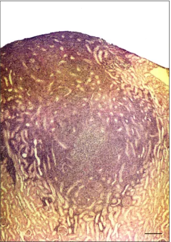
Widespread granulomas located in multiple foci were observed in the mediastinal lymph nodes. The granulomas revealed necrosis in the centre, and cellular infiltration comprising mainly of lymphocytes, macrophages, epithelioid histiocytes, neutrophil leukocytes, and a few plasma cells (Fig. 2). Lymph follicles were observed to be partly hyperplasic, and the cortical and medullar sinuses were filled with sloughed endothelial cells and a few number of neutrophil leukocytes.
Focal granulomas with irregular distribution and borders of ambiguous distinction were found in the meninges of the cerebrum, particularly in the basis of the fourth ventricle and pons cerebri. Some of the lesions were found out to extend to the parenchyma. Slightly eosinophilic necrotic areas were located in the centre of the granulomas, whereas inflammation involving mainly lymphocytes and neutrophil leukocytes, and a few numbers of histiocytes and plasma cells were noticed in the periphery (Fig. 3). In the region surrounding this area, a perivascularly located mononuclear cell infiltration, mainly comprising of lymphocytes, was observed. Similar findings were noted in the meninges of the cerebellum. Causative
Mycobacterium sp. was not found out in granuloma
tissues, stained with ZN technique. However, tubercuolosis positive antigens were detected by immunoperoxidase technique in the cytoplasm of macrophages and in the free state in the same tissues (Fig. 4).
Fig. 1. Tuberculous foci in the cortex of kidney. HE,
533
Fig. 2. Tuberculous foci in the mediastinal lymph node
(arrow). HE, Bar=200 µm
Fig. 3. Inflammatory infiltration cells in the meninges
and cerebellum. HE, Bar=200 µm
Fig. 4. Tuberculous agent in the cytoplasm of
macrophage (thin arrow). ABC-P, Bar=50 µm
Discussion
Tuberculosis is considered to be one of the most significant infectious diseases with regard to public health and economy. It is still widespread in Turkey, despite being either under control or completely eradicated in many countries (16).
Mycobacteria are found in the saliva, faeces, urine, or open abscess material of infected animals (4, 21). The infection is generally acquired through alimentary intake of contaminated food such as milk and meat, and to a less extent, through inhalation or wounds of the skin. Tuberculosis is reported to be generally observed as a generalised infection in cats (1, 2, 5, 11, 16). The presence of tuberculous lesions in the mediastinal lymph nodes, despite the absence of pulmonary lesions (incomplete primary complex) in this study, suggested the acquirement of the disease through inhalation. Lesions located in the kidneys and central nervous system, were considered to have developed following generalisation of the disease.
Occasionally, M. tuberculosis infections have been reported to be passed onto cats from humans (9). In this study, the owner of the animal was found to be infected with M. tuberculosis and carrier of the disease for 24 years, and therefore, the infection was considered to have passed onto the cat from the owner.
In a study carried out by Gunn-Moore et al. (8), amongst 19 cats diagnosed with tuberculosis, 12 were observed to display skin lesions (face, thorax, paws, base of tail and perineum), 9 were determined to have submandibular lymphadenopathy, 4 displayed generalised lymphadenopathy (popliteal and mediastinal lymphadenopathy), and 2 were demonstrated to suffer from localised arthritis. Despite the determination of generalised lymphadenopathy (mesenteric lymphadenopathy) in our study, in conformity with the literature, skin lesions and joint related findings were not encountered.
In the above-mentioned study of Gunn-Moore
et al. (8), thoracic radiography findings comprised
lymphadenopathy and dyspnoea associated with diffuse pulmonary infiltration in one cat, symptoms of bilateral submandibular lymphadenopathy and cough associated with miliary increase in lung density in another cat, and arthritis associated with diffuse increase in lung density again in one cat. In conformity with the study, numerous foci displaying increase in the opacity in lung radiography were observed also in this study. However, small foci were not detected in pathological examination.
Macroscopic findings of the infection have been observed mainly in the lymph nodes (12 cases) and lungs (9 cases), and then in the liver (4 cases), kidneys (3 cases), spleen (3 cases), testes (1 case), and adrenal glands (1 case) (1-6, 11, 16, 18). Infected lymph nodes have been determined to be enlarged 1-5 times, and caseous foci of an approximate diameter of 2 mm have been observed to be located mainly in the cortex in cross sections (1, 2, 5, 11, 16). Furthermore, irregular miliary caseous foci of greyish colour and with a diameter of 0.5-2 cm and borders of ambiguous distinction have been found to be located in the paranchyma of the lungs (3, 5, 16, 18). The liver has been observed to be swollen and to display yellowish red multiple, irregular foci, smaller than 1 mm (1, 5, 6, 14). Numerous white foci with a diameter of 1-3 mm have been determined mainly in the cortex of the kidneys (16). The spleen was enlarged and displayed whitish spots located in the
534
parenchyma (1, 6, 13). Furthermore, white coloured multiple granulomatous foci, resembling a bunch of grapes in shape, have been reported in the pericardium and mediastinal pleura (16). Findings similar to those reported in the mentioned literature were also observed in the mediastinal lymph nodes and kidneys in this study. In addition to the aforementioned observations, findings specific to tuberculosis were revealed in microscopic examination in cats with nervous symptoms and without any macroscopic finding in the cerebrum and cerebellum (17). In this study, macroscopical and microscopical findings were observed in the cerebrum and cerebellum.
Immunohistochemical staining methods have been reported to be more sensitive for Mycobacteria in comparison to Ziehl-Neelsen staining (10, 15). In addition, positive mycobacterium antigens were detected by immunoperoxidase technique in 45 d old calf (20). In this study, we were unable to identify the causative agent by means of ZN staining despite the demonstration of positive results upon the application of immunohistochemical staining methods. The utilisation of immunohistochemical staining methods as more sensitive can be suggested in the diagnosis of cases suspected of tuberculosis.
References
1. Barry M., Taylor J., Woods J.P.: Disseminated
Mycobacterium avium infection in a cat. Cand Vet J
2002, 43, 369-371.
2. Blunden A.S., Smith K.C.: A pathological study of a mycobacterial infection in a cat caused by a variant with cultural characteristics between Mycobacterium
tuberculosis and M bovis. Vet Rec 1996, 138, 87-88.
3. Buergelt C.D., Fowler J.L., Wright P.J.: Disseminated avian tuberculosis in a cat. California Vet 1982, 10, 13-15.
4. Dietrich U., Arnold P., Guscetti F., Peyffer G.E., Spiess B.: Ocular manifestation of disseminated
Mycobacterium simiae infection in a cat. J Small Anim
Pract 2003, 44, 121-125.
5. Dorlet R.: Disseminated tuberculosis caused by
Mycobacterium avium in a cat. J Am Vet Med Assoc
1986, 189, 1336-1337.
6. Griffin A., Newton A.L., Aronson L.R., Brown D.C., Hess R.S.: Disseminated Mycobacterium avium complex infection following renal transplantation in cat. J Am Vet Med Assoc 2003, 222, 1097-1100.
7. Gunn-Moore D.A., Jenkins P.A.: Tuberculosis in cats. Vet Rec 1994, 134, 395.
8. Gunn-Moore D.A., Jenkins P.A., Lucke V.M.: Feline tuberculosis: a literature review and discussion of 19 cases caused by an unusual mycobacterial variant. Vet Rec 1996, 138, 53-58.
9. Gunn-Moore D.A.: www.fabcats.org/tuberculosis.html . 10. Gutièrrez C.M.M., García M.J.F.: Comparison of
Ziehl-Neelsen staining and immunohistochemistry for detection Mycobacterium bovis in bovine and caprine tuberculosis lesions. J Comp Path 1993, 109, 361-370. 11. Hix J.W., Jones T.C., Karlson A.: Avian tubercle
bacillus infection in the cat. J Am Vet Med Assoc 1961,
138, 641-647.
12. Jennings A.R.: The distribution of tuberculous lesions in the dog and cat. Vet Rec 1949, 61, 380-384.
13. Jordan H.L., Chon L.A., Armstrong J.: Disseminated
Mycobacterium avium complex infection in the three
Siamese cats. J Am Vet Med Assoc 1994, 204, 90-93. 14. Kaneene J. B., Bruning-Fann C., Dunn J., Mullaney T.
P., Berry D., Massey J.P., Thoen C. O., Halstead S., Schwartz K.: Epidemiologic investigation of
Mycobacterium bovis in a population of cats. Am J Vet
Res 2002, 63, 1507-1511.
15. Massone A.R., Itagaki S., Ibargoyen G.S., Martin A.A., Doi K., Gimeno E.J.: Demonstration of Mycobacterium
paratuberculosis in tissue section: comparative of
histopathological and immunohistochemical methods. Isr J Vet Med 1991, 46, 48-50.
16. Monies R.J., Cranwell M.P., Palmer N., Inwald J., Hewinson R.G., Rule B.: Bovine tuberculosis in domestic cats. Vet Rec 2000, 146, 407-408.
17. Paulsen D.B., Kern M., Weigand C.M.: Mycobacterial neuritis in a cat. J Am Vet Med Assoc 2000, 10, 1589-1591.
18. Perez J., Calzada J., Leon-Vizcaino L., Cubero M.J., Velarde J., Mozos E.: Tuberculosis an Iberian lynx (Lynx pardina). Vet Rec 2001, 31, 414-415.
19. Orr C.M., Kelly D.F., Lucke V.M.: Tuberculosis in cats: a report of two cases. J Small Anim Pract 1980,
21, 247-253.
20. Vural Atalay S., Tunca R.: Generalized tuberculosis in a 45 days old calf. Dtsch Tierarztl Wochenschr 2001,
108, 568-470.
21. Wilesmith J.W., Clifton Hadley R.S.: Tuberculosis in cats. Vet Rec 1994, 134, 359.