Nilay Sahin,1 Ali Yavuz Karahan2
1Department of Physical Therapy and Rehabilitation, Balikesir University Faculty of Medicine, Balikesir, Turkey 2Department of Physical Therapy and Rehabilitation, Usak University Faculty of Medicine, Usak, Turkey
ABSTRACT
OBJECTIVE: The aim of this study was to examine how much recovery upper obstetric brachial plexus palsy (OBPP) patients showed with exercises when they were 3, 6, and 12 months old and to evaluate whether the exercise treatment given at different frequencies contributes to this recovery or not.
METHODS: This study was designed as a randomized controlled trial. Sixty cases who were referred to Pediatric Rehabili-tation and Pediatric Orthopedic Clinics with the diagnosis of having Groups I and II OBPP according to Narakas classification were included in the study. Subjects were randomly divided into two treatment groups. The first group had intense exercise program 3 times daily, and the second group had a standard exercise program once in a day. The subjects were assessed using a passive-active range of motion (ROM) and hospital for sick children muscle grading system at their first clinic visit and every month after until they became 12 months old.
RESULTS: In both groups, a significant recovery was observed in ROM and muscle strength of all movements of the shoul-der, elbow flexion, and forearm supination at the 3rd, 6th, and 12th-month reassessments, whereas a significant difference was
not achieved on both parameters between two groups.
CONCLUSION: According to the results, exercise frequency did not affect the recovery rate and results in the cases with OBPP and exercises were influential against possible complications that may occur.
Keywords: Brachial plexus lesion; exercise; functional recovery; muscle strength.
Received: July 08, 2017 Accepted: December 28, 2017 Online: August 07, 2018 Correspondence: Dr. Ali Yavuz KARAHAN. Usak Universitesi Tip Fakultesi, Fiziksel Tedavi ve Rehabilitasyon Anabilim Dali, 64200 Usak, Turkey. Tel: +90 276 221 22 33 e-mail: [email protected]
© Copyright 2018 by Istanbul Provincial Directorate of Health - Available online at www.northclinist.com
North Clin Istanb 2019;6(1):1-6 doi: 10.14744/nci.2017.29200
Effect of exercise doses on functional recovery
in neonatal brachial plexus palsy: A randomized
controlled study
O
bstetric brachial plexus palsy (OBPP) is a flaccid paralysis mostly seen on arm due to the injury of brachial plexus during birth. It occurs as a result of dis-tention and tear in the body of brachial plexus and avul-sion of spinal roots [1, 2]. At every 1000 birth, OBPP incidence is 0.3–1.56%, recovery rate is 84%, and perma-nent damage rate is 0.5–25%. Hence, OBPP is an impor-tant neonatal morbidity cause [3-5]. The most observed form of OBPP is the involvement of upper brachial plexus (C5-6-7). In C5-6 injuries, shoulder abductors,internal rotators, and forearm flexor muscles are mostly affected, and the decline in forearm supination move-ment can be seen, but hand functions are retained. If an injury to C7 is added, then triceps and wrist extensors are also affected [4, 6, 7].
Although an enough spontaneous recovery to use ex-tremity functionally can be gained in most of the upper OBPP patients, all these patients are given an exercise program at the first stage. [2, 8-10]. The aim to give ex-ercises is mostly to prevent contracture formation and
Cite this article as: Sahin N, Karahan AY. Effect of exercise doses on functional recovery in neonatal brachial plexus palsy: A randomized controlled study. North Clin Istanb 2019;2019;6(1):1-6.
to avoid muscle imbalance [4, 7]. There are no accurate data on the effect of any particular exercise which is more than the other, or one has additional effects on recovery than others to prevent complication; besides, there is no consensus on what frequency, intensity, or duration should exercise therapy be given. While some authors claimed that exercises do not have any effect on the recovery of the patients, others suggested that exercises could contribute to recovery by positively affecting regenerative process [4].
It was hypothesized that intense exercise programs given in cases with peripheral nerve or spinal cord in-jury may accelerate recovery by contributing to plasticity development [11-13]. When no improvement is gained in biceps muscle function in patients with OBPP in the first 3 months during follow-up, shoulder dysfunction or residual upper extremity incapability often develops. Therefore, some researchers recommend going further to surgical interventions in cases that recovery is not observed in biceps muscle function within 3–6 months [9]. Therefore, the aim of this study was to examine how much recovery upper OBPP patients showed with ex-ercises when they were 3, 6, and 12 months old and to evaluate whether the exercise treatment given at different frequencies contributes to this recovery or not.
MATERIALS AND METHODS
This study conducted in Physical Medicine and Reha-bilitation Department of Balıkesir University between June and December 2014. The study was approved by the local research ethics committee of Selcuk University’s Faculty of Medicine (No: 276/2014). Written informed consent was obtained from parents of all patients for be-ing included in the study. 60 cases that were referred to Pediatric Rehabilitation and Pediatric Orthopedic Clin-ics with the diagnosis of Groups I and II OBPP accord-ing to Narakas classification were included in the study following a multidisciplinary approach, physical exami-nation, and electromyography (EMG) assessments. The patients in Groups III and IV according to Narakas clas-sification 2 and the ones who had another comorbid neu-rological problem, contracture, and/or fracture on the af-fected arm were excluded from the study. The parents of patients were elucidated with precise information about the study, and all of them signed approval forms.
Sixty patients were divided into two groups randomly (for the randomization-allocation concealment, all the randomization numbers were concealed in separate en-velopes and marked by the patient number on the outer
envelope. For randomization, the patients admitted into the program were divided into groups); the first group had intense exercise program 3 times daily (intensive ex-ercise group [IEG] as a Group I), and the second group defined as a control group and had a standard exercise program once in a day (EG as Group II).
Exercise program
Passive range of motion (ROM) exercises were given to all joints of the upper extremities at first, and in the fol-lowing months, active ROM and muscle enhancement exercises (according to the cooperation level of the chil-dren) were started. The duration time of exercises was related to the children’s cooperation. Since biceps muscle force of 3 patients in the Group 1 and 1 patient in the Group II was weak, they were excluded from the study. The subjects were assessed using passive-active ROM degrees and hospital for sick children muscle grading sys-tem on their first clinic visit and every month after until they became 12 months old [5].
Assessment Criteria
1. Narakas classification
This classification is composed of four groups: Group I C5-6 shoulder and biceps paralysis, Group II C5-7 paralysis of shoulder, biceps, and forearm extensors, Group III C5-Th1 complete paralysis of the extremity, and Group IV C5-Th1 complete paralysis of extremity accompanying Horner’s syndrome [5].
2. Range of motion
ROM was evaluated both passively and actively with joint-specific movements: Shoulder ROM: Flexion, ab-duction, extension, and external/internal rotations; el-bow ROM: Flexion, extension, supination, and prona-tion; and wrist ROM: Extension and flexion. Active ROM was measured on 3 different days following each other and calculated by averaging these measures by the same observer, which proved intraobserver reliability.
3. Muscle testing
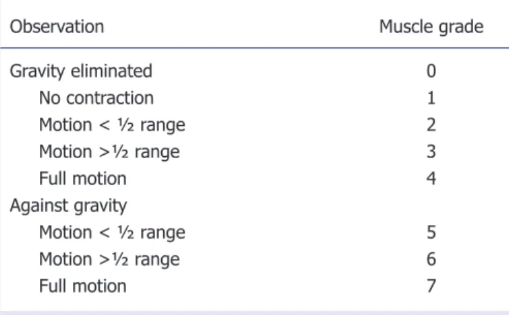
Shoulder flexors and abductors, elbow flexor, and forearm supinator muscles were evaluated using hospital for sick children muscle grading system (Clarke and Curtis). This classification system is appropriate for children younger than 3 years old, which measures all joints without any commands. The hospital for sick children scale is not a “muscle grading system” but is an assessment of sponta-neous active movement. There are seven stages totally, in
which the gravity is eliminated until stage 4 (Table 1). The families were educated in the hospital by a tal-ented physiotherapist with a 5-day oral, visual, and applied training program, in which the parents were then tested. The patients were examined and exercise programs were checked by the same physician every month until they be-came 12 months old. It was ordered to make the exercises 3 times daily for the IEG and once daily for the EG. Statistical Analysis
Statistical evaluation was performed using SPSS 16 (SPSS Inc. Released 2007. SPSS for Windows, Ver-sion 16.0. Chicago, SPSS Inc.). The one-sample Kol-mogorov–Smirnov test was applied for non-parametric values that comply with a normal distribution in inner-group assessments. Analysis of variance (ANOVA) was used in two-way repeated (time and groups) measure-ments to compare data from the parameters repeatedly measured in the inner group. In cases in which the vari-ance analysis test result was significant, Bonferroni cor-rection was performed to compare between groups as a post hoc test. The Bonferroni student t-test was used to detect different groups. The student t-test was used for independent groups to analyze data that complied with a normal distribution. P<0.05 was considered to be statis-tically significant (p<0.05).
RESULTS
Since mean biceps muscle force of four patients was 1.42 when they were 4 months old, these children were re-ferred to surgery and excluded from the study. Hence, a
total of 62 patients (38 boys and 24 girls), with a mean age of 7.50±5.18 days, were included in the study. The affected side was the left in 42 cases and right in 20 cases. All infants were born vaginally, and 52 cases had diffi-cult labor history. The average birth weight was 4388 g (Table 2). 97% of the subjects were in Group I, while 3% were in Group II according to Narakas classification.
Statistical analysis inside the groups: In terms of ac-tive ROM, while a statistically significant improvement was gained in both groups between 1st–3rd months, 3rd–
6th months, and 6th–12th months in shoulder abduction,
external rotation and flexion, and elbow flexion and fore-arm supination and between 1st–3rd months and 3rd–
6th months in shoulder internal rotation and extension
(p<0.05); there was no significant difference in elbow extension, forearm pronation, and wrist extension since they already had nearly normal values at the beginning of the treatment (p>0.05). Active ROM measures of the patients are shown in Table 3. There was a statistically significant improvement in the muscle strength in both groups between 1st–6th months and 6th–12th months
in shoulder abduction, between 1st–3rd and 6th–12th
months in shoulder flexion, and between 1st, 3rd, 6th, and
12th months in elbow flexion and forearm supination
(p<0.05) (Table 4).
Statistical analysis between the groups: No statisti-cally significant improvement was observed between the groups in EMP measures and muscle force assessments (p>0.05).
DISCUSSION
In upper brachial plexus OBPP patients, there was no significant difference between the groups who had in-tensive exercise program and those who did not. Only four of the cases were further referred to surgery. In all the cases except for these four cases, good arm functions were observed while they were 3 months old, and nearly normal functions were found while they were 12 months
Table 1. Hospital for sick children muscle grading system
Observation Muscle grade
Gravity eliminated 0 No contraction 1 Motion < ½ range 2 Motion >½ range 3 Full motion 4 Against gravity Motion < ½ range 5 Motion >½ range 6 Full motion 7
Table 2. Characteristics of patients of obstetric brachial plexus palsy
Age (days) 7.50±5.18
Sex (male/female) 38/15
Weight (gr) 4388
old. Hence, almost exact recovery was gained in 94% of the cases with OBPP in our study. The patients with OBPP at the C5–6 level, generally, display improvement in a period from 3 months to 9 months of age [3-5, 7]. It is reported that improvement period may continue until 12 months in some studies [5].
The diagnosis of OBPP is based on a precise physical examination with a support of EMG study. The valid pa-rameters used to evaluate improvement in OBPP cases are ROM measurements, EMG studies, and Clarke and Curtis muscle classification system for children un-der the age of [3-5]. If a common development cannot
Birth 3 months 6 months 12 months
Group I Group II Group I Group II Group I Group II Group I Group II
Shoulder (°) Abduction 11.92 (4.38) 12.82 (3.63) 38.71 (3.38)* 38.04 (3.61)ǂ 62.92 (2.46)* 62.82 (2.53)ǂ 87.63 (2.53)* 87.40 (2.55)ǂ External rotation 0 0 18.07 (2.46)* 18.03 (2.24)ǂ 23.92 (2.48)* 23.47 (2.35)ǂ 28.20 (2.42)* 28.04 (2.49)ǂ Internal rotation 39.74 (3.79) 39.56 (3.60) 60.38 (4.35)* 60.21 (3.19)ǂ 78.33 (2.38)* 77.60 (2.56)ǂ 79.23 (1.82)* 78.96 (2.24)ǂ Extension 0 0 18.84 (2.13)* 17.60 (2.55)ǂ 22.15 (2.13)* 22.17 (2.53)ǂ 22.53 (2.33)* 22.04 (2.38)ǂ Flexion 19.87 (3.34) 19.13 (3.88) 51.53 (2.33)* 51.39 (2.55)ǂ 66.53 (2.33)* 65.43 (2.21)ǂ 83.58 (2.27)* 82.60 (2.45)ǂ Elbow (°) Flexion 0 0 68.07 (2.46)* 67.60 (2.55)ǂ 77.92 (2.46)* 77.39 (2.54)ǂ 102.82 (4.55)* 102.78 (5.10)ǂ Extension 103.07 (4.67) 103.47 (4.86) 115.89 (4.98)* 115.65 (5.06)ǂ 118.46 (3.65)* 117.82 (4.21)ǂ 118.46 (3.65)* 117.82 (4.21)ǂ Forearm (°) Pronation 118.46 (2.33) 118.69 (2.24) 121.38 (5.05)* 121.21 (5.10)ǂ 121.38 (5.05)* 121.21 (5.10)ǂ 121.38 (5.05)* 121.21 (5.10)ǂ Supination 0 0 36.79 (2.42)* 37.60 (2.55)ǂ 72.79 (2.42)* 72.60 (2.55)ǂ 106.66 (4.77)* 105.21 (5.10)ǂ Wrist (°) 81.79 (2.42) 81.52 (2.35) 88.20 (2.42)* 88.47 (2.35)ǂ 88.20 (2.42)* 88.47 (2.35)ǂ 88.20 (2.42)* 88.47 (2.35)ǂ Flexion 71.46 (2.33) 71.95 (2.49) 71.46 (2.33)* 71.95 (2.49)ǂ 71.46 (2.33)* 71.95 (2.49)ǂ 71.46 (2.33)* 71.95 (2.49)ǂ *: Statistically significant differences according to the Birth value inner Group I evaluations (p<0.05). ǂ: Statistically significant differences according to the Birth value inner Group II evaluations (p<0.05). p>0.05 for each between group evaluations; (°): Degree of movement at a joint.
Table 3. Measurements of active range of motion in patients with obstetric brachial plexus palsy
Birth 3 months 6 months 12 months
Group I Group II Group I Group II Group I Group II Group I Group II
Shoulder Abduction 3.50 (0.46) 3.52 (0.51) 4.07 (0.57)* 4.01 (0.73)ǂ 5.51 (0.50)* 5.56 (0.50)ǂ 6.64 (0.48)* 6.56 (0.50)ǂ Flexion 3.61 (0.49) 3.65 (0.48) 5.40 (0.46)* 5.47 (0.51)ǂ 5.43 (0.48)* 5.48 (0.52)ǂ 6.46 (0.42)* 6.30 (0.47)ǂ Elbow Flexion 0.56 (0.50) 0.52 (0.51) 4.71 (0.64)* 4.82 (0.77)ǂ 5.45 (0.64)* 5.43 (0.50)ǂ 6.38 (0.45)* 6.39 (0.49)ǂ Forearm Supination 0.41 (0.50) 0.43 (0.50) 2.58 (0.52)* 2.60(0.49)ǂ 5.53 (0.49)* 5.52 (0.51)ǂ 6.30 (0.40)* 6.34 (0.48)ǂ
*: Statistically significant differences according to the Birth value inner Group I evaluations (p<0.05). ǂ: Statistically significant differences according to the Birth value inner Group II evaluations (p<0.05). p>0.05 for each between group evaluations.
be gained in the shoulder and biceps muscles while the child is in 3 months and 6 months old, then he/she is referred to surgery [4, 7]. While 70–95% of the patients improve almost completely, 10% may require surgery [4, 7, 14]. Likely 6% of our subjects had operations, 94% displayed a nearly complete improvement between 6 and 12 months. The increase in biceps and shoulder muscles showed a rapid incline in the 3rd month and became
al-most normal in 6th–12th months.
Physiotherapy program is the recommended treat-ment option for patients with C5-7 OBPP at the first stage. This conservative treatment includes exercise, splint, and electrical stimulation. The purpose of con-servative treatment is mainly to prevent contracture for-mation, hypoplasic arm development, dislocation of the radial head, and negative psychosocial consequences [1, 2, 4, 7]. Although a spontaneous recovery can be seen in most of the cases, contracture formation in muscles and dislocation in shoulder and elbow may prevent functional recovery [7]. At this point, exercise program substantially contributes to the recovery of most of the patients.
However, there are no accurate data about the fre-quency, intensity, and time-planning of exercise pro-grams. It is suggested that permanent disability may develop in some of the cases despite physiotherapy treat-ment [7]. This situation has raised more questions about the place of exercise in OBPP. Some authors suggested that exercises did not have much effect on recovery [4]. However, the other some have defended that intensive exercise program has a positive impact on recovery. However, they have still not clearly explained how this effect occurs [10].
When we examine the processes in the recovery of OBPP, we will observe that not only peripheral recon-struction but also central and spinal cord plasticity play a significant role in OBPP recovery [6, 8, 15]. It is hy-pothesized that physical exercises increase plasticity, es-pecially, in the spinal cord and central area by inclining ependymal cell proliferation and thus ensure functional recovery [11-13, 16]. Therefore, the representation of the body parts in the motor cortex can be altered after injury and during motor learning, and changes in the brain may be related to activity [15]. Plasticity increases due to the increased sensory input after exercises _ENREF_6(6). Cortical remodeling was shown to be increased after hand injuries through exercises. Hence, it was suggested that exercises are critical after peripheral nerve or central
nervous system injuries [6]. Thus, our study looked at the effects of two different exercise programs for the up-per brachial plexus lesions.
This study proved that there was no significant recov-ery difference between the groups in terms of time and degree and showed that intensive exercises did not have an additional contribution on peripheral nerve injuries, which meant that the frequency of the exercises did not have a significant effect on recovery. No complications developed in the patients during their follow-ups. How-ever, further studies may be planned with some biochem-ical parameters as well as cranial and spinal cord imaging so that the effects of exercises on the recovery of cases with OBPP, especially, on the development of plasticity could be determined more accurately. Besides, further studies could be made on the effects of exercises on re-covery with different intensity and duration as well as different frequencies.
Clinical Messages
1. Exercises positively affect recovery rate and results and prevent possible complications that may occur in cases with OBPP.
2. Intensive exercises did not have an additional contri-bution on peripheral nerve injuries, which means that the frequency of the exercises that the patients have daily did not have a significant effect on recovery.
Conflict of Interest: The authors declare no conflict of interest. Financial Disclosure:The authors declared that this study has re-ceived no financial support.
Authorship Contributions: Concept – N.S.; Design – N.S.; Su-pervision – N.S., A.Y.K.; Materials – N.S., A.Y.K.; Data collection &/ or processing – N.S., A.Y.K.; Analysis and/or interpretation – N.S., A.Y.K.; Writing – N.S., A.Y.K.; Critical review – N.S., A.Y.K.
REFERENCES
1. Yang LJ. Neonatal brachial plexus palsy-management and prognostic factors. Semin Perinatol 2014;38:222–34.
2. Chauhan SP, Blackwell SB, Ananth CV. Neonatal brachial plexus palsy: Incidence, prevalence, and temporal trends. Semin Perinatol 2014;38:210–8.
3. Backe B, Magnussen EB, Johansen OJ, Sellaeg G, Russwurm H. Ob-stetric brachial plexus palsy: A birth injury not explained by the known risk factors. Acta Obstet Gynecol Scand 2008;87:1027–32.
4. Bahm J, Ocampo-Pavez C, Disselhorst-Klug C, Sellhaus B, Weis J. Ob-stetric brachial plexus palsy: Treatment strategy, long-term results, and prognosis. Dtsch Arztebl Int 2009;106:83–90.
A, et al. Congenital brachial palsy: Incidence, causes, and outcome in the United Kingdom and republic of Ireland. Arch Dis Child Fetal Neonatal Ed 2003;88:F185–9.
6. Strömbeck C, Krumlinde-Sundholm L, Remahl S, Sejersen T. Long-term follow-up of children with obstetric brachial plexus palsy I: Func-tional aspects. Dev Med Child Neurol 2007;49:198–203.
7. Zafeiriou DI, Psychogiou K. Obstetrical brachial plexus palsy. Pediatr Neurol 2008;38:235–42.
8. Buesch FE, Schlaepfer B, de Bruin ED, Wohlrab G, Ammann-Reiffer C, Meyer-Heim A, et al. Constraint-induced movement therapy for children with obstetric brachial plexus palsy: Two single-case series. Int J Rehabil Res 2010;33:187–92.
9. Smith NC, Rowan P, Benson LJ, Ezaki M, Carter PR. Neonatal brachial plexus palsy. Outcome of absent biceps function at three months of age. J Bone Joint Surg Am 2004;86-A:2163–70.
10. Terzis JK, Papakonstantinou KC. Management of obstetric brachial plexus palsy. Hand Clin 1999;15:717–36.
11. Asensio-Pinilla E, Udina E, Jaramillo J, Navarro X. Electrical stimula-tion combined with exercise increase axonal regenerastimula-tion after periph-eral nerve injury. Exp Neurol 2009;219:258–65.
12. Foret A, Quertainmont R, Botman O, Bouhy D, Amabili P, Brook G, et al. Stem cells in the adult rat spinal cord: Plasticity after injury and treadmill training exercise. J Neurochem 2010;112:762–72.
13. Kao T, Shumsky JS, Murray M, Moxon KA. Exercise induces cor-tical plasticity after neonatal spinal cord injury in the rat. J Neurosci 2009;29:7549–57.
14. Bae DS, Waters PM, Zurakowski D. Correlation of pediatric outcomes data collection instrument with measures of active movement in chil-dren with brachial plexus birth palsy. J Pediatr Orthop 2008;28:584– 92.
15. Strömbeck C, Remahl S, Krumlinde-Sundholm L, Sejersen T. Long-term follow-up of children with obstetric brachial plexus palsy II: Neu-rophysiological aspects. Dev Med Child Neurol 2007;49:204–9. 16. Molnar GE, Alexander MA. Pediatric Rehabilitation. Philadelphia,