Çelik Gül G. JOTCSA. 2018; 5(2): 881-884. RESEARCH ARTICLE
881
(This article was produced from 6th National Congress of Inorganic Chemistry and was sent to the JOTCSA
editorial board for publication)
CrVMoO
7: MICROWAVE SYNTHESIS AND STRUCTURAL
CHARACTERIZATION
Gülşah Çelik Gül 11University of Balıkesir, 10145, Balıkesir, Turkey
Abstract: In this study, microwave synthesis method was operated to obtain CrVMoO7. Structural
characterization of the compound was realized by powder X-ray diffraction (XRD) and Fourier transform infrared spectrometry (FTIR). Morphological property and elemental composition were determined via scanning electron microscopy (SEM) and energy dispersive X-ray analysis (EDS). Thermal nature of the sample was identified by thermogravimetric analyzer (TGA).
Keywords: CrVMoO7; X-ray diffraction; Rietveld refinement method; Microwave synthesis.
Submitted: May 22, 2017. Accepted: July 16, 2018.
Cite this: Çelik Gül G. CrVMoO7: MICROWAVE SYNTHESIS AND STRUCTURAL CHARACTERIZATION. JOTCSA. 2018;5(2):881-4.
DOI: http://dx.doi.org/10.18596/jotcsa.315212.
*Corresponding author. E-mail: [email protected], Tel.: +90 266 612 1067/2012. INTRODUCTION
Catalysis for most industrial processes has significance due to saving time, energy, and money. The catalysts used in these processes are generally transition metal oxides which are applied either alone or mixtures. The basic
mixtures contain V2O5 and
MoO3/WO3/Fe2O3/Cr2O3. Among them, Cr2O3‒
V2O5‒MoO3 three-component system has been
studied in early times by many researchers (1-4). The formula CrVMoO7 as unknown phase has
occurred by the reaction of Cr2O3‒V2O5‒MoO3
system in the solid-state form (5-7). The properties of this phase have been limitedly known , except melting point at 820 °C to form solid Cr2O3 (2). According to the other
investigations, the related phase has been formed via incorporation of MoO3 into the Cr2V4O13 lattice
(6, 7). It has not been examined in previous reports what the nature of CrVMoO7 is and how
the structure occurs (3, 8, 9). There are only a few documents about crystallographic morphology and thermal properties of CrVMoO7.
The prominent one is about indexing of powder X-ray diffraction pattern and calculation of unit cell parameters of CrVMoO7 resulting a=5.53346 Å,
b=6.58901 Å and c=7.86551 Å in triclinic system
(8). The infrared spectrum of the compound point out that VO4, MoO4 and CrO6 subgroups exist in
the structure (10, 11). As a result of these, to the best of our knowledge, microwave synthesis, crystallographic, morphologic and thermal properties of CrVMoO7 have been studied for the
first time with this paper which has not been reported previously.
MATERIALS AND METHODS
Cr2O3, V2O5 and MoO3 compounds have been used
as analytical grade and supplied by Merck. Oxide types starting materials have been weighed in 0.5:0.5:1 molar ratio and ground in an agate mortar followed by microwave treatment in a domestic microwave oven (2.45 GHz, 850 W power) for 20 min. The final sample has been washed three times with hot pure water and ethanol. The washed material has been treated at 400 °C for 2 hours to get the best crystals. The powder X-ray diffraction (XRD) measurement has been completed by Panalytical X’Pert Pro Diffractometer and CuKα radiation (λ=1.54056 Ǻ,
40 mA, 50 kV) with a scan rate of 1°/min with a step size 0.02°. The Rietveld analysis of the sample has been calculated by using powder
Çelik Gül G. JOTCSA. 2018; 5(2): 881-884. RESEARCH ARTICLE
882
diffraction data via High Score Plus (HS+) Program (License number: 92000029). Fourier transform infrared spectrum (FTIR) has been formed on a Perkin Elmer Spectrum 100 FTIR Spectrometer from 4000 to 650 cm-1. Scanning
electron microscopy/energy dispersive X-ray analysis has been achieved in SEM JEOL 6390-LV/EDX. Thermal property of the sample has been checked by Perkin Elmer thermogravimetric analyzer TGA. A Siemens V12 domestic microwave oven has been used as the microwave source.
RESULTS AND DISCUSSION
Figure 1 displays the XRD pattern of the synthesized material. The XRD pattern of the sample corresponds to CrVMoO7 with the ICSD
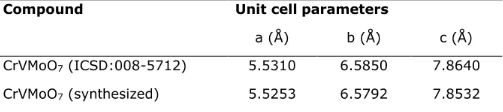
card number 008-5712 (Fig. 2). The unit cell parameters of the observed diffraction data has been calculated by Rietveld Refinement Program. The findings are completely in accordance with database given in Table 1. There is no other phase as an impurity or starting material.
Figure 1. The XRD pattern of CrVMoO7.
Figure 2. The comparison of powder XRD pattern between CrVMoO7 (ICSD:008-5712) and CrVMoO7
(synthesized).
Table 1. The comparison of unit cell parameters calculated and database values.
Compound Unit cell parameters
a (Å) b (Å) c (Å)
CrVMoO7 (ICSD:008-5712) 5.5310 6.5850 7.8640
CrVMoO7 (synthesized) 5.5253 6.5792 7.8532
Figure 3 shows FTIR spectrum of CrVMoO7. The
four wavenumbers in the range of 600-1000 cm
-1 correspond to vibrations of M‒M and Mo‒O
Çelik Gül G. JOTCSA. 2018; 5(2): 881-884. RESEARCH ARTICLE
883
Figure 3. The FTIR spectrum of CrVMoO7.
Figure 4 exhibits the SEM micrograph of CrVMoO7.
The homogeneous view of the sample confirm the
formation of chromium vanadium molybdate. The particle size distribution of CrVMoO7 is in 2-5 μm.
Figure 4. The SEM image of CrVMoO7.
The EDS graph of CrVMoO7 is given in Figure 5.
The elemental composition of the compounds has been calculated as 3:3:3.2:6.8 by EDS results
which are in accordance with the molecular formula.
Figure 5. The EDS graph of CrVMoO7.
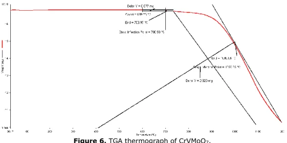
The graph of thermogravimetric analysis is represented in Figure 6. The first smaller thermal loss starts nearly at 700 °C, and the other is in
the range of 1050‒1200 °C. The material lost totally 40% of its mass.
4000 3500 3000 2500 000 1500 1000 600 100 39 40 50 60 70 80 90 Wave number, cm-1 %T
Çelik Gül G. JOTCSA. 2018; 5(2): 881-884. RESEARCH ARTICLE
884
Figure 6. TGA thermograph of CrVMoO7.
As a conclusion, CrVMoO7 has been synthesized
for the first time with microwave method at 850 W power for 20 minutes. The unit cell parameters of the compound have been calculated by Rietveld Refinement Program benefiting from XRD data. The homogeneous morphology and similar elemental composition have been confirmed via SEM and EDS results. High thermal stability of the compound has been determined from TGA.
ACKNOWLEDGMENTS
We want to thank The Scientific and Technological Research Council of Turkey and Scientific Research Project Fund of Balikesir University for financial supports and Dr. Sevim Demirözü Şenol for scientific support.
REFERENCES
1. Walczak J, Filipek E. In Sofia, Bulgaria: IUPAC; 1987. p. 171.
2. Walczak J, Filipek E. The CrVO4-MoO3 system. Thermochimica Acta. 1989 Sep;150(1):125–31.
3. Walczak J, Filipek E. Phase equilibria in the Cr2V4O13-CrVMoO7 system. Thermochimica Acta. 1990 May;161(2):239–45.
4. Walczak J, Filipek E. Studies on the V2O5-CrVMoO7 system. Thermochimica Acta. 1990 Dec;173:235–40.
5. Walczak J, Filipek E. Zeszyty Naukowe Politechniki Slaskiej. Seria: Chemia. 1988;119(958):380.
6. Walczak J, Filipek E. Reactivity of MoO3 towards Cr2V4O13. Thermochimica Acta. 1988 Oct;133:67–72.
7. Kuriata J, Sadłowski L, Walczak J, Filipek E. Temperature Dependence of the EPR Linewidth of CrVO4. physica status solidi (b). 1987 Jul 1;142(1):K73–7.
8. Walczak J, Filipek E, Tabero P. CrVMoO7 and phase equilibria in the V9Mo6O40-CrVMoO7 system. Thermochimica Acta. 1992
Sep;206:279–84.
9. Walczak J, Filipek E. Studies on the CrVMoO7-Cr2(MoO4)3 system. Thermochimica Acta. 1993 Nov;228:127–30.
10. Walczak J, Rychiowska-Himmel I, Tabero P. Iron(III) tungstate and its modifications. Journal of Materials Science. 1992;27(13):3680–4. 11. Plasova L, Kefeli L. Inorganic Materials. Vol. 3. 1967. 906 p.
12. Baykal A, Kizilyalli M, Kniep R. X-ray powder diffraction and IR study of NaMg (H2O) 2 [BP2O8]· H2O and NH4Mg (H2O) 2 [BP2O8]· H2O. Journal of materials science.
2000;35(18):4621–4626. 13. Gözel G, Kizilyalli M, Kniep R. Characterization of a new calcium
ultraphosphate, Ca3 (P5O14) 2. Journal of Solid State Chemistry. 1997;129(2):196–199. 14. Gözel G, Baykal A, Kizilyalli M, Kniep R. Solid-State Synthesis, X-ray Powder
Investigation and IR Study of α-Mg3[BPO7]. Journal of the European Ceramic Society. 1998 Dec;18(14):2241–6.