Introduction
The diseases caused by Campylobacter species constitute major problems for animal husbandry and public health all over the world as well as in Turkey. Common presence of
Campylobacter species in the environment and sufficiency
of very small dose for causing infection increase the risk of catching an infection. It has been reported that, Campylobacter species have been isolated from many domestic and wild animals and caused gastroenteritis in some animals and humans [2,17]. Particularly, feces of the poultry and cattle have a potential role for contamination of the environment [19]. In Turkey, the most common agents of bacterial diarrhea are Salmonella, Shigella and Campylobacter. In studies of acute gastroenteritis cases in Turkey, the isolation rate of Campylobacter has been reported to vary between 1.4% and 14.6%. Predominantly isolated species of Campylobacter from these infections has been shown to be C. jejuni. C.
coli and C. lari were also detected in gastroenteritis cases
of humans, though to a lesser extent when compared to C.
jejuni [3,4]. In recent years, C. lanienae has been determined
in higher proportions than C. jejuni in humans, cattle and pigs [6,8]. There is no data available about the presence of C.
lanienae in other animal species. In addition, in the limited
number of previous studies, the agent has only been isolated
in fecal samples [7,8]. Hence, it is unknown whether this agent is situated in the internal organs of animals and if yes, at what proportions. This study was conducted to investigate the presence of Campylobacter species, especially C. lanienae, in the internal organs of clinically healthy cattle, sheep and chickens in eastern Turkey.
Material and Methods
SAMPLE COLLECTION
In this study, a total number of 2700 internal organ samples were collected from clinically healthy cattle, sheep and chickens slaughtered in three provinces located in the east of Turkey between May 2011 and February 2012. The samples contained 150 gall bladder and 150 intestinal contents from each of the animal species in each province. While gall bladder samples were taken by sterile disposable syringes, intestinal content samples were taken from small intestine eviscerated immediately after slaughter with the help of swab. The samples were transferred to tubes containing 0.9% NaCl and delivered to the laboratory under suitable conditions (at 4 ° C) within a short time for routine bacteriological examination.
SUMMARY
The aim of the present study was to determine the prevalence of Campylobacter jejuni, C. coli, C. lari and C. lanienae which are the most common causes of acute bacterial gastroenteritis in humans, in clinically healthy cattle, sheep and chickens. A total number of 2700 abattoir samples containing 150 intestinal contents and 150 gall bladders from each of the animal species in each of the three provinces located in the east of Turkey. Following the inoculation of the samples onto modified Charcoal Cefoperazone Deoxycholate Agar (mCCDA), Campylobacter spp. was isolated from 24.7% (667/2700). In the analysis of the isolates with species-specific Polymerase Chain Reaction (PCR), the identification percentages of Campylobacter species were determined as 78.1% for C. jejuni, 23.7% for C. coli, 1.2% for C. lanienae and 1.0% for C. lari. To the authors’ knowledge, this study reports the isolation of C. lanienae in animals for the first time in Turkey and in chickens species for the first time in the world.
Keywords: Campylobacter lanienae, C. jejuni, C. coli, C.
lari, Sheep, Cattle, Chicken
RÉSUMÉ
Premier isolement de Campylobacter lanienae chez le poulets
Le but de la présente étude était de déterminer la prévalence de Campylobacter jejuni, C. coli, C. lari et C. lanienae qui sont les causes les plus fréquentes de gastro-entérite bactérienne aiguë chez l’homme, chez des bovins, des moutons et des poulets cliniquement sains. Un total de 2700 échantillons contenant 150 contenu intestinal et 150 vésicules biliaires de chacune des espèces animales a été collecté à l’abattoir dans chacune des trois provinces situées à l’est de la Turquie. Suite à l’inoculation des échantillons sur milieu Charbon Cefoperazone Agar Deoxycholate modifiée (mCCDA), des Campylobacter spp. ont été isolés à partir de 24,7% (667/2700) des prélèvements. L’analyse d’espèce a été réalisée par PCR. Les pourcentages d’identification respectifs suivants ont été obtenus: 78,1% pour C. jejuni, 23,7% pour C. coli, 1,2% pour C. lanienae et 1,0% pour C. lari.
Mots-clés: Campylobacter lanienae, C. jejuni, C. coli, C.
lari, ovins, bovins, poulet
The First Isolation of Campylobacter
lanienae from chickens
M. N. ACIK1, M. KARAHAN2, H. ONGOR3, B. KARAGULLE3, B. CETİNKAYA3
1 Department of Microbiology, Faculty of Veterinary Medicine, Bingol University, 12000 Bingol, TURKEY 2 Department of Microbiology, Faculty of Veterinary Medicine, Cumhuriyet University, 58000 Sivas, TURKEY 3 Department of Microbiology, Faculty of Veterinary Medicine, Firat University, 23119 Elazig, TURKEY
Revue Méd. Vét., 2013, 164, 7, 368-373
THE FIRST ISOLATION OF CAMPYLOBACTER LANIENAE FROM CHICKENS
369
ISOLATION OF
CAMPYLOBACTER
Gall bladder samples were inoculated in two ways onto medium: pre-enrichment and direct inoculation. In the first method, pre-enrichment was performed by taking 2 ml fluid from gall bladder samples and transferring under aseptic conditions to 10 ml of Brucella broth (Difco,Detroit, MI, USA) containing 7% laked horse blood (SR0048C;Oxoid, Basingstoke, UK), Preston Campylobacter Selective Supplement (SR117E; Oxoid) and Campylobacter Growth Supplement (SR0048;Oxoid). The broths were incubated at 42 ° C in microaerobic atmosphere (5%O2, 10%CO2 and 85% N2) for 24-48 hours. Following incubation, a loop-full of broth was inoculated onto modified Cefaperazone Charcoal Deoxycholate Agar (mCCDA, Oxoid) and incubated at 42 ° C in microaerobic atmosphere for 48-72 hours.
In the direct inoculation method, a loop-full of the gall bladder fluid samples were inoculated onto MCCDA and incubated under the same conditions. Swab samples were inoculated onto medium by using the method of pre-enrichment mentioned above. The isolates were presumptively identified as Campylobacter spp. by colony appearance, microscopic morphology and motility. The five colonies from each positive sample were stored at -20 °C in Nutrient broth number 2 (CM067B; Oxoid) containing 15% glycerol.
DNA EXTRACTION
The method described by Acık and Cetinkaya [1] was used for DNA extraction. Briefly, bacterial suspension was treated with TNES (Tris-EDTA-NaCl-SDS) and proteinase K, and then was subjected to phenol extraction. Following the precipitation process with alcohol and sodium acetate, the pellet was diluted with 100 µl of sterile distilled water.
DETECTION OF
CAMPYLOBACTER
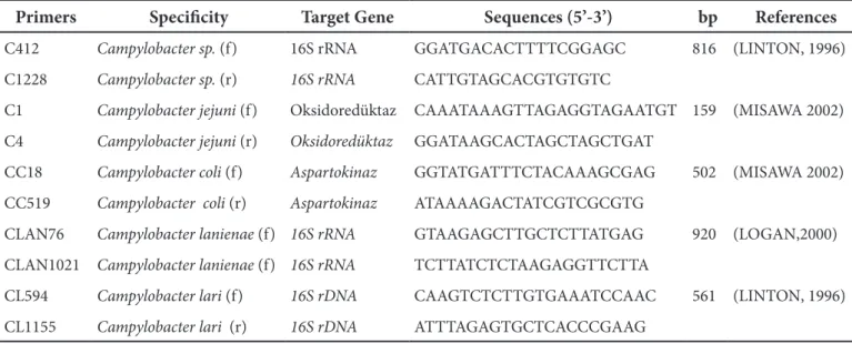
SPECIES. BY POLYMERASE CHAIN REACTIONThe isolates identified as Campylobacter spp. by culture method were confirmed using a pair of primers specific to
Campylobacter spp. (Table I) Species-specific primers were
used to identify Campylobacter isolates at species level (Table I). The assays were performed in a TC 512 Temperature Cycling System (Techne, Staffordshire, United Kingdom) in a total reaction volume of 50 µl, containing 5 µl 10 · PCR buffer (750 mM Tris HCl, pH 8.8, 200 mM (NH4)2SO4, 0.1% Tween20), 5 µl of 25 mM MgCl, 250 µM of each dNTP, 1.25 U Taq DNA Polymerase (MBI Fermentas, St Leon-Rot, Germany), 20 pmol of each primer (Table I), and 5 µl of template DNA. Reference strains of C. jejuni [NCTC-National Collection of Typing Cultures, London, UK- 11 322], C. coli [NCTC 11 366], C. lari [NCTC11352] and C.
lanienae [NCTC 13004] were used as positive controls in the
PCR assays.
SEQUENCE ANALYSIS
Two C. lanienae isolates, one originated from sheep and the other from chickens, were subjected to sequence analysis. The primers described by Logan et al. [9] were used for sequence analysis which was performed by RefGen Gene Research and Biotechnology Limited Company.
STATISTICAL ANALYSIS
Fisher’s exact test was used to evaluate the differences between various parameters. P < 0.05 was considered as statistically significant.
Primers Specificity Target Gene Sequences (5’-3’) bp References
C412 Campylobacter sp. (f) 16S rRNA GGATGACACTTTTCGGAGC 816 (LINTON, 1996)
C1228 Campylobacter sp. (r) 16S rRNA CATTGTAGCACGTGTGTC
C1 Campylobacter jejuni (f) Oksidoredüktaz CAAATAAAGTTAGAGGTAGAATGT 159 (MISAWA 2002)
C4 Campylobacter jejuni (r) Oksidoredüktaz GGATAAGCACTAGCTAGCTGAT
CC18 Campylobacter coli (f) Aspartokinaz GGTATGATTTCTACAAAGCGAG 502 (MISAWA 2002)
CC519 Campylobacter coli (r) Aspartokinaz ATAAAAGACTATCGTCGCGTG
CLAN76 Campylobacter lanienae (f) 16S rRNA GTAAGAGCTTGCTCTTATGAG 920 (LOGAN,2000) CLAN1021 Campylobacter lanienae (f) 16S rRNA TCTTATCTCTAAGAGGTTCTTA
CL594 Campylobacter lari (f) 16S rDNA CAAGTCTCTTGTGAAATCCAAC 561 (LINTON, 1996) CL1155 Campylobacter lari (r) 16S rDNA ATTTAGAGTGCTCACCCGAAG
Re vu e M éd. V ét., 2013, 164
ACIK (M. N.) AND COLLABORATORS
Samples samples (%)
positive by culture and PCR
C.jejuni C.coli C.lanienae C.lari Other
campylobacters Intestinal content Cattle Gall Bladder Malatya 150 57(38) 54 (94.7) 0 0(0) 3(5.3) 0(0) Bingol 150 39(26) 33(84.6) 2(5.1) 0(0) 0(0) 4(10.3) Elazığ 150 48(32) 40(83.3) 7(14.6) 1(2.1) 0(0) 0(0) Total 450 144(32) 127(88.2) 9(6.2) 1(0.7) 3(2.1) 4(2.8) Malatya 150 19 (12.7) 19 (100) 0(0) 0(0) 0(0) 0(0) Bingol 150 20 (13.3) 18 (90) 2(10) 0(0) 0(0) 0(0) Elazığ 150 31 (20.7) 30(96.8) 1(3.2) 0(0) 0(0) 0(0) Total 450 70 (15.6) 67(95.7) 3(4.3) 0(0) 0(0) 0(0) Intestinal content Sheep Gall Bladder Malatya* 150 58(38.7) 35(60.3) 48(82.8) 0(0) 0(0) 0(0) Bingol 150 40 (26.7) 18(45) 21(52.5) 0(0) 0(0) 1(2.5) Elazığ* 150 91 (56.7) 63(69.2) 42(46.2) 4(4.4) 2(2.2) 0 Total 450 189 (42) 116(61.4) 111(58.7) 4(2.1) 2(1.1) 1(0.5) Malatya 150 48 (32) 36(75) 6(12.5) 0(0) 0(0) 6(12.5) Bingol 150 24(16) 24(100) 0(0) 0(0) 0(0) 0(0) Elazığ 150 64 (42.7) 45(70.3) 18(28.1) 1(1.7) 0(0) 0(0) Total 450 136(30.2) 105(77.2) 24(17.7) 1(0.7) 0(0) 6(4.4) Intestinal content Chicken Gall Bladder Malatya 150 47 (31.3) 36(76.6) 6(12.8) 2(4.25) 1(2.1) 2(4.25) Bingol 150 17(11.3) 12(70.6) 2(11.75) 0(0) 1(5.9) 2(11.75) Elazığ 150 12 (8) 12(100) 0(0) 0(0) 0(0) 0 Total 450 76(16.9) 60(78.9) 8(10.5) 2(2.65) 2(2.65) 4(5.3) Malatya 150 18(12) 18(100) 0(0) 0(0) 0(0) 0(0) Bingol 150 28(18.7) 22(78.6) 3(10.7) 0(0) 0(0) 3(10.7) Elazığ 150 6(4) 6(100) 0(0) 0(0) 0(0) 0(0) Total 450 52(11.6) 46(88.5) 3(5.8) 0(0) 0(0) 3(5.7) Overall 2700 667(24.7) 521(78.1) 158(23.7) 8(1.2) 7(1.0) 15(2.2)
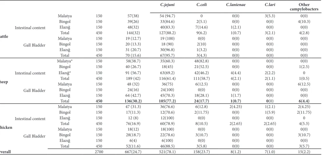
* Both C. jejuni and C. coli were identified in 20 and 25 intestinal content samples of sheep in Elazig and Malatya provinces. respectively. Table II: Identification of Campylobacter spp. isolates obtained from intestinal content and gall bladder of animals by PCR
Revue Méd. Vét., 2013, 164, 7, 368-373
THE FIRST ISOLATION OF CAMPYLOBACTER LANIENAE FROM CHICKENS
371
Results
ISOLATION AND IDENTIFICATION OF
CAMPYLOBACTERS
Of the 2700 samples tested by conventional culture and PCR, 667 (24.7%) were detected to be positive for
Campylobacter spp. The isolation rate of Campylobacter spp.
was determined to be the highest in the intestinal content samples of sheep with 42% (183/450) and the lowest in the gall bladder samples of chickens with 11.6% (52/450). The distribution of isolation rates by sample, animal species and province is presented in Table II.
IDENTIFICATION AT SPECIES LEVEL BY PCR
In the analysis of 659 isolates with species-specific PCR,
the identification percentages of Campylobacter species were determined as 78.1% (521/667) for C. jejuni, 23.7% (158/667) for C. coli, 1.2% (8/667) for C. lanienae and 1.0% (7/667) for
C. lari. Both C. jejuni and C. coli were detected in 24.6%
(45/183) of the Campylobacter spp. isolates obtained from the intestinal content samples of sheep. On the other hand 15 (2.3%) isolates could not be identified at species level. C.
lanienae was detected in seven intestinal content and one
gall bladder samples of animals. The agent was found in two intestinal content samples of chicken for the first time in the world (Table II).
SEQUENCE ANALYSIS RESULTS
The sequence analysis of two randomly selected isolates revealed homology with C. lanienae at the rates of 99-100%.
Discussion
The recent studies demonstrate that prevalence of campylobacters which are the most common agents of the acute bacterial gastroenteritis in humans and animals are in increase which maintains their importance. In the study performed by European Food Safety Authority (EFSA) in 28 countries; 26 of which being in Europe, the prevalence of campylobacters were reported to be almost five times higher than that of Salmonella in poultry [15]. Nichols et al. [11] have showed that Campylobacter cases increased in course of time following the examination of one million cases during 23 years in England and Wales [11]. Also in the present study, the prevalence of Campylobacter sp., especially of C. jejuni, in cattle, sheep and chickens has been demonstrated to be in increase when compared with the results of earlier studies performed in the same region. The isolation rates of C. jejuni and C. coli in the intestinal content samples of chickens were reported as 20% and 9%, respectively in a study conducted by Ertas et al. [5], whereas these rates were calculated as 78.9% and 10.5% in the present study. Similarly, while Acık and Cetinkaya [1] reported the isolation rates of 39.5% and 26.1% for C. jejuni and C. coli in the examination of intestinal content samples of clinically healthy sheep, this study revealed
much higher rates (61.4% for C. jejuni and 58.7% for C. coli). These results indicate that gradual increase in the prevalence of Campylobacter has occurred in Turkey as well as in some parts of the world like the UK. On the other hand, many factors such as the number of samples examined, season, the culture media and the methods used might be responsible for these differences. In particular, the inoculation method used for isolation of campylobacter from the samples (direct plating-enrichment) and the types of medium may lead to the differences. mCCDA selective broth used in recent years for the isolation of Campylobacter has been shown to be superior to the other media. Rodgers et al. [14] reported that direct inoculation of chicken cecum samples onto mCCDA was more effective than Skirrows and Preston Agar [14]. On the other hand,, Stanley et al. [18] reported that, the isolation rate of Campylobacter was increased by inoculating after pre- enrichment process which suppresses the growth of other bacteria in contaminated samples such as feces [18]. In this study, while intestinal content samples were subjected to pre-enrichment in order to minimize the contamination, gall bladder samples were subjected to inoculation after both direct and pre-enrichment process and the same isolation rates were obtained in both methods.
Campylobacter jejuni was isolated at very high
proportions in internal organ samples of all animal species examined. Interestingly, C. coli was found at significantly higher frequencies in both intestinal content and gall bladder samples of sheep when compared to the samples of chickens and cattle (P<0.05). Stanley et al. [18] reported that; the average rate of Campylobacter obtained from the intestinal contents of sheep was 10-fold higher than that of cattle, but lower than that of chickens In Turkey, cattle are predominantly reared at small family premises and are therefore subjected to breeding within the premise instead of grazing on pasture, whereas hundreds of sheep flocks are bred on pasture which may increase the risk of transmission of Campylobacter agents from one animal to another. The data of this study demonstrate that, sheep may play an important role in the contamination of the environment by C. coli and subsequently in its transmission to humans. In order to prove this, it is necessary to reveal the source of origin and genetic relationship between sheep and human isolates by using molecular typing methods such as Pulsed Field Gel Electrophoresis (PFGE) and Multi Locus Sequence Typing (MLST).
C. lanienae, which has been identified for the first time in the feces of people working in a pig abattoir in Switzerland, has been isolated in cattle, pigs and sheep [6,9,12]. However, the pathogenicity of this agent in humans and animals has not been established yet. To the authors’ knowledge, the presence of C. lanienae either in humans or in animals has not been reported in Turkey so far. Inglis et al. [6] reported the isolation of C. lanienae at a rate of 56% in the examination of cattle feces by direct PCR. This rate is significantly higher than that obtained in the present study. The methodologies used in both studies were thought to be effective on this difference.
Hence, it has been put forward that, most of culture media used for the isolation of C. jejuni and C. coli inhibited the growth of C. lanienae and direct detection by PCR from feces might therefore be more useful [6]. On the other hand, it has been reported that, also direct PCR was problematic because of the inhibitors in feces such as polypholic components which may cause false negative results. In order to minimize or eliminate the effects of these inhibitors, the QIAamp DNA stool kits containing polysaccharide mixture can be used [7]. In addition, subjecting samples to direct inoculation or pre-enrichment has a significant impact on the isolation rate of C. lanienae. Shin and Lee [16] subjected intestinal content samples of pigs to direct inoculation and pre-enrichment and reported 8% isolation rate of C. lanienae with pre-enrichment and 16% isolation rate with direct inoculation [16]. However, this situation is vice versa for the isolation of C. coli, as the same researchers obtained higher isolation rate for C. coli when the samples were subjected to pre-enrichment. C. lanieanae has been shown to be phylogenetically related to C. fetus subsp.
fetus, C. hyointesitnalis subsp. hyointesitnalis and C. mucosalis
[9]. However, it may be distinguished biochemically from
C. fetus subsp. fetus by inability to grow at 25 ° C, and from
C. hyointesitnalis subsp. hyointesitnalis and C. mucosalis by producing H2S at Triple Sugar Iron Agar (TSI) and growth at 1% glycine. Logan et al. [9] defined C. lanieanae as a new species, but did not consider C. hyointestinalis subsp. lawsonii in the phylogenetic analysis. On the other hand, Inglis et al. [6] reported that C. lanienae was related more closely with
C. hyointestinalis subs. lawsonii on the base of 16S rDNA.
These researchers amplified DNA of C. hyointestinalis subsp.
lawsonii successfully by employing a pair of primers specific
to C. lanienae which was described by Logan et al. [9] and then tried to modify these primers [6]. In our study, both primer pairs produced by Logan et al. [9] and by Inglis et al. [6] were used and the PCR products at the molecular size of 920-bp which is indicative for the presence of C. lanienae were obtained from eight samples by both primer sets. In addition, the sequence analysis of two randomly selected isolates revealed homology with C. lanienae at the rates of 99-100%. In order to distinguish the C. lanienae isolates from Campylobacter mucosalis and C. hyoeintestinalis subsp.
hyointestinalis, H2S production capacity at TSI agar was checked and none of the eight isolates were detected to produce H2S.
C. lanienae was isolated in the intestinal contents of
chickens for the first time in the world in the present study. However, this is not sufficient to prove that chickens are the actual hosts of C. lanienae. Although it is possible to suggest that chickens are among the actual hosts of C. lanienae and contaminate the environment throughout their feces which may pose risk for humans, the fact that chicken breeders in Turkey also raise other domestic animals like cattle, sheep and goats within the same farm indicates that C. lanienae might be transmitted horizontally to chickens thru ruminants. In order to determine the origin of C. lanienae in humans, isolates obtained from different animals and humans should be examined by molecular typing methods.
Campylobacter lari exists in gastrointestinal tracts of
human and many animals as a commensal and spreads around with the feces of animals. C. lari has been identified in low rates in both animals and humans. This agent has been isolated frequently in chickens, but at low proportions in other animals such as cattle and sheep [18]. Stanley et al. [18] reported the isolation rate of 0.2% for C. lari in intestines of lambs at slaughter. In the present study, similar proportions (2.1% for cattle, 1.1 % for sheep and 2.6% for chickens) were obtained from the animal species examined. All the C.
lari isolates were detected in the intestinal content samples
of the animals. As mentioned above, the large majority of nutrient broths commercially available and widely used for the isolation of campylobacters have been produced primarily for isolation of C. jejuni and C. coli. Because these media usually inhibit the growth of other Campylobacter species, there is a need to develop new media which are suitable for the better growth of Campylobacter agents like C. lari. It is therefore believed that the real prevalence of Campylocater species such as C. lari would be revealed by the direct PCR application from feces.
In conclusion, this is the first study that reports the isolation of C. lanienae in chickens. Also, this agent was shown in animal species for the first time in Turkey. When compared with previous studies conducted in the same geographical area, it can be said that the prevalence of
Campylobacter species has increased steadily in animal
populations in Turkey. The potential role of sheep for the environmental contamination and human cases is thought to be higher than that of the other animals.
Acknowledgements
This study was funded by The Scientific and Technical Research Council of Turkey (TUBITAK, TOVAG-110O356).
References
1. ACIK, M.N., CETINKAYA B.: The heterogeneity of
Campylobacter jejuni and Campylobacter coli strains
isolated from healthy cattle. Lett. Appl. Microbiol., 2005, 41, 397-403.
2. ACIK, M.N., CETINKAYA B.: Heterogeneity of
Campylobacter jejuni and Campylobacter coli strains
from healthy sheep. Vet. Microbiol., 2006, 115, 370-375. 3. AKGUN Y., USTUNEL M.E., BOLATLI T.: Eskişehir
bölgesi’nde C. jejuni’nin gastroenterit etyolojisindeki yeri. İnf. Derg., 1989, 3, 365-373.
4. ATES-YILMAZ A., TUGRUL, H.M.: Edirne’de ishal etkenleri arasında Campylobacter türlerinin yerinin ve antimikrobiklere duyarlılıklarının araştırılması. İnf.
Derg., 2005, 19, 53-59.
5. ERTAS H.B., CETINKAYA B., MUZ A., ONGOR H.: Genotyping of broiler-originated Campylobacter jejuni and Campylobacter coli isolates using fla typing and random amplified polymorphic DNA methods. Int. J.
Revue Méd. Vét., 2013, 164, 7, 368-373
THE FIRST ISOLATION OF CAMPYLOBACTER LANIENAE FROM CHICKENS
373
6. INGLIS G.D., KALISCHUK L.D.: Use of PCR for direct detection of Campylobacter species in bovine feces. Appl.
Environ. Microbiol., 2003, 69, 3435-3447.
7. INGLIS G.D., KALISCHUK L.D.: Direct quantification of Campylobacter jejuni and Campylobacter lanienae in feces of cattle by real-time quantitative PCR. Appl.
Environ. Microbiol., 2004, 70, 2296–2306.
8. LINTON D., OWEN R.J., STAMLEY J.: Rapid identification by PCR of the genus Campylobacter and of five Campylobacter species enteropathogenic for man and animals. Res. Microbiol. 1996, 147, 707-718. 9. LOGAN J.M., BURNENS A., LINTON D., LAWSON A.
J., STANLEY J.: Campylobacter lanienae sp. nov., a new species isolated from workers in an abattoir. Int. J. Syst.
Evol. Microbiol., 2000, 2, 865-872.
10. MISAWA N., KAWASHIMA K., KAWAMOTO H., KONDO F.: Development of a combined filtration-enrichment culture followed by a one-step duplex PCR technique for the rapid detection of Campylobacter jejuni and C. coli in human faecal samples. J. Med.
Microbiol., 2002, 51, 86-89.
11. NICHOLS G.L., RICHARDSON J.F., SHEPPARD S.K., LANE C., SARRAN C.: Campylobacter epidemiology: a descriptive study reviewing 1 million cases in England and Wales between 1989 and 2011. BMJ
Open, 2012, 12, 2(4).
12. OPORTO B., HURTADO A.: Emerging thermotolerant
Campylobacter species in healthy ruminants and swine. Foodborne Pathog. Dis., 2011, 8, 807-813.
13. OYARZABAL O.A., WESLEY I.V., HARMON K.M., SCHROEDER-TUCKER L., BARBAREE J.M., LAUERMAN L.H., BACKERT S., CONNER D.E.: Specific identification of Campylobacter fetus by PCR targeting variable regions of the 16S rDNA. Vet.
Microbiol., 1997, 58, 61-71.
14. RODGERS J.D., CLIFTON-HADLEY F.A., MARIN C., VIDAL A.B.: An evaluation of survival and detection of Campylobacter jejuni and C. coli in broiler caecal contents using culture-based methods. J. Appl.
Microbiol., 2010, 109, 1244-1252.
15. SCIENTIFIC REPORT OF EFSA.: Analysis of the baseline survey on the prevalence of Campylobacter in broiler batches and of Campylobacter and Salmonella on broiler carcasses in the EU, 2008 Part A: Campylobacter and Salmonella prevalence estimates. EFSA J., 2010, 8, 1503.
16. SHIN E., LEE Y.: Comparison of three different methods for Campylobacter isolation from porcine intestines. J.
Microbiol. Biotechnol., 2009, 19, 647-650.
17. SKIRROW M.B.: Diseases due to Campylobacter,
Helicobacter and related bacteria. J. Comp. Pathol. 1994,
111, 113–149.
18. STANLEY K.N., WALLACE J.S., CURRIE J.E., DIGGLE P.J., JONES K.: Seasonal variation of thermophilic campylobacters in lambs at slaughter. J. Appl. Microbiol., 1998, 84, 1111-1116.
19. TAUXE R.V.: Epidemiyology of Campylobacter jejuni infections in the United States and other industrialized nations, p.9-19. In I. NACHAMKIN, M.J. BLASER, L.S.TOMPKINS(ed.), Campylobacter jejuni: current status and future trends. American Society for Microbiology, Washington, D.C., (1992).