Clinical Features and Spectral-Domain
Optical Coherence Tomography Findings of
Complete Congenital Stationary
Night Blindness Patients
AABBSSTTRRAACCTT OObbjjeeccttiivvee:: To describe clinical findings and spectral domain optical coherence tomog-raphy (SD-OCT) features of our complete congenital stationary night blindness (CSNB) patients. M
Maatteerriiaall aanndd MMeetthhooddss:: This retrospective study included 12 eyes of six patients diagnosed with com-plete type CSNB. Patients were evaluated with SD-OCT, fundus autofluorescence (FAF) and elec-trophysiological tests. Segmentation of retinal layers was performed on SD-OCT images of all CSNB patients and compared with 6 age matched, normal myopic controls. RReessuullttss:: All patients' anterior segments findings were normal and none of our patients had nystagmus or strabismus. Pale optic disc was observed in two patients. On FAF imaging eyes with CSNB demonstrated unremarkable fundi including normal distribution of autofluorescence. Full-field electroretinography (ERG) demon-strated b-wave to a-wave amplitude ratio of less than one in the combined rod–cone response which describes electronegative ERG. SD-OCT segmentation revealed statistically significant thinning of the total retina, retinal nerve fiber layer (RNFL), inner plexiform layer (IPL) and inner retinal thick-nesses in the CSNB group compared to control group (respectively p=0.015, p=0.011, p=0.017 and p=0.021). In the other layers of retina, we observed thinning in the CSNB group, but this difference was not statistically significant. CCoonncclluussiioonn:: In our study we observed selective thinning of RNFL and IPL in our patients. We thought that thinning of the IPL and RNFL and possibly optic disc paleness in complete CSNB patients suggests bipolar cell dysfunction or synaptogenesis defect between bipo-lar cells and ganglion cells and possibly reduced number of these cells.
KKeeyywwoorrddss:: Congenital stationary night blindness; optical coherence tomography; full-field electroretinography
Ö
ÖZZEETT AAmmaaçç:: Konjenital durağan gece körlüğü (KDGK) hastalarımızın klinik bulguları ve spekt-ral domain optik koherens tomografi (SD-OKT) bulgularını tanımlamak. GGeerreeçç vvee YYöönntteemmlleerr:: Bu retrospektif çalışmaya, komplet tip KDGK tanısı alan altı hastanın 12 gözü dahil edildi. Has-talar SD-OKT, fundus otofloresans (FOF) ve elektrofizyolojik testlerle değerlendirildi. Tüm KDGK hastalarının SD-OKT görüntülerinde retinal tabakaların segmentasyonu yapıldı ve altı sağlıklı miyop kontrol ile karşılaştırıldı. BBuullgguullaarr:: Tüm hastaların anterior segment bulguları normaldi ve hastaların hiçbirinde nistagmus veya şaşılık yoktu. İki hastada optik disk soluk ola-rak gözlendi. FOF görüntülemede KDGK’lı gözlerde otofloresansın normal dağılımı ile birlikte normal fundus bulguları saptandı. Tam alan elektroretinografi (ERG), kombine rod-kon cevap-larında b dalgasının amplitüdünün a dalgasına oranının birden küçük olduğu elektronegatif ERG bulguları gösterdi. KDGK hastaları normal grupla karşılaştırıldığında SD-OKT segmentasyo-nunda total retina, retina sinir lifi tabakası (RSLT), iç pleksiform tabaka (İPT) ve iç retinal ta-bakalarda istatistiksel olarak anlamlı incelme olduğu görüldü (sırasıyla p=0,015, p=0,011, p=0,017 ve p=0,021 idi). Diğer retina tabakalarında KDGK grubunda incelme tespit edildi ancak bu fark istatistiksel olarak anlamlı değildi. SSoonnuuçç:: Çalışmamızda hastalarımızda RSLT ve İPT tabaka-larında incelme olduğu tespit edildi. Komplet tip KDGK hastatabaka-larında İPT ve RSLT'nin incel-mesi ve optik disk solukluğunun, bipolar ve gangliyon hücreler arasındaki bipolar hücre fonksiyon bozukluğu, sinaptogenez defekti ve bu hücrelerin sayısındaki muhtemel azalmaya bağlı olduğu düşünüldü.
AAnnaahhttaarr KKeelliimmeelleerr:: Konjenital durağan gece körlüğü; optik koherens tomografi; tam alan elektroretinografi
Rukiye AYDIN,a Merve ÖZBEK,b Fevzi ŞENTÜRKb
aDepartment of Ophthalmology,
Columbia University College of Physicians and Surgeons Edward Harkness Eye Institute, New York
bDepartment of Ophthalmology,
İstanbul Medipol University Faculty of Medicine, İstanbul
Re ce i ved: 20.12.2017
Received in revised form: 06.02.2018 Ac cep ted: 19.02.2018
Available online: 25.10.2018 Cor res pon den ce:
Rukiye AYDIN Columbia University
College of Physicians and Surgeons Edward Harkness Eye Institute, Department of Ophthalmology, New York, USA
Cop yright © 2018 by Tür ki ye Kli nik le ri
ongenital stationary night blindness (CSNB) refers to a group of disorders characterized by night blindness and non-progressive retinal dysfunction caused by defective signal transmission between photoreceptors and bipolar cells.1,2 Onset of disease occurs in infancy, and pa-tients often have accompanying symptoms of nys-tagmus and strabismus.1
Congenital stationary night blindness has been classified into complete and incomplete types, based on electroretinogram (ERG) findings and clinical characteristics.3
In the complete form, full-field ERG testing reveals normal to mildly subnormal cone function and complete absence of rod function. In contrast, patients with the incomplete form have greater dis-turbances of cone function, but they maintain some rod function.4
Both complete CSNB and incomplete CSNB demonstrate the classic negative ERG pattern, or Schubert Bornschein type, in which the b-wave is smaller than the a-wave during maximal response.5,6 Previously, optical coherence tomography findings in incomplete CSBN has been defined by Chen et al1and recently Al Oreany et al7reported relative thinning in inner nuclear layer (INL) com-pared to other retinal layers in affected twin broth-ers of a family diagnosed with autosomal recessive complete CSNB.
In this study we aimed to describe our com-plete CSNB patients clinical findings and spectral domain optical coherence tomography (SD-OCT) features.
MATERIAL AND METHODS
This retrospective study included 12 eyes of six pa-tients diagnosed with complete type CSNB. Pa-tients with any retinal diseases that could influence ERG responses such as retinal detachment, uveitis, macular diseases or history of consumption of oral treatment or topical eye drops that affect retinal function were also excluded. Informed consent was obtained. Institutional Review Board (IRB)/Ethics Committee approval was obtained. The research
adhered to the tenets of the Declaration of Helsinki.
The patients underwent complete ophthalmic examination, including corrected visual acuity measurement (with Snellen chart), slit lamp bio-microscopy and indirect ophthalmoscopy.
The clinical diagnosis of CSNB was established by retina specialists and confirmed by full-field scotopic and photopic ERGs performed according to the International Society for Clinical Electro-physiology of Vision (ISCEV) standards.8
Optical coherence tomography was performed with the Spectralis (Heidelberg Engineering, Hei-delberg, Germany) which has software that allows the segmentation of individual layers of the retina including the total retina, RNFL, ganglion cell layer (GCL), INL, inner plexiform layer (IPL), outer plex-iform layer (OPL), outer nuclear layer (ONL) and retina pigment epithelium (RPE). Results for these layers were compared between CSNB and control groups.
An angiography device (HRA-2, Heidelberg Retinal Angiography, Dossenheim, Germany) was used to obtain fundus autofluorescence (FAF) im-ages. After instilling 1% tropicamide and achieving sufficient midriyasis, FAF images were obtained.
All ERG measurements were performed at the Medipol University Ophthalmology Department. Baseline standard ERG was provided by one expe-rienced technician with RETIport32 device. Stan-dardized full-field ERGs were elicited with Ganzfeld stimuli using the commercial ERG system (Retiport32; Roland Consult) according ISCEV guidelines.8 ISCEV standard ERG responses in-clude: (1) dark-adapted 0.01 ERG (formerly ‘‘rod response’’); (2) dark-adapted 3.0 ERG (formerly ‘‘maximal or standard combined rod–cone re-sponse’’); (3) dark-adapted 3.0 oscillatory potentials (formerly ‘‘oscillatory potentials’’); (4) light-adapted 3.0 ERG (formerly ‘‘single-flash cone re-sponse’’); (5) light-adapted 3.0 flicker ERG (formerly ‘‘30-Hz flicker’’).
For the recordings, pupils of both eyes were maximally dilated with 0.5 % tropicamide and 0.5
% phenylephrine, and the other eye was occluded. Silver/ nylon fiber electrodes (DTL, Laird Tech-nologies, Sauquoit Inc., Scranton, USA) were used. The active electrode was inserted into inferior fornix of each eye. ERG recordings were obtained on both eyes. The ISCEV-ERG GF program, which is an integrated part of the system (Roland Consult, Electrophysiologic Diagnostic Systems, Wiesbaden, Germany), was used to record standard ERGs. Stimulation was performed using a full-field flash Ganzfeld stimulator (Roland Consult). All re-sponses were differentially amplified, displayed on an oscilloscope, digitized and stored on a compact disc. An adjustable voltage window was used to re-ject records contaminated by artifacts. The refer-ence and ground electrodes were placed near the temporal orbital rim and on the forehead, respec-tively. Dark-adapted ERGs were performed after 20 min of dark adaptation, and after 10 min of light adaptation before recording light-adapted ERGs. For the light-adapted ERGs, the background lumi-nance was set at 30 cd/m2. Stimulus strength of 0.01 cd s/m2was used for rod stimulation and strength of 3.0 cd s/m2for all other standard responses. The band pass of the amplifiers was 1–300 Hz. The out-come measures were the difference between the mean rod response, standard combined response, single-flash cone response and 30 Hz flicker wave amplitude of the patients and normal population.
Statistical analysis was performed using SPSS software (Statistical Package for Social Sciences, version 20, SPSS Inc., Chicago, IL, USA). Qualita-tive variables were expressed as percentages, and quantitative data were expressed as mean values with standard deviations (SD) and/or confidence intervals (CI). Normal distributions of quantitative data were assessed using the Kolmogorov–Smirnov test. P values less than 0.05 were regarded as sta-tistically significant. The p values were adjusted by Bonferroni correction in order to avoid the possible inflation of p values owing to multiple compar-isons.
RESULTS
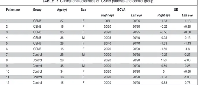
The clinical characteristics of the six CSNB patients and control group are summarized in the (Table 1). There were no significant differences in the patient ages and spherical equivalents values. Snellen best-corrected visual acuity for patients with CSNB ranged from 20/40 to 20/20 and patients ranged in age from 15 to 36 years and 15 to 45 years in control groups. All study patients had unmarkable fundus biomicroscopic examination re-sults. All patients’ anterior segments findings were normal, none of patients had nystagmus or strabis-mus. Pale optic disc was observed in two patients. On FAF imaging eyes with CSNB demonstrated
Patient no Group Age (y) Sex BCVA SE
Right eye Left eye Right eye Left eye
1 CSNB 27 F 20/4 20/25 -1.38 -1.13 2 CSNB 16 F 20/20 20/20 +0.25 +0.25 3 CSNB 35 F 20/20 20/25 +0.50 +0.50 4 CSNB 36 M 20/25 20/40 -0.25 -0.13 5 CSNB 28 F 20/40 20/40 -1.63 `-1.13 6 CSNB 15 F 20/20 20/20 -1.50 -1.8 7 Control 25 M 20/20 20/20 +0.25 -0.25 8 Control 28 F 20/20 20/20 1.50 -2.00 9 Control 45 M 20/20 20/20 -0.50 -0.25 10 Control 34 F 20/20 20/20 0 +0.50 11 Control 18 F 20/20 20/20 -1.88 -1.38 12 Control 15 F 20/20 20/20 -0.63 -0.75
TABLE 1: Clinical characteristics of CSNB patients and control group.
unremarkable fundi including normal distribution of autofluorescence in the (Figure 1).
Central 30° visual fields showed sensitivities within normal range values throughout the visual field for all patients.
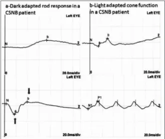
Full-field ERG demonstrated scotopic ERG a-wave was normal but the b-a-wave severely reduced and b-wave to a-wave amplitude ratio of less than one in the combined rod–cone response which de-scribes electronegative ERG. Light adapted cone re-sponses and 30 Hz flicker rere-sponses were normal. Representative ERG waveforms of Patient 4 and a normal subject are shown in the (Figure 2 and Fig-ure 3).
All CSNB patients demonstrated a selective re-duction of the b-wave that produces a b-wave to a-wave amplitude ratio of less than one in the scotopic bright-flash combined rod–cone full-field ERG response. This ERG finding describes elec-tronegative response on maximal full-field scotopic ERG.
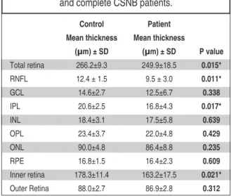
SD-OCT segmentation reveals a decrease in the total retina, RNFL, IPL and inner retinal thick-nesses in the CSNB group (Figure 4). In the other layers of retina, we observe a thinning in the CSNB group, but this difference was not statistically sig-nificant. In the control and patient groups respec-tively the total retinal thickness was 266.2 ± 9.3μm and 249.9 ± 18.5 μm, the RNFL thickness was 12.4 ± 1.5μm and 9.5 ± 3.0μm, the GCL thickness was 14.6±2.7µm and 12.5±6.7µm, the IPL thickness was 20.6 ± 2.5μm and 16.8 ± 4.3μm, the INL thickness was 18.4±3.1µm and 17.5±5.8µm, the OPL thick-ness was 23.4±3.7µm and 22.0±4.8µm, the ONL
thickness was 90.0 ±4.8 μm and 86.4 ± 8.8 μm, RPE thickness was 16.8 ± 1.5 μm and 16.4 ± 2.3 μm, the inner retinal thickness was 178.3 ± 11.4 μm and 163.2 ± 17.5 μm, the outer retinal thickness was 88.0±2.7μm and 86.9±2.8μm. Retinal thicknesses of all patients were summarized in the (Table 2).
DISCUSSION
In this study, we aim to describe six complete type CSNB patients’ clinical findings, electrophysiolog-ical tests and OCT findings and compare with the healthy subjects. According to the full field ERG results, all of our patients diagnosed with complete type CSNB. There was no detectable b-wave in the FIGURE 1: Color fundus photograph (left) and fundus autofluorescence
image (right) from right eye of Patient 4. There are normal distribution of au-tofluorescence and normal optic disc and fundus appearance.
FIGURE 2: Normal electroretinography of control patient 10.The amplitude of b-wave is larger than a-wave.
FIGURE 3: Full field electroretinography of patient 4. There are an electro-negative configuration in the combined rod-cone response. A-wave is larger than the b wave. This is consistent with the electronegative ERG.
rod-specific ERG, whereas the cone and rod-cone a-waves were normal in all CSNB patients.
In this study we observed significant thinning in total retina, RNFL, IPL and inner retinal thick-nesses in complete CSNB patients compared to con-trols. The control group used for this study consisted of healthy mild myopic subjects designed to match myopic patients in CSNB group.
Formerly, in incomplete CSNB patients, Schatz et al..and Chen at al..were described outer retinal thinning especially in the outer plexiform and photoreceptor layers.1,9
Al Oreany et al. were analyzed SD-OCT find-ings of two affected twin brothers diagnosed with complete CSNB with TRPM1 mutations and com-pared them with five myopic control eyes.7They did not find significant thinning in total retinal thickness compared to control group; however, they observed relative thinning of INL compared to other retinal layers.
Godora et al. were analyzed Cirrus HD-OCT ( Carl Zeiss Meditec, Inc, Dublin, California, USA) findings of three patients with complete CSNB caused by mutations in GRM6 and compared them with 93 healty subjects.10They observed reduced total retinal thickness in complete congenital sta-tionary night blindness patients. These three pa-tients had normal outer retinal layer thicknesses however they had reduced GCL+IPL thicknesses compare to control eyes.
Based on our SD-OCT segmentation results we suppose that the thinning in RNFL and IPL cause to decreased total retinal and the inner retinal thick-nesses. Of our patients had pale optic disc and al-most all of our patients had thinner RNFL and IPL thicknesses than age and myopia matched controls. The reason of pale optic disc is not known in CSNB. Al Oreany et al. were related optic disc hypoplasia with possibly disturbed synaptogenesis problem between bipolar cells and ganglion cells.7
Our report had some limitations. We could not make molecular genetic screening to our patients because of financial inadequacy of families how-FIGURE 4: Spectral-domain optical coherence tomography segmentation image of right eye of Patient 4. RNFL: Retinal nerve fiber layer; GCL: Ganglion cell layer; IPL: Inner plexiform layer; INL: Inner nuclear layer; OPL: Outer plexiform layer; ONL: Outer nuclear layer; ELM: External limiting membrane; RPE: Re-tina pigment epithelium.
Control Patient
Mean thickness Mean thickness
(µm) ± SD (µm) ± SD P value Total retina 266.2±9.3 249.9±18.5 0.015* RNFL 12.4 ± 1.5 9.5 ± 3.0 0.011* GCL 14.6±2.7 12.5±6.7 0.338 IPL 20.6±2.5 16.8±4.3 0.017* INL 18.4±3.1 17.5±5.8 0.639 OPL 23.4±3.7 22.0±4.8 0.429 ONL 90.0±4.8 86.4±8.8 0.235 RPE 16.8±1.5 16.4±2.3 0.609 Inner retina 178.3±11.4 163.2±17.5 0.021* Outer Retina 88.0±2.7 86.9±2.8 0.312
TABLE 2: The retinal layer thicknesses in the control and complete CSNB patients.
RNFL: Retinal nerve fiber layer GCL: Ganglion cell layer IPL: Inner plexiform layer INL: Inner nuclear layer OPL: Outer plexiform layer ONL: Outer nuclear layer RPE: Retina pigment epithelium.
ever we did not take any complain about night blindness in none of our patients family members. Today, TRPM1, GPR179, GRM6, LRIT3, and NYX genes have been identified as associated with com-plete type CSNB.11
All are caused by defects in visual signal trans-duction within rod photoreceptors or in defective photoreceptor to bipolar cell signaling.
In our study we observed selective thinning of RNFL and IPL in our patients. The IPL consists of synaptic connections between the axons of bipolar cells and dendrites of ganglion cells. As a result of our study, we thought that thinning of the IPL and RNFL and possibly optic disc paleness in complete CSNB patients suggests bipolar cell dysfunction or synaptogenesis defect between bipolar cells and ganglion cells and reduced number of these cells.
S
Soouurrccee ooff FFiinnaannccee
During this study, no financial or spiritual support was received neither from any pharmaceutical company that has a direct
connection with the research subject, nor from a company that provides or produces medical instruments and materials which may negatively affect the evaluation process of this study.
C
Coonnfflliicctt ooff IInntteerreesstt
No conflicts of interest between the authors and / or family members of the scientific and medical committee members or members of the potential conflicts of interest, counseling, ex-pertise, working conditions, share holding and similar situa-tions in any firm.
A
Auutthhoorrsshhiipp CCoonnttrriibbuuttiioonnss
I
Iddeeaa//CCoonncceepptt:: Fevzi Şentürk, Rukiye Aydın, Merve Özbek; DDee--s
siiggnn:: Rukiye Aydın, Fevzi Şentürk, Merve Özbek; CCoonnttrrooll//SSuu--p
peerrvviissiioonn:: Merve Özbek, Rukiye Aydın, Fevzi Şentürk; DDaattaa C
Coolllleeccttiioonn aanndd//oorr PPrroocceessssiinngg:: Merve Özbek, Rukiye Aydın, Fevzi Şentürk; AAnnaallyyssiiss aanndd//oorr IInntteerrpprreettaattiioonn:: Fevzi Şentürk, Rukiye Aydın, Merve Özbek; LLiitteerraattuurree RReevviieeww:: Rukiye Aydın, Fevzi Şentürk, Merve Özbek; WWrriittiinngg tthhee AArrttiiccllee:: Merve Özbek, Rukiye Aydın, Fevzi Şentürk; CCrriittiiccaall RReevviieeww:: Rukiye Aydın, Fevzi Şentürk, Merve Özbek; RReeffeerreenncceess aanndd F
Fuunnddiinnggss:: Rukiye Aydın, Fevzi Şentürk, Merve Özbek; M Maattee--r
riiaallss:: Fevzi Şentürk, Rukiye Aydın, Merve Özbek.
1. Chen RW, Greenberg JP, Lazow MA, Ra-machandran R, Lima LH, Hwang JC, et al. Autofluorence imaging and spectral domain optical coherence tomography in incomplete congenital stationary night blindness and com-parison with retinitis pigmentosa. Am J Oph-thalmol 2012;153(1):143-54.e2.
2. Schubert G, Bornschein H. [Analysis of the human electroretinogram]. Ophthalmologica 1952;123(6):396-413.
3. Tan X, Aoki A, Yanagi Y. Color vision abnor-mality as an initial presentation of the com-plete type of congenital stationary night blindness. Clin Ophthalmol 2013;7:1587-90. 4. Miyake Y, Yagasaki K, Horiguchi M, Kawase
Y, Kanda T. Congenital stationary night blind-ness with negative electroretinogram: a new classification. Arch Ophthalmol 1986;104(7): 1013-20.
5. Kabanarou SA, Holder GE, Fitzke FW, Bird AC, Webster AR. Congenital stationary night blindness and a “Schubert-Bornschein” type electrophysiology in a family with dominant in-heritance. Br J Ophthalmol 2004;88(8):1018-22.
6. Miyake Y, Yagasaki K, Horiguchi M, Kawase Y. On- and off-responses in photopic elec-troretinogram in complete and incomplete types of congenital stationary night blindness. Jpn J Ophthalmol 1987;31(1):81-7. 7. Al Oreany AA, Al Hadlaq A, Schatz P.
Con-genital stationary night blindness with hy-poplastic discs, negative electroretinogram and thinning of the inner nuclear layer. Grae-fes Arch Clin Exp Ophthalmol 2016;254(10): 1951-6.
8. Marmor MF, Fulton AB, Holder GE, Miyake Y, Brigell M, Bach M. ISCEV Standard for
full-field clinical electroretinography (2008 update). Doc Ophthalmol 2009;118(1):69-77.
9. Schatz P, Abdalla Elsayed MEA, Khan AO. Multimodal imaging in CABP4-related reti-nopathy. Ophthalmic Genet 2017;38(5):459-64.
10. Godara P, Cooper RF, Diederichs MA, Ser-gouniotis P, Genead MA, Webster AR, et al. Assessing photoreceptor reflectance and structure in congenital stationary night blind-ness. Invest Ophthalmol Vis Sci 2012;53(14): 5256.
11. Malaichamy S, Sen P, Sachidanandam R, Arokiasamy T, Lancelot ME, Audo I, et al. Molecular profiling of complete conge-nital stationary night blindness: a pilot study on an Indian cohort. Mol Vis 2014;20:341-51.